

Hyperconnectivity of the ventromedial prefrontal cortex in obsessive-compulsive disorder
- PMID: 31742235
- PMCID: PMC6861127
- DOI: 10.1177/2398212818808710
Hyperconnectivity of the ventromedial prefrontal cortex in obsessive-compulsive disorder
Abstract
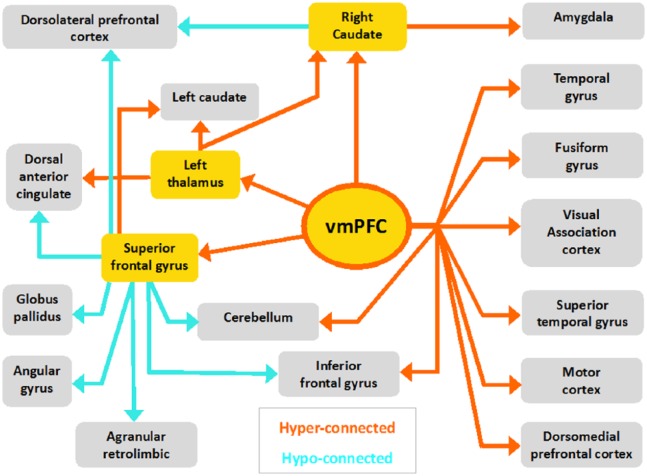
Neuroimaging research has highlighted maladaptive thalamo-cortico-striatal interactions in obsessive-compulsive disorder as well as a more general deficit in prefrontal functioning linked with compromised executive functioning. More specifically, dysfunction in the ventromedial prefrontal cortex, a central hub in coordinating flexible behaviour, is thought to be central to obsessive-compulsive disorder symptomatology. We sought to determine the intrinsic alterations of the ventromedial prefrontal cortex in obsessive-compulsive disorder employing resting-state functional connectivity magnetic resonance imaging analyses with a ventromedial prefrontal cortex seed region of interest. A total of 38 obsessive-compulsive disorder patients and 33 matched controls were included in our analyses. We found widespread ventromedial prefrontal cortex hyperconnectivity during rest in patients with obsessive-compulsive disorder, displaying increased connectivity with its own surrounding region in addition to hyperconnectivity with several areas along the thalamo-cortico-striatal loop: thalamus, caudate and frontal gyrus. Obsessive-compulsive disorder patients also exhibited increased functional connectivity from the ventromedial prefrontal cortex to temporal and occipital lobes, cerebellum and the motor cortex, reflecting ventromedial prefrontal cortex hyperconnectivity in large-scale brain networks. Furthermore, hyperconnectivity of the ventromedial prefrontal cortex and caudate correlated with obsessive-compulsive disorder symptomatology. Additionally, we used three key thalamo-cortico-striatal regions that were hyperconnected with our ventromedial prefrontal cortex seed as supplementary seed regions, revealing hypoconnectivity along the orbito- and lateral prefrontal cortex-striatal pathway. Taken together, these results confirm a central role of a hyperconnected ventromedial prefrontal cortex in obsessive-compulsive disorder, with a special role for maladaptive crosstalk with the caudate, and indications for hypoconnectivity along the lateral and orbito pathways.
Keywords: Ventromedial prefrontal cortex; functional magnetic resonance imaging; neuroimaging; obsessive-compulsive disorder; prefrontal cortex; resting state.
Conflict of interest statement
Declaration of conflicting interests The author(s) declared no potential conflicts of interest with respect to the research, authorship and/or publication of this article.
Figures


References
-
- Abramovitch A, Dar R, Schweiger A, et al. (2011) Neuropsychological impairments and their association with obsessive-compulsive symptom severity in obsessive-compulsive disorder. Archives of Clinical Neuropsychology 26(4): 364–376. - PubMed
-
- Abramovitch A, Mittelman A, Tankersley AP, et al. (2015) Neuropsychological investigations in obsessive-compulsive disorder: A systematic review of methodological challenges. Psychiatry Research 228(1): 112–120. - PubMed
-
- Alexander GE, DeLong MR, Strick PL. (1986) Parallel organization of functionally segregated circuits linking basal ganglia and cortex. Annual Review of Neuroscience 9(1): 357–381. - PubMed
-
- American Psychiatric Association (2013) Diagnostic and Statistical Manual of Mental Disorders. 5th ed. Washington, DC: American Psychiatric Association.
Grants and funding
LinkOut - more resources
Full Text Sources
