

Case Reports
doi: 10.2340/00015555-3371.
A Case of Four Synchronous Cutaneous Melanomas: Melanocortin 1 Receptor Polymorphisms and Excessive Sun Exposure
Affiliations
- PMID: 31742646
- PMCID: PMC9128902
- DOI: 10.2340/00015555-3371
Item in Clipboard
Case Reports
A Case of Four Synchronous Cutaneous Melanomas: Melanocortin 1 Receptor Polymorphisms and Excessive Sun Exposure
Acta Derm Venereol.
.
No abstract available
Figures
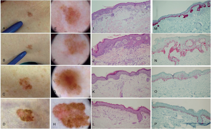
Clinical, dermoscopic, histopathological and immunohistochemical Melan-A staining of the 4 synchronous melanomas. (A, E, I, M) Melanoma of the left dorsum; (B, F, J, N) melanoma of the left shoulder; (C, G, K, O) melanoma of the right leg; and (D, H, L, P) melanoma of the right thigh. (A–D) All lesions shared similar clinical findings, presenting as light-brown macules with irregular borders and red-pink-white. (E–H) Dermoscopy revealed common features in all 4 lesions. (E) Multicomponent pattern, milky red areas, a regression area, shiny white streaks. (F) Multicomponent pattern, brownish homogenous areas, regression areas, and polymorphic vessels. (G) Multicomponent pattern, central brownish homogenous area, regression area with peppering. (H) Multicomponent milky red area, shiny streaks and polymorphic vessels. (I–P) Histopathological and immunohistochemical images (haematoxylin-eosin stain, Melan-A stain; original magnification ×10). All 4 melanocytic lesions were large (>10 mm of maximum diameter). They consisted of epidermal atypical melanocytic proliferation with a predominant lentiginous growth pattern and focally with pagetoid extension of atypical melanocytes up to the granular layer. For both the melanocytic atypia and the proliferation pattern, a diagnosis of melanoma was confirmed. Immunohistochemical assay for Melan-A helped in displaying the proliferation pattern.
Similar articles
-
Phenotypic and histologic characteristics of cutaneous melanoma in patients with melanocortin-1 receptor polymorphisms.Actas Dermosifiliogr. 2012 Jan;103(1):44-50. doi: 10.1016/j.adengl.2011.04.006. Epub 2012 Mar 29. Actas Dermosifiliogr. 2012. PMID: 22464597
-
High incidence of primary melanomas in an MC1R RHC homozygote/CDKN2A mutant genotype patient.Arch Dermatol Res. 2015 Oct;307(8):741-5. doi: 10.1007/s00403-015-1582-y. Epub 2015 Jun 24. Arch Dermatol Res. 2015. PMID: 26103950
-
MC1R germline variants confer risk for BRAF-mutant melanoma.Science. 2006 Jul 28;313(5786):521-2. doi: 10.1126/science.1127515. Epub 2006 Jun 29. Science. 2006. PMID: 16809487
-
Melanocortin-1 receptor: loss of function mutations and skin cancer.Dermatol Online J. 2006 Sep 8;12(5):13. Dermatol Online J. 2006. PMID: 16962028 Review.
-
[MAP-Kinase pathway abnormalities in melanoma: B-RAF is not the sole cause].Ann Dermatol Venereol. 2012 Oct;139(10):691-2. doi: 10.1016/j.annder.2012.04.157. Epub 2012 May 31. Ann Dermatol Venereol. 2012. PMID: 23122387 Review. French. No abstract available.
Cited by
-
Synchronizing the Nomenclature Surrounding Synchronous Primary Cutaneous Melanomas: A Systematic Review.J Clin Aesthet Dermatol. 2024 Aug;17(8):44-49. J Clin Aesthet Dermatol. 2024. PMID: 39148963 Free PMC article. Review.
References
-
- De Giorgi V, Salvini C, Sestini S, Vignoli M, Sestini R, Papi F, et al. . Triple synchronous cutaneous melanoma: a clinical, dermoscopic, and genetic case study. Dermatol Surg 2007; 33: 488–489. - PubMed
-
- Goggins WB, Tsao H. A population-based analysis of risk factors for a second primary cutaneous melanoma among melanoma survivors. Cancer 2003; 97: 639–643. - PubMed
-
- Hwa C, Price LS, Belitskaya-Levy I, Ma MW, Shapiro RL, Berman RS, et al. . Single versus multiple primary melanomas: old questions and new answers. Cancer 2012; 118: 4148–4192. - PubMed
-
- Zell D, Kim N, Olivero M, Elgart G, Rabinovitz H. Early diagnosis of multiple primary amelanotic/hypomelanotic melanoma using dermoscopy. Dermatol Surg 2008; 34: 1254–1257. - PubMed
-
- Aoude LG, Wadt KA, Pritchard AL, Hayward NK. Genetics of familial melanoma: 20 years after CDKN2A. Pigment Cell Melanoma Res 2015; 28: 148–160. - PubMed
Publication types
MeSH terms
Substances
LinkOut - more resources
Full Text Sources
Medical
