. 2020 Jan;24(1):886-898.
doi: 10.1111/jcmm.14798. Epub 2019 Nov 19.

N-acetylcysteine prevents oxidized low-density lipoprotein-induced reduction of MG53 and enhances MG53 protective effect on bone marrow stem cells

Affiliations

N-acetylcysteine prevents oxidized low-density lipoprotein-induced reduction of MG53 and enhances MG53 protective effect on bone marrow stem cells

Xin Li et al. J Cell Mol Med. 2020 Jan.

Abstract

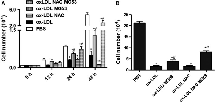
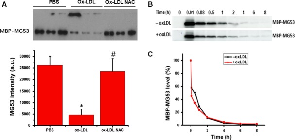
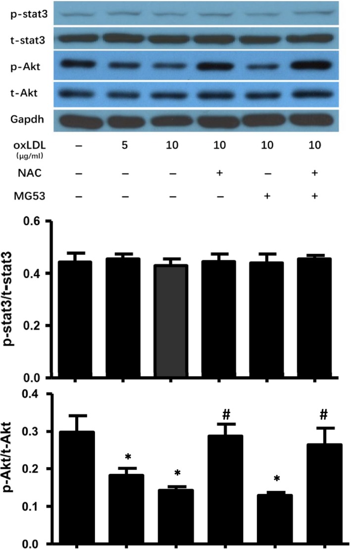
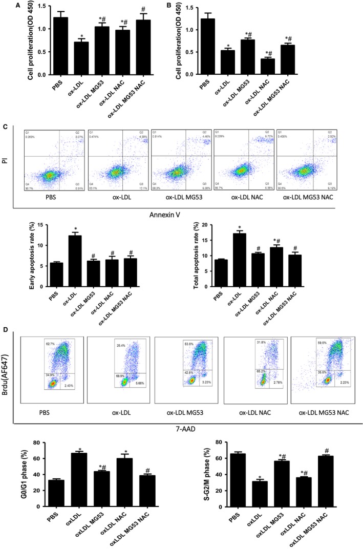
MG53 is an important membrane repair protein and partially protects bone marrow multipotent adult progenitor cells (MAPCs) against oxidized low-density lipoprotein (ox-LDL). The present study was to test the hypothesis that the limited protective effect of MG53 on MAPCs was due to ox-LDL-induced reduction of MG53. MAPCs were cultured with and without ox-LDL (0-20 μg/mL) for up to 48 hours with or without MG53 and antioxidant N-acetylcysteine (NAC). Serum MG53 level was measured in ox-LDL-treated mice with or without NAC treatment. Ox-LDL induced significant membrane damage and substantially impaired MAPC survival with selective inhibition of Akt phosphorylation. NAC treatment effectively prevented ox-LDL-induced reduction of Akt phosphorylation without protecting MAPCs against ox-LDL. While having no effect on Akt phosphorylation, MG53 significantly decreased ox-LDL-induced membrane damage and partially improved the survival, proliferation and apoptosis of MAPCs in vitro. Ox-LDL significantly decreased MG53 level in vitro and serum MG53 level in vivo without changing MG53 clearance. NAC treatment prevented ox-LDL-induced MG53 reduction both in vitro and in vivo. Combined NAC and MG53 treatment significantly improved MAPC survival against ox-LDL. These data suggested that NAC enhanced the protective effect of MG53 on MAPCs against ox-LDL through preventing ox-LDL-induced reduction of MG53.

Keywords: MG53; N-acetylcysteine; bone marrow stem cell; membrane damage; multipotent adult progenitor cells; oxidized low-density lipoprotein.


Conflict of interest statement

None.

Figures

Figure 1
Figure 1
Interaction between N‐acetylcysteine (NAC) and rhMG53 on MAPCs in the presence of ox‐LDL. Normal growth of MAPCs was observed under the standard culture conditions. The number of MAPCs in the culture system was significantly decreased in the presence of ox‐LDL. When ox‐LDL concentration was at 10 μg/mL, NAC treatment (1 mmol/L) did not prevent ox‐LDL‐induced reduction of the number of MAPCs. Treatment with rhMG53 (at EC50 level) significantly improved the cell number in the presence of 10 μg/mL ox‐LDL, but not completely restored the cell number to the control level. Treatment with rhMG53 in combination with NAC almost completely restored the cell number to the control level (BSA). NAC treatment did not prevent ox‐LDL‐induced reduction in cell number at 24 h, and yet, NAC further decreased ox‐LDL‐induced reduction in cell number at 48 h (A). When ox‐LDL concentration was increased to 20 μg/mL, the number of MAPCs was only about 1/10 of that cultured with PBS or Sat‐LDL (control) after 24 h. Treatment with NAC did not improve ox‐LDL‐induced reduction of cell number at 24 h. However, compared with BSA control, treatment with rhMG53 doubled the cell number and further increased the cell number when both NAC and MG53 were present (B). PBS: cells treated with PBS or sat‐LDL (control); ox‐LDL: cells treated with ox‐LDL; ox‐LDL + NAC: cells treated with ox‐LDL and NAC; ox‐LDL + MG53: cells treated with ox‐LDL and MG53; and ox‐LDL + NAC+MG53: cells treated with ox‐LDL and NAC as well as MG53. Data were presented as means ± SEM. *P < .01 as compared with control, # P < .01 as compared with ox‐LDL (n = 3 independent experiments)
Figure 2
Figure 2
Effect of N‐acetylcysteine (NAC) on the level of rhMG53 protein in vitro and in vivo with and without ox‐LDL. MG53 level was determined in culture system in the presence of ox‐LDL with and without NAC. After 12 h of incubation in the culture environment for MAPCs, a detectable amount of rhMG53 protein was present in the culture media. Ox‐LDL (10 μg/mL) significantly reduced the level of rhMG53 protein in the culture system with or without MAPCs. The presence of NAC (1 mmol/L) effectively prevented ox‐LDL‐induced reduction of rhMG53 protein level in the culture system (A). Serum levels of MG53 protein in wild‐type (WT) mice were determined with Western blotting after three consecutive days of tail vein injection of ox‐LDL with or without NAC treatment. Serum MG53 was readily detectable in WT mice and was significantly decreased in mice treated with ox‐LDL. Pre‐treatment of the mice with NAC effectively prevented ox‐LDL‐induced decrease in serum MG53 level (as pointed with the arrow, B). Of note, serum MG53 level in male hyperlipidemic LDLR(‐/‐) mice after 8 wk of high‐fat diet was significantly lower than the serum MG53 level in male age‐matched WT mice (C). PBS: cells treated with PBS or sat‐LDL (control); ox‐LDL: cells treated with ox‐LDL; ox‐LDL + NAC: cells treated with ox‐LDL and NAC; NAC: cells treated with NAC (control); WT: control mice treated with PBS or sat‐LDL; WT + ox‐LDL: mice treated with ox‐LDL; and WT + ox‐LDL + NAC: mice treated with ox‐LDL and NAC. LDLR(‐/‐): hyperlipidemic LDL receptor knockout mice with 8 wk of high‐fat diet. Data were presented as means ± SEM. *P < .01 as compared with control, # P < .01 as compared with ox‐LDL (n = 3 independent experiments)
Figure 3
Figure 3
Effect of ox‐LDL on MG53 clearance in vivo. MBP‐MG53 was generated to determine MG53 clearance in vivo. Prior to injection into mice, in vitro experiments showed that ox‐LDL (10 μg/mL) significantly decreased the concentration of MBP‐MG53 after 12 h of incubation, and NAC treatment completely prevented ox‐LDL‐induced reduction of MBP‐MG53 protein (A). After injection through tail vein, serum concentration of MBP‐MG53 was decreased rapidly over time. At 30 min after injection, less than half of the injected MG53 protein was present in the circulation. After 2 h, only 10% of the injected MG53 protein remained in the blood. By 6 h, the injected protein was completely removed from the blood. Treating mice with ox‐LDL (once a day for 3 d via tail vein) had no significant effect on MG53 clearance from circulation (B). MBP‐MG53: maltose‐binding protein‐conjugated MG53; PBS: cells treated with PBS or sat‐LDL (control); ox‐LDL: cells treated with ox‐LDL; ox‐LDL + NAC: cells treated with ox‐LDL and NAC; ox‐LDL: wild‐type mice treated with ox‐LDL; and ‐ox‐LDL: control mice without ox‐LDL treatment. Data were presented as means ± SD. *P < .01 as compared with control, # P < .01 as compared with ox‐LDL (n = 3 independent experiments)
Figure 4
Figure 4
Effect of NAC on ox‐LDL‐induced membrane damage of bone marrow stem cells in vitro. Membrane integrity was evaluated by monitoring the entry of fluorescent FM1‐43 dye into the cells in the presence of ox‐LDL (10 μg/mL) with or without NAC treatment. A significant amount of FM1‐43 dye was detected 6 hours after incubation with ox‐LDL using confocal microscope. There was no significant difference in FM1‐43 dye entry into the cells when exposed to ox‐LDL with or without NAC treatment (A). Quantitative and dynamic analysis with a quantitative live cell imaging assay showed that exposure to ox‐LDL (10 μg/mL) dramatically increased FM1‐43 dye accumulation inside the cells that was not significantly changed with NAC treatment. The dynamic of FM1‐43 dye entry was analysed by imageJ (more than 200 cells were analysed for each condition and each experiment) (B). On the other hand, treatment with rhMG53 (either alone or with NAC) significantly reduced FM1‐43 dye entry into and accumulation inside the cells (A and B). PBS: cells treated with PBS or sat‐LDL (control); ox‐LDL: cells treated with ox‐LDL; ox‐LDL + NAC: cells treated with ox‐LDL and NAC; ox‐LDL + MG53: cells treated with ox‐LDL and MG53; and ox‐LDL + NAC + MG53: cells treated with ox‐LDL and NAC as well as MG53. Data were presented as means ± SEM. *P < .01 as compared with control, # P < .01 as compared with ox‐LDL (n = 3‐4 independent experiments, scale bar: 10 μm)
Figure 5
Figure 5
Effect of NAC and MG53 treatment on Akt phosphorylation in bone marrow stem cells in the presence of ox‐LDL in vitro. Intracellular levels of total and phosphorylated Akt and STAT3 were evaluated in MAPCs after exposure to ox‐LDL with and without NAC and/ or MG53 treatment. Western blotting analysis showed that ox‐LDL selectively decreased Akt phosphorylation without change in total Akt and total or phosphorylated STAT3 in MAPCs. NAC treatment completely prevented ox‐LDL‐induced inhibition of Akt phosphorylation in MAPCs. On the other hand, MG53 treatment did not change the levels of Akt or STAT3 expression in MAPCs exposed to ox‐LDL. PBS: cells treated with PBS or sat‐LDL (control); ox‐LDL: cells treated with ox‐LDL; ox‐LDL + NAC: cells treated with ox‐LDL and NAC; ox‐LDL + MG53: cells treated with ox‐LDL and MG53; and ox‐LDL + NAC+MG53: cells treated with ox‐LDL and NAC as well as MG53. Data were presented as means ± SEM. *P < .01 as compared with control, # P < .01 as compared with ox‐LDL (n = 3 independent experiments)
Figure 6
Figure 6
Effects of NAC on cell proliferation, apoptosis and cell cycle in the presence of ox‐LDL. MAPCs were incubated with ox‐LDL for 24 hours in the presence of MG53 with or without NAC. Ox‐LDL significantly inhibited the proliferation of MAPCs (A and B), induced apoptosis of MAPCs (C) and arrested the cell cycle at G0/G1 phase (D). Treatment with MG53 partially reversed ox‐LDL‐induced inhibition of cell proliferation (A and B), effectively prevented ox‐LDL‐induced apoptosis (C) and largely reversed ox‐LDL‐induced cell cycle arrest (D). NAC treatment significantly prevented ox‐LDL‐induced inhibition of cell proliferation and blocked ox‐LDL‐induced early apoptosis when ox‐LDL concentration was at 5 μg/mL. However, when ox‐LDL concentration was increased to 10 μg/mL, NAC treatment further increased ox‐LDL‐induced inhibition of cell proliferation (A) while having no effect on ox‐LDL‐induced cell cycle arrest (D). No significant differences in proliferation, apoptosis and cell cycle of MAPCs were observed when the cells were treated with MG53 alone or with combination of NAC and MG53 in the presence of ox‐LDL at 10 μg/mL (A‐D). PBS: cells treated with PBS or sat‐LDL (control); ox‐LDL: cells treated with ox‐LDL; ox‐LDL + NAC: cells treated with ox‐LDL and NAC; ox‐LDL + MG53: cells treated with ox‐LDL and MG53; and ox‐LDL + NAC+MG53: cells treated with ox‐LDL and NAC as well as MG53. Data were presented as means ± SEM. *P < .01 as compared with control, # P < .01 as compared with ox‐LDL (n = 3‐4 independent experiments)

Similar articles

Cited by

References

    1. Mundra V, Gerling IC, Mahato RI. Mesenchymal stem cell‐based therapy. Mol Pharm. 2013;10:77‐89. - PMC - PubMed
    1. Yi T, Song SU. Immunomodulatory properties of mesenchymal stem cells and their therapeutic applications. Arch Pharm Res. 2012;35(2):213‐221. - PubMed
    1. Lalu MM, Mclntyre L, Pugliese C, et al. Safety of cell therapy with mesenchymal stromal cells (SafeCell): a systematic review and meta‐analysis of clinical trials. PLoS ONE. 2012;7(10):e47559. - PMC - PubMed
    1. Abdelwahid E, Kalvelyte A, Stulpinas A, et al. Stem cell death and survival in heart regeneration and repair. Apoptosis. 2016;21(3):252‐268. - PMC - PubMed
    1. Chen Z, Chen L, Zeng C, et al. Functionally improved mesenchymal stem cells to better treat myocardial infarction. Stem Cells Int. 2018;2018:1‐14. - PMC - PubMed

Publication types

MeSH terms