. 2020 Mar 2;130(3):1336-1349.
doi: 10.1172/JCI130808.

GABA interneurons are the cellular trigger for ketamine's rapid antidepressant actions

Affiliations

GABA interneurons are the cellular trigger for ketamine's rapid antidepressant actions

Danielle M Gerhard et al. J Clin Invest. .

Abstract

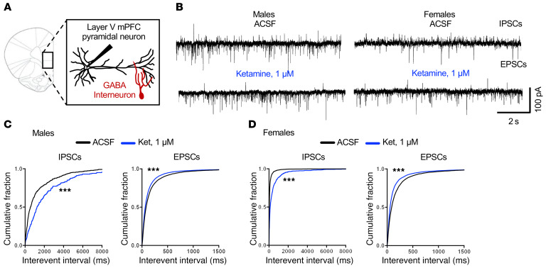
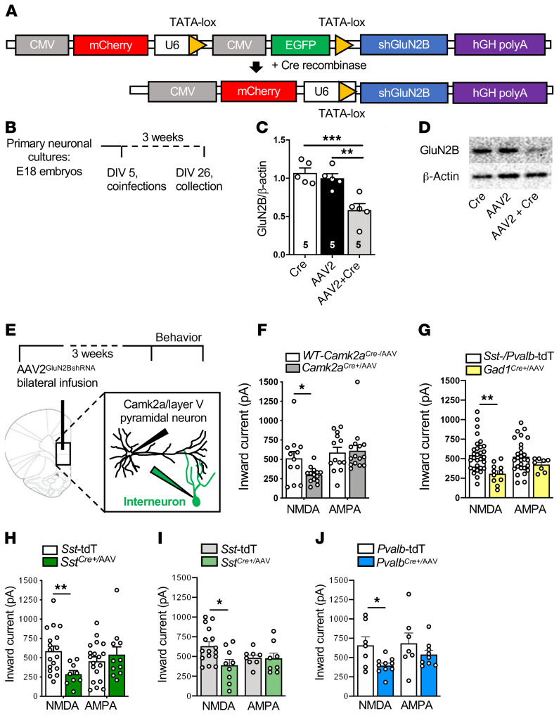
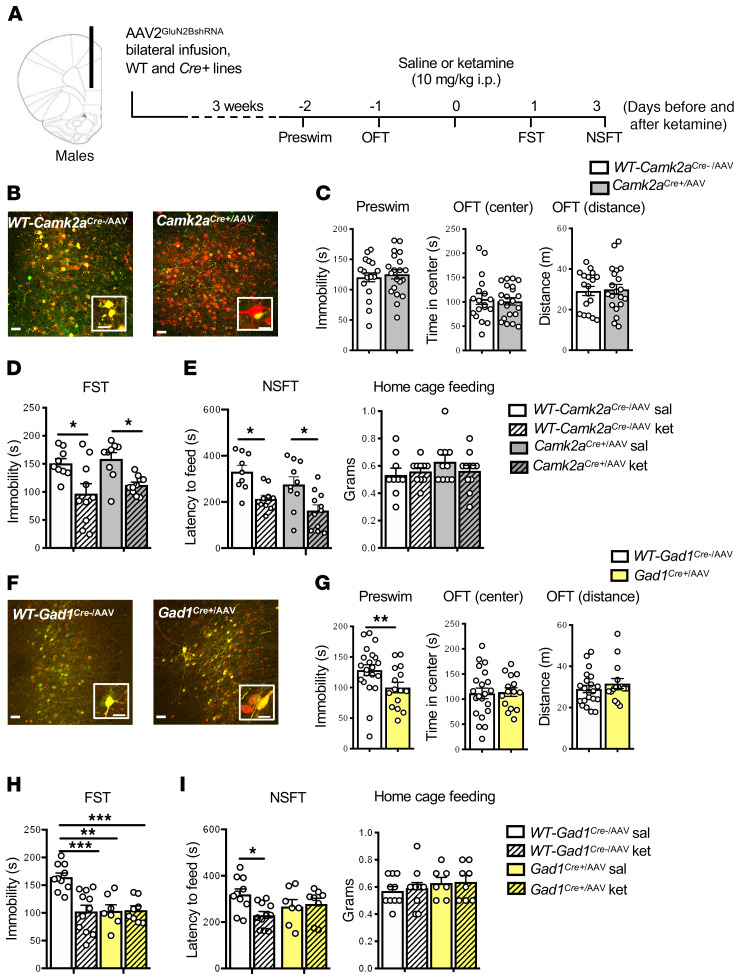
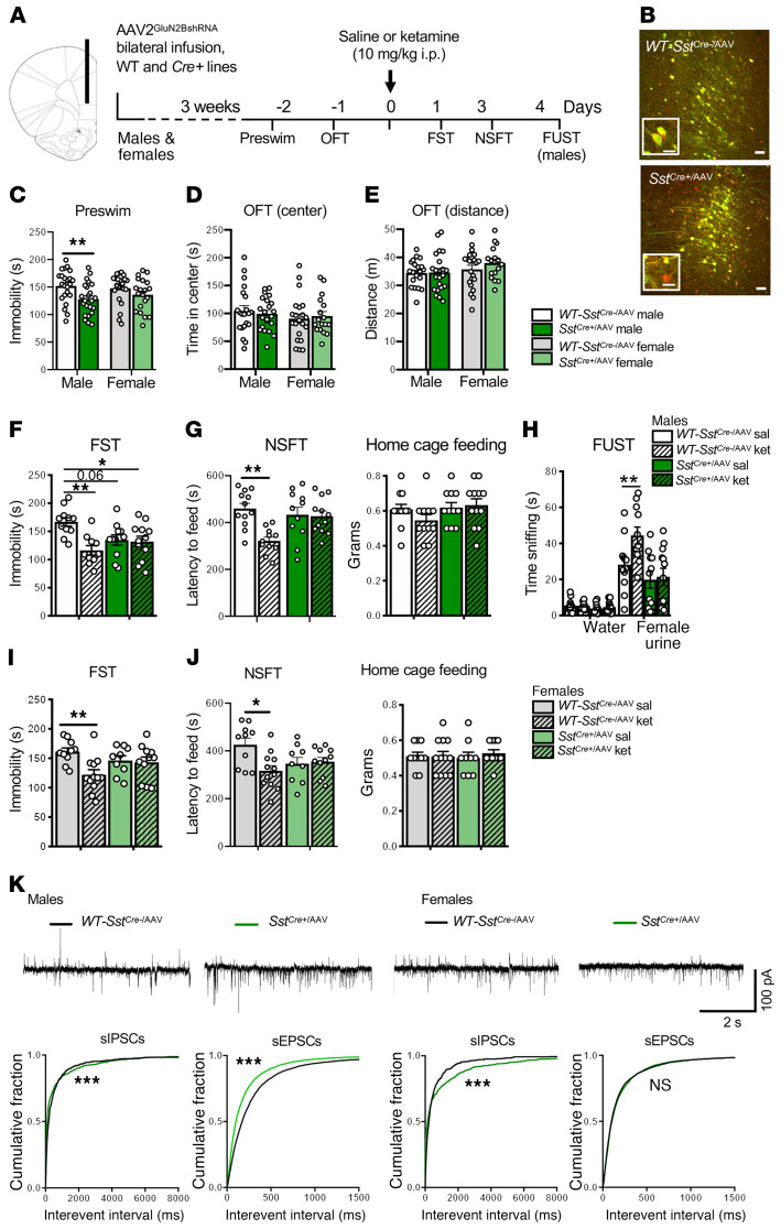
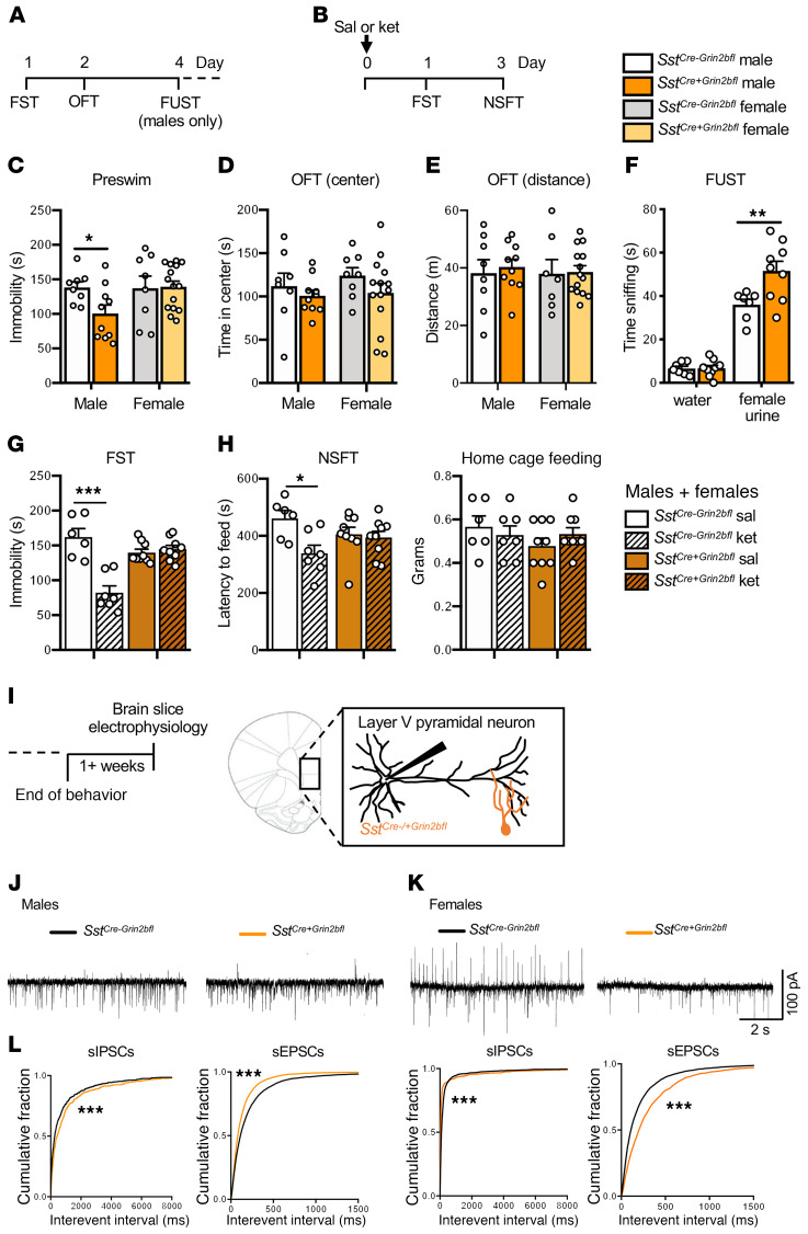
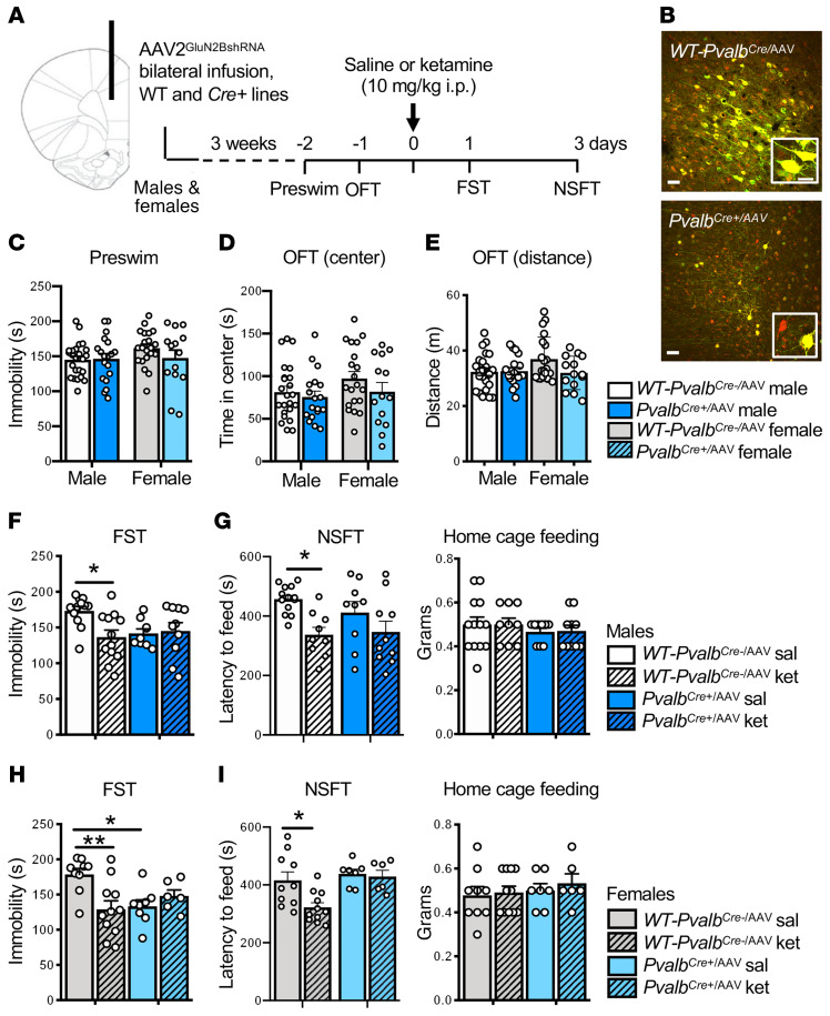
A single subanesthetic dose of ketamine, an NMDA receptor (NMDAR) antagonist, produces rapid and sustained antidepressant actions in depressed patients, addressing a major unmet need for the treatment of mood disorders. Ketamine produces a rapid increase in extracellular glutamate and synaptic formation in the prefrontal cortex, but the initial cellular trigger that initiates this increase and ketamine's behavioral actions has not been identified. To address this question, we used a combination of viral shRNA and conditional mutation to produce cell-specific knockdown or deletion of a key NMDAR subunit, GluN2B, implicated in the actions of ketamine. The results demonstrated that the antidepressant actions of ketamine were blocked by GluN2B-NMDAR knockdown on GABA (Gad1) interneurons, as well as subtypes expressing somatostatin (Sst) or parvalbumin (Pvalb), but not glutamate principle neurons in the medial prefrontal cortex (mPFC). Further analysis of GABA subtypes showed that cell-specific knockdown or deletion of GluN2B in Sst interneurons blocked or occluded the antidepressant actions of ketamine and revealed sex-specific differences that are associated with excitatory postsynaptic currents on mPFC principle neurons. These findings demonstrate that GluN2B-NMDARs on GABA interneurons are the initial cellular trigger for the rapid antidepressant actions of ketamine and show sex-specific adaptive mechanisms to GluN2B modulation.

Keywords: Depression; Neuroscience.


Conflict of interest statement

Conflict of interest: The authors have declared that no conflict of interest exists.

Figures

Figure 1
Figure 1. Ketamine reduces spontaneous inhibitory postsynaptic currents in mPFC slices.
(A) Brain slice electrophysiology schematic. (B) Representative traces of spontaneous inhibitory postsynaptic currents (sIPSCs) and spontaneous excitatory postsynaptic currents (sEPSCs) from male and female mice before and after application of 1 μM ketamine. (C) In male mice, 1 μM ketamine significantly decreased sIPSCs and increased sEPSCs (n = 9–10 cells, 10 mice, ***P < 0.001). (D) In female mice, 1 μM ketamine significantly decreased sIPSCs and increased sEPSCs (n = 7–8 cells, 5 mice, ***P < 0.001). Kolmogorov-Smirnov 2-sample test was used. Data represented as the cumulative probability of the interevent interval. Abbreviations: ACSF, artificial cerebrospinal fluid; Ket, ketamine.
Figure 2
Figure 2. AAV2GluN2BshRNA knocks down GluN2B and reduces NMDA inward currents in a Cre-dependent manner.
(A) Schematic of the pGluN2BshRNA knockdown construct before and after introduction of Cre recombinase to generate the active construct. (B) Schedule for cell culture experiments. (C) There was a significant reduction in GluN2B protein in the AAV2GluN2BshRNA+AAV1-Cre (AAV2-Cre) group compared with AAV2GluN2BshRNA (AAV2) or AAV2-Cre alone (Cre) (n = 5 per group, 1-way ANOVA with Tukey’s multiple-comparisons test, [F2,12 = 14.1, P < 0.001]). (D) Representative images of Western blots. Lanes were run on the same gel and were contiguous. (E) Schematic of the schedule for brain slice electrophysiology experiments. (FJ) The influence of AAV2GluN2BshRNA on NMDAR and AMPAR inward currents was tested across genotypes. There was a significant reduction in NMDA-induced inward currents in all genotypes compared with their controls. Unpaired 2-tailed t tests were performed. (F) Camk2aCre+/AAV (Camk2aCre+/AAV: n = 3 mice, 14–15 cells; WT-Camk2aCre+/AAV: n = 2 mice, 12–13 cells; t24 = 2.510, P = 0.019). (G) Gad1Cre+/AAV (Gad1Cre+/AAV: n = 5 mice, 8–11 cells; Sst-/Pvalb-Td-Tomato [TdT]: n = 10 mice, 28–30 cells; t39 = 3.447, P = 0.0014). (H) SstCre+/AAV males (SstCre+/AAV: n = 2 mice, 9–11 cells; Sst-TdT: n = 8 mice, 16–17 cells; t25 = 2.976, P = 0.006). (I) SstCre+/AAV females (SstCre+/AAV: n = 3 mice, 8–9 cells; Sst-TdT: n = 9 mice, 8–16 cells; t23 = 2.686, P = 0.013). (J) PvalbCre+/AAV males (PvalbCre+/AAV: n = 3 mice, 9–10 cells; Pvalb-TdT: n = 5 mice, 7 cells; t15 = 2.696, P = 0.017). There were no significant differences in AMPA-induced inward currents in any genotype. *P < 0.05; **P < 0.01; ***P < 0.001. All data are represented as mean ± SEM. Abbreviations: tdT, td-Tomato.
Figure 3
Figure 3. Infusion of AAV2GluN2BshRNA into mPFC of Gad1Cre+, but not Camk2aCre+, male mice occludes the antidepressant effects of ketamine.
(A) Procedure schematic. (B and F) Representative images of AAV2GluN2BshRNA-mediated expression and recombination in the mPFC of (B) WT-Camk2aCre–, Camk2aCre+, (F) WT-Gad1Cre–/AAV, and Gad1Cre+/AAV male mice. Scale bars: 50 μm and 20 μm (insets). (C) In Camk2aCre+/AAV mice, there was no effect of GluN2B knockdown on baseline immobility (preswim) or time spent in center and distance traveled in the open field test (OFT; n = 18–21 per group). (D) WT-Camk2aCre–/AAV-ket and Camk2aCre+/AAV-ket mice showed significantly reduced immobility in the forced swim test (FST) compared with saline controls (n = 12–14 per group, treatment: F1,34 = 18.18, P = 0.0002). (E) WT-Camk2aCre-/AAV-ket and Camk2aCre+/AAV-ket mice showed significantly reduced latency to feed in the novelty-suppressed feeding test (NSFT; n = 9–11 per group, treatment: F1,36 = 19.89, P < 0.0001, genotype: F1,36 = 4.186, P = 0.0481). No significant differences were found in home cage feeding. (G) There was a significant reduction in preswim immobility in Gad1Cre+/AAV mice, but no effect on time spent in center and distance traveled in the OFT (n = 15–21 per group, t34 = 2.226, P = 0.0327). (H) Only WT-Gad1Cre-/AAV-ket mice showed significantly reduced immobility in FST compared with their saline controls (n = 7–11 per group, genotype: F1,32 = 8.662, P = 0.006, treatment: F1,32 = 9.313, P = 0.0045 genotype times treatment: F1,32 = 10.04, P = 0.0034). (I) Only WT-Gad1Cre-/AAV-ket mice showed significantly reduced latency to feed in the NSFT (n = 7–11 per group, genotype times treatment: F1,32 = 4.716, P = 0.0374). Preswim and OFT: unpaired 2-tailed t test. FST and NSFT: 2-way ANOVA with Tukey’s multiple-comparisons test. *P < 0.05; **P < 0.01; ***P < 0.001. All data are represented as mean ± SEM. Abbreviations: sal, saline; ket, ketamine.
Figure 4
Figure 4. AAV2GluN2BshRNA into the mPFC of SstCre+ mice produces sex differences in baseline behavior and blocks the antidepressant effects of ketamine.
(A) Procedure schematic. (B) Representative images of viral expression. Scale bars: 50 μm and 20 μm (insets). (C) GluN2B knockdown in SstCre+/AAV mice reduced baseline immobility (preswim) in males compared with sex-matched controls (n = 23 males, 20–22 females/group, males: t44 = 2.806, P = 0.0075), but did not affect time spent in center or distance traveled in the OFT (D and E; n = 21–22 males, 17–22 females/group). (F and I) Only WT-SstCre–/AAV-ket mice showed reduced immobility in the FST compared with controls: n = 10–12 (F) and 9–12 (I) per group (males — treatment: F1,40 = 9.248, P = 0.0041, genotype times treatment: F1,40 = 7.453, P = 0.0094; females — treatment: F1,38 = 6.567, P = 0.0145, genotype times treatment: F1,38 = 4.744, P = 0.0357). (G and J) Only WT-SstCre-/AAV-ket mice showed reduced latency to feed in the NSFT: n = 11–12 (G) and 9–12 (J) per group (males — treatment: F1,42 = 9.171, P = 0.0042, genotype times treatment: F1,42 = 7.716, P = 0.0081; females — treatment: F1,38 = 4.454, P = 0.0415, genotype times treatment: F1,38 = 6.176, P = 0.0175). (G and J) No differences were observed in home cage feeding. (H) Only male WT-SstCre–/AAV-ket mice showed increased time sniffing female urine in the FUST compared with controls (n = 11–12 per group, genotype: F1,84 = 102.6, P < 0.0001, treatment: F3,84 = 6.199, P = 0.0007, genotype times treatment: F3,84 = 5.065, P = 0.0028), with no differences in time sniffing water. (K and L) Representative traces of sIPSCs or sEPSCs in layer V pyramidal neurons. (K) SstCre+/AAV males had decreased sIPSCs and increased sEPSCs compared with controls (n = 25–36 cells, 8–9 mice) (L) SstCre+ females had decreased sIPSCs, but no differences on sEPSCs compared with controls (n = 12–19 cells, 5 mice). Behavioral data are represented as mean ± SEM. Preswim, OFT: unpaired 2-tailed t test. FST, NSFT: 2-way ANOVA with Tukey’s multiple-comparisons test. FUST: 2-way ANOVA with Sidak’s multiple-comparisons test. Electrophysiology data are represented as cumulative probability of the interevent interval (IEI). IEIs: Kolmogorov-Smirnov 2-sample test. *P < 0.05; **P < 0.01; ***P < 0.001. NS, nonsignificant.
Figure 5
Figure 5. Genetic deletion of Grin2b from Sst+ interneurons: behavioral and electrophysiological responses.
(A and B) Procedure schematics for baseline (A) and postketamine (B) behaviors. (C) Only SstCre+Grin2bfl male mice had reduced baseline immobility (preswim) when compared with sex-matched controls (SstCre–Grin2bfl; n = 8–10 males, 8–15 females/group, males: t16 = 2.421, P = 0.0277). (D and E) There were no effects in SstCre+Grin2bfl mice on baseline time spent in center or distance traveled in the OFT compared with sex-matched controls (n = 8–10 males, 8–15 females/ group). (F) Male SstCre+Grin2bfl mice showed significantly more time sniffing female urine compared with controls in the FUST (n = 7–9/group, test: F1,28 = 163.3, P < 0.0001, genotype: F1,28 = 7.244, P = 0.0119, test times genotype: F1,28 = 7.069, P = 0.0128). (G and H) In a naive group of male and female mice, only control SstCre-Grin2bfl-ket mice showed a significant reduction in (G) time spent immobile (n = 6–9/ group, genotype: F1,27 = 6.67, P = 0.02, treatment: F1,27 = 23.1, P < 0.0001, genotype times treatment: F1,27 = 29.5, P < 0.0001) and in (H) latency to feed in the novelty-suppressed feeding test (NSFT), when compared with controls (n = 6–9/group, treatment: F1,27 = 6.54, P = 0.02, treatment times genotype: F1,27 = 4.56, P = 0.04). No significant differences were observed in home cage feeding. (I) Brain slice electrophysiology schematic. (J) Representative traces of sIPSCs and sEPSCs. (K) Male SstCre+Grin2bfl mice had significantly decreased sIPSCs and increased sEPSCs compared with controls (n = 20–23 cells, 7–9 mice). (L) Female SstCre+Grin2bfl mice had both significantly decreased sIPSCs and sEPSCs compared with controls (n = 17–22 cells, 6–8 mice). Behavioral data are represented as mean ± SEM. Preswim and OFT: unpaired 2-tailed t test. FUST: 2-way ANOVA with Sidak’s multiple-comparisons test. FST and NSFT: 2-way ANOVA with Tukey’s multiple-comparisons test. Electrophysiology data are represented as the cumulative probability of the interevent interval (IEI). IEIs: Kolmogorov-Smirnov 2-sample test. *P < 0.05; **P < 0.01; ***P < 0.001. Abbreviations: sal, saline; ket, ketamine.
Figure 6
Figure 6. Infusion of AAV2GluN2BshRNA into the mPFC of PvalbCre+ male and female mice blocks the antidepressant effects of ketamine.
(A) Procedure schematic. (B) Representative images of virus-mediated expression and recombination in the mPFC of WT-PvalbCre–/AAV and PvalbCre+/AAV mice. Scale bars: 50 μm and 20 μm (insets). (C) GluN2B knockdown in PvalbCre+/AAV mice had no effect on baseline immobility (preswim; n = 19–24 males and 14–21 females per group). (D and E) GluN2B knockdown in PvalbCre+/AAV mice had no effect on baseline time spent in center or distance traveled in the open field test (OFT); however, there was a strong trend toward reduced distance traveled in PvalbCre+/AAV females (n = 18–23 males and 14–20 females per group). (F and H) Only WT-PvalbCre–/AAV-ket mice showed significantly reduced immobility in the forced swim test (FST) compared with their saline controls for male (F) and female (H) mice; however, PvalbCre+/AAV-sal female mice showed a significant reduction in immobility compared with WT-PvalbCre–/AAV-sal females: n = 9–12 (F) and 8–11 (H) per group; males — genotype times treatment: F1,39 = 5.1, P = 0.0291; females — genotype times treatment: F1,30 = 9.094, P = 0.0052. (G and I) Only WT-PvalbCre–/AAV-ket mice showed significantly reduced latency to feed in the novelty-suppressed feeding test (NSFT) compared with controls for (G) males and (I) females: n = 9–13 (G) and 6–11 (I) per group; males — treatment: F1,37 = 10.91, P = 0.0021; females — genotype: F1,30 = 7.782, P = 0.0091, treatment: F1,30 = 4.926, P = 0.0342. All data are represented as mean ± SEM. Preswim and OFT: unpaired 2-tailed t tests performed within sex. FST and NSFT: 2-way ANOVA with Tukey’s multiple-comparisons test. *P < 0.05; **P < 0.01.

References

    1. Berman RM, et al. Antidepressant effects of ketamine in depressed patients. Biol Psychiatry. 2000;47(4):351–354. doi: 10.1016/S0006-3223(99)00230-9. - DOI - PubMed
    1. Li N, et al. mTOR-dependent synapse formation underlies the rapid antidepressant effects of NMDA antagonists. Science. 2010;329(5994):959–964. doi: 10.1126/science.1190287. - DOI - PMC - PubMed
    1. Li N, et al. Glutamate N-methyl-D-aspartate receptor antagonists rapidly reverse behavioral and synaptic deficits caused by chronic stress exposure. Biol Psychiatry. 2011;69(8):754–761. doi: 10.1016/j.biopsych.2010.12.015. - DOI - PMC - PubMed
    1. Autry AE, et al. NMDA receptor blockade at rest triggers rapid behavioural antidepressant responses. Nature. 2011;475(7354):91–95. doi: 10.1038/nature10130. - DOI - PMC - PubMed
    1. Moghaddam B, Adams B, Verma A, Daly D. Activation of glutamatergic neurotransmission by ketamine: a novel step in the pathway from NMDA receptor blockade to dopaminergic and cognitive disruptions associated with the prefrontal cortex. J Neurosci. 1997;17(8):2921–2927. doi: 10.1523/JNEUROSCI.17-08-02921.1997. - DOI - PMC - PubMed

Publication types

MeSH terms