

scafSLICR: A MATLAB-based slicing algorithm to enable 3D-printing of tissue engineering scaffolds with heterogeneous porous microarchitecture
- PMID: 31743350
- PMCID: PMC6863524
- DOI: 10.1371/journal.pone.0225007
scafSLICR: A MATLAB-based slicing algorithm to enable 3D-printing of tissue engineering scaffolds with heterogeneous porous microarchitecture
Abstract
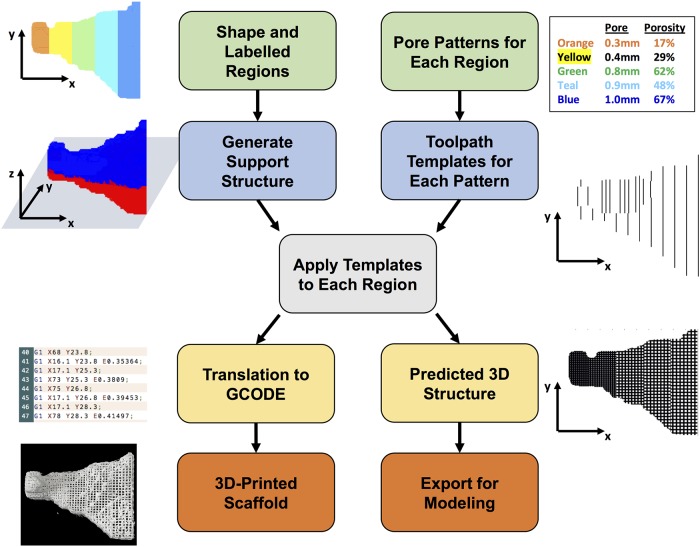
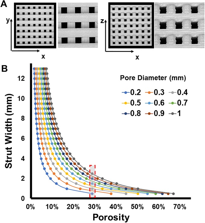
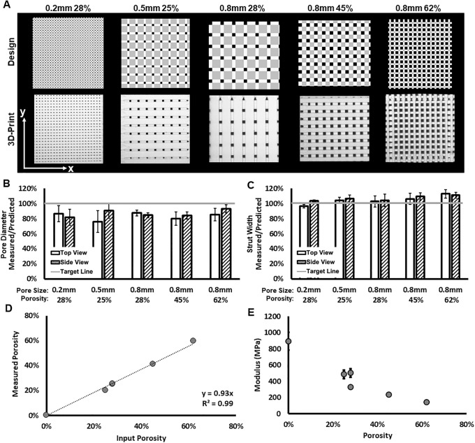
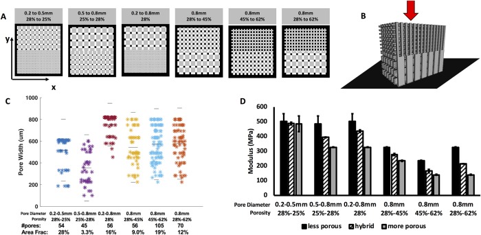
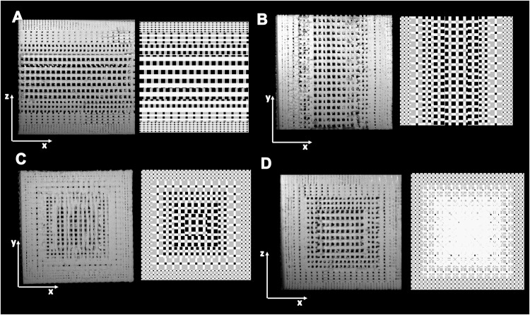
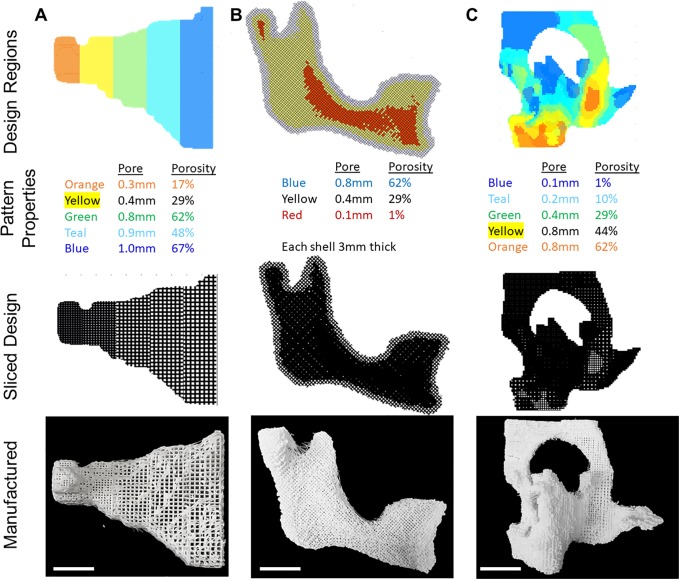
3D-printing is a powerful manufacturing tool that can create precise microscale architectures across macroscale geometries. Within biomedical research, 3D-printing of various materials has been used to fabricate rigid scaffolds for cell and tissue engineering constructs with precise microarchitecture to direct cell behavior and macroscale geometry provides patient specificity. While 3D-printing hardware has become low-cost due to modeling and rapid prototyping applications, there is no common paradigm or platform for the controlled design and manufacture of 3D-printed constructs for tissue engineering. Specifically, controlling the tissue engineering features of pore size, porosity, and pore arrangement is difficult using currently available software. We have developed a MATLAB approach termed scafSLICR to design and manufacture tissue-engineered scaffolds with precise microarchitecture and with simple options to enable spatially patterned pore properties. Using scafSLICR, we designed, manufactured, and characterized porous scaffolds in acrylonitrile butadiene styrene with a variety of pore sizes, porosities, and gradients. We found that transitions between different porous regions maintained an open, connected porous network without compromising mechanical integrity. Further, we demonstrated the usefulness of scafSLICR in patterning different porous designs throughout large anatomic shapes and in preparing craniofacial tissue engineering bone scaffolds. Finally, scafSLICR is distributed as open-source MATLAB scripts and as a stand-alone graphical interface.
Conflict of interest statement
The authors have declared that no competing interests exist.
Figures
Similar articles
-
Fabrication and mechanical characterization of 3D printed vertical uniform and gradient scaffolds for bone and osteochondral tissue engineering.Acta Biomater. 2019 May;90:37-48. doi: 10.1016/j.actbio.2019.03.041. Epub 2019 Mar 21. Acta Biomater. 2019. PMID: 30905862 Free PMC article.
-
Three-dimensional (3D) printed scaffold and material selection for bone repair.Acta Biomater. 2019 Jan 15;84:16-33. doi: 10.1016/j.actbio.2018.11.039. Epub 2018 Nov 24. Acta Biomater. 2019. PMID: 30481607 Review.
-
Design and Structure-Function Characterization of 3D Printed Synthetic Porous Biomaterials for Tissue Engineering.Adv Healthc Mater. 2018 Apr;7(7):e1701095. doi: 10.1002/adhm.201701095. Epub 2017 Dec 27. Adv Healthc Mater. 2018. PMID: 29280325 Review.
-
3D printed dual macro-, microscale porous network as a tissue engineering scaffold with drug delivering function.Biofabrication. 2019 Apr 26;11(3):035014. doi: 10.1088/1758-5090/ab14ff. Biofabrication. 2019. PMID: 30933941
-
Permeability and mechanical properties of gradient porous PDMS scaffolds fabricated by 3D-printed sacrificial templates designed with minimal surfaces.Acta Biomater. 2019 Sep 15;96:149-160. doi: 10.1016/j.actbio.2019.06.040. Epub 2019 Jun 25. Acta Biomater. 2019. PMID: 31252172
Cited by
-
A robust, autonomous, volumetric quality assurance method for 3D printed porous scaffolds.3D Print Med. 2022 Apr 6;8(1):9. doi: 10.1186/s41205-022-00135-x. 3D Print Med. 2022. PMID: 35384521 Free PMC article.
-
New Poly(lactic acid)-Hydrogel Core-Shell Scaffolds Highly Support MSCs' Viability, Proliferation and Osteogenic Differentiation.Polymers (Basel). 2023 Dec 6;15(24):4631. doi: 10.3390/polym15244631. Polymers (Basel). 2023. PMID: 38139883 Free PMC article.
-
An architecturally rational hemostat for rapid stopping of massive bleeding on anticoagulation therapy.Proc Natl Acad Sci U S A. 2024 Jan 30;121(5):e2316170121. doi: 10.1073/pnas.2316170121. Epub 2024 Jan 22. Proc Natl Acad Sci U S A. 2024. PMID: 38252814 Free PMC article.
-
Novel 3D Hybrid Nanofiber Scaffolds for Bone Regeneration.Polymers (Basel). 2020 Mar 2;12(3):544. doi: 10.3390/polym12030544. Polymers (Basel). 2020. PMID: 32131525 Free PMC article.
-
Reasoning on Pore Terminology in 3D Bioprinting.Gels. 2024 Feb 19;10(2):153. doi: 10.3390/gels10020153. Gels. 2024. PMID: 38391483 Free PMC article. Review.
References
-
- Kelly CN, Miller AT, Hollister SJ, Guldberg RE, Gall K. Design and Structure–Function Characterization of 3D Printed Synthetic Porous Biomaterials for Tissue Engineering. Adv Healthc Mater. 2018;7(7):1–16. - PubMed
-
- Jammalamadaka U, Tappa K. Recent Advances in Biomaterials for 3D Printing and Tissue Engineering. J Funct Biomater [Internet]. 2018;9(1):22 http://www.mdpi.com/2079-4983/9/1/22 - PMC - PubMed
-
- Cox SC, Thornby JA, Gibbons GJ, Williams MA, Mallick KK. 3D printing of porous hydroxyapatite scaffolds intended for use in bone tissue engineering applications. Mater Sci Eng C Mater Biol Appl [Internet]. 2015;47:237–47. http://www.sciencedirect.com/science/article/pii/S0928493114007255 - PubMed
Publication types
MeSH terms
Grants and funding
LinkOut - more resources
Full Text Sources
