Review
. 2020 Jun;68(6):1085-1099.
doi: 10.1002/glia.23753. Epub 2019 Nov 19.

Microglia and sexual differentiation of the developing brain: A focus on ontogeny and intrinsic factors

Affiliations
Review

Microglia and sexual differentiation of the developing brain: A focus on ontogeny and intrinsic factors

Evan A Bordt et al. Glia. 2020 Jun.

Abstract

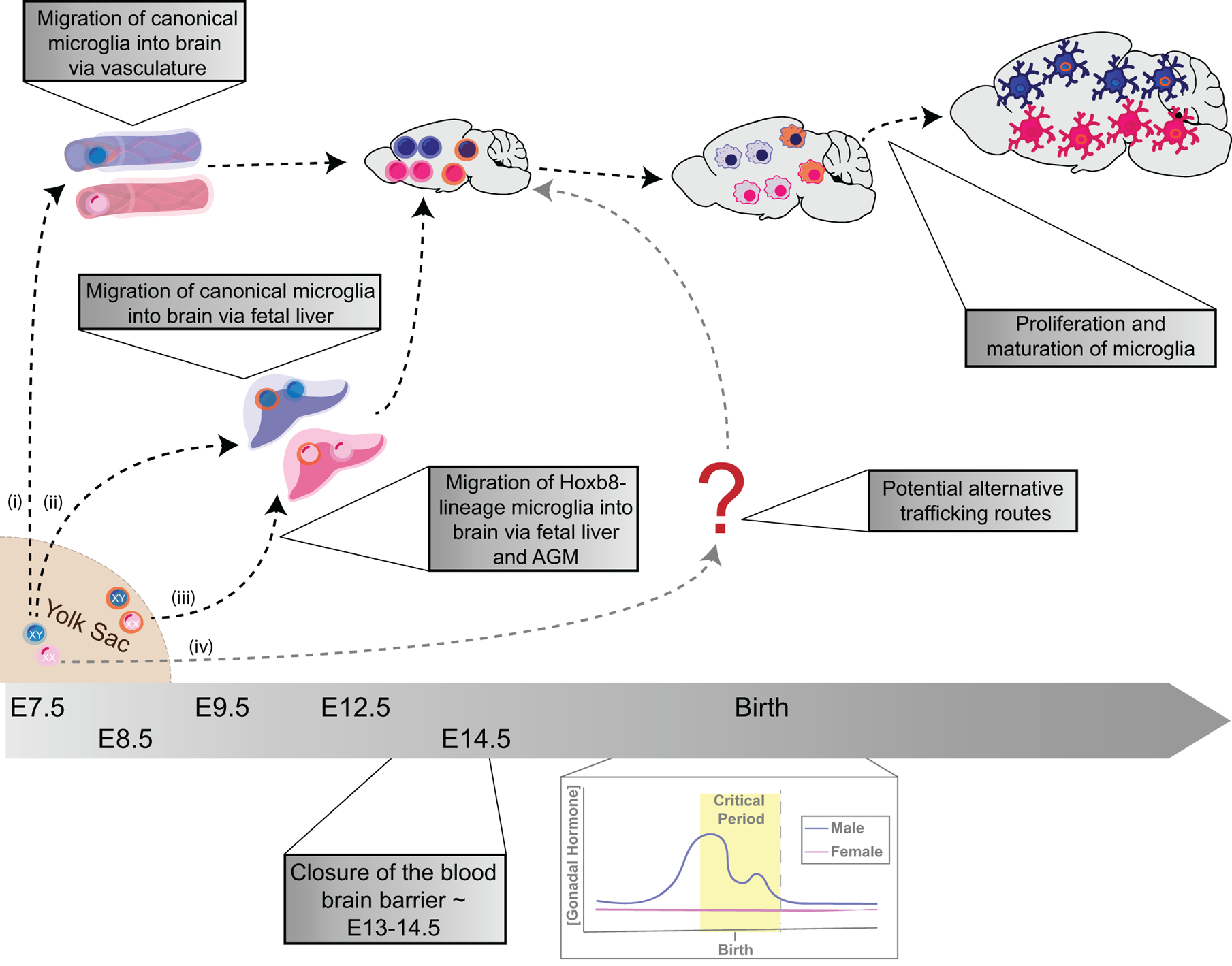
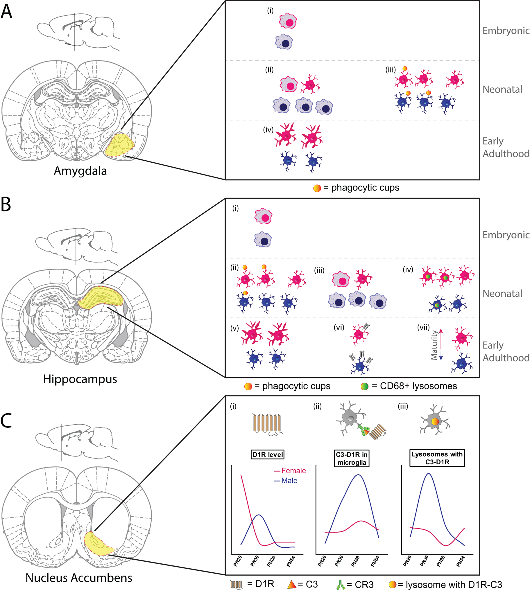
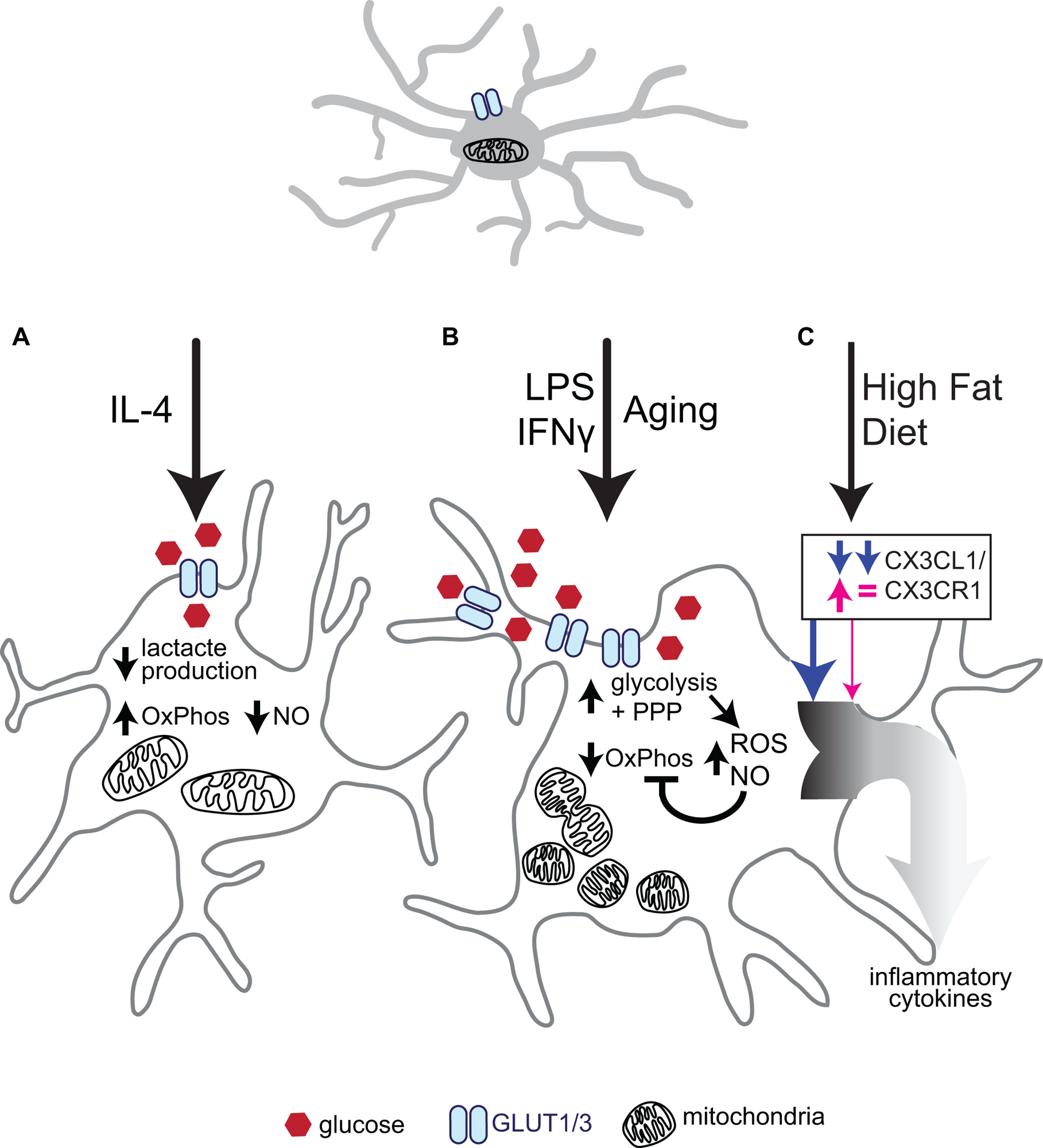
Sexual differentiation of the brain during early development likely underlies the strong sex biases prevalent in many neurological conditions. Mounting evidence indicates that microglia, the innate immune cells of the central nervous system, are intricately involved in these sex-specific processes of differentiation. In this review, we synthesize literature demonstrating sex differences in microglial number, morphology, transcriptional state, and functionality throughout spatiotemporal development as well as highlight current literature regarding ontogeny of microglia. Along with vanRyzin et al. in this issue, we explore the idea that differences in microglia imparted by chromosomal or ontogeny-related programming can influence microglial-driven sexual differentiation of the brain, as well as the idea that extrinsic differences in the male and female brain microenvironment may in turn impart sex differences in microglia.


Figures

Figure 1.
Figure 1.. Ontogeny-specific programming may lead to sex-specific differences in microglial development.
Canonical microglia appear in the fetal yolk sac ~E7.5 in rodents, whereas Hoxb8-lineage microglia appear in the fetal yolk sac ~E8.5. (i) Canonical microglia traffic through the vasculature or (ii) through the fetal liver and aorto-gonad mesonephros, whereas (iii) Hoxb8-lineage microglia traffic through the fetal liver and aorto-gonad mesonephros on the way to the brain. (iv) There is a potential for alternative routes for microglia to traffic from the fetal yolk sac to the brain. The blood brain barrier closes around E13-E14.5, cutting off further migration of monocytes into the healthy developing brain. Around birth, during the time frame of the male-specific gonadal sex hormone surge, there are more canonical microglia in the brain than Hoxb8-lineage microglia. While both subsets of microglia proliferate and mature following birth, the first postnatal week is characterized by a higher rate of proliferation of Hoxb8-lineage microglia than is observed in canonical microglia. This figure is based on data presented in (Ginhoux et al. 2010; Chen et al. 2010; De et al. 2018; VanRyzin, Pickett, and McCarthy 2018; Hoeffel and Ginhoux 2015)
Figure 2.
Figure 2.. Sex- and spatial- heterogeneity of microglia early in development.
A. (i) There are no sex differences in microglial number or morphology in the E17 amygdala, with the majority of both male and female microglia showing amoeboid morphology. (ii) By PN4 male amygdala had more microglia and a higher proportion of microglia with amoeboid morphology than females. (iii) On PN2 and PN4, male microglia within the amygdala had more microglia with phagocytic cups than did females. (iv) By PN30, microglial morphology within the amygdala had shifted towards a more ramified/complex state. At this time females had more microglia with long, thick processes than males. This figure is based on data presented in (Schwarz, Sholar, and Bilbo 2012; VanRyzin et al. 2019) B. (i) There are no sex differences in microglial number or morphology in CA1 or CA3 regions of the hippocampus at E17, with the majority of both male and female microglia showing amoeboid morphology. (ii) At PN3, female rat hippocampus had more microglia with phagocytic cups than male hippocampus. (iii) By PN4 male hippocampus had more microglia and a higher proportion of microglia with amoeboid morphology than females. (iv) Females in PN8 hippocampus had more microglia and more CD68 intensity within microglia than males. (v) By PN30, microglial morphology within the hippocampus had shifted towards a more ramified/complex state. At this time females had more microglia with long, thick processes than males. (vi) 3-week old male mice had more MHC I expression on microglia (CD11b+CD45high cells) isolated from hippocampus than females. (vii) Microglial maturation (determined by microglial developmental index) was lower in microglia isolated from the hippocampus of a PN60 male mouse than from a female mouse. This figure is based on data presented in (Schwarz, Sholar, and Bilbo 2012; Hanamsagar et al. 2017; Nelson, Warden, and Lenz 2017; Weinhard et al. 2018; Guneykaya et al. 2018) C. (i) Dopamine D1 receptor levels in the nucleus accumbens peak at PN20 in females and are downregulated by PN30, whereas D1R levels peak at PN30 in males, and are then downregulated. (ii) Co-localization of microglial C3 and D1R peak in males at PN30 and PN38, with only a small upregulation seen by PN38 in females. (iii) CD68+ microglial lysosomes containing co-localized C3 and D1R peak in males at PN30, with a minor dip seen in females at PN38. This figure is based on data presented in (Kopec et al. 2018).
Figure 3.
Figure 3.. Effects of microglial activation on metabolism.
(A) Stimulation with the classic anti-inflammatory interleukin, IL-4, decreases glucose uptake and lactate production. IL-4 stimulated microglia also downregulate Nitric Oxide (NO) and TNF-ɑ production as glucose is preferentially metabolised through oxidative phosphorylation (OxPhos) to increase ATP production. (B) The microglial inflammatory response is stimulated in a number of ways, such as infection (classically simulated with LPS and/or IFNɣ) or naturally through aging. These stressors have been shown to increase levels of the glucose transporter GLUT1 and thereby increase glycolytic and pentose phosphate pathway (PPP) flux, resulting in increased NADPH and ribose 5-phosphate availability to favor nucleotide synthesis versus ATP production. This push towards glycolysis and PPP is coupled with a decrease in mitochondrial respiration (OxPhos) and increased mitochondrial fission, resulting in increased mitochondrial number, but decreased mitochondrial size. Reactive oxygen species (ROS) production through NOX2 and Nitric Oxide (NO) production are increased in a manner dependent on glucose uptake. Prolonged increases in NO can inhibit oxidative phosphorylation further. (C) Interestingly, the inflammatory response is also activated following High Fat Diet (HFD) in hypothalamic microglia only in males. HFD in males diminished CX3CR1 signaling and led to an enhanced inflammatory response, while in females CXCL1 protein was increased, but overall CXC3R1 signaling remained relatively unaffected, and no increased inflammation was observed. This figure is based on data presented in (Orihuela, McPherson, and Harry 2016; Jaber et al. 2017; Voloboueva et al. 2013; L. Wang et al. 2019; Dorfman et al. 2017)

References

    1. Aanerud Joel, Borghammer Per, Rodell Anders, Jónsdottir Kristjana Y., and Gjedde Albert. 2017. “Sex Differences of Human Cortical Blood Flow and Energy Metabolism.” Journal of Cerebral Blood Flow and Metabolism: Official Journal of the International Society of Cerebral Blood Flow and Metabolism 37 (7): 2433–40. - PMC - PubMed
    1. Ajami Bahareh, Samusik Nikolay, Wieghofer Peter, Ho Peggy P., Crotti Andrea, Bjornson Zach, Prinz Marco, Fantl Wendy J., Nolan Garry P., and Steinman Lawrence. 2018. “Single-Cell Mass Cytometry Reveals Distinct Populations of Brain Myeloid Cells in Mouse Neuroinflammation and Neurodegeneration Models.” Nature Neuroscience 21 (4): 541–51. - PMC - PubMed
    1. Amen Daniel G., Trujillo Manuel, Keator David, Taylor Derek V., Willeumier Kristen, Meysami Somayeh, and Raji Cyrus A.. 2017. “Gender-Based Cerebral Perfusion Differences in 46,034 Functional Neuroimaging Scans.” Journal of Alzheimer’s Disease: JAD 60 (2): 605–14. - PubMed
    1. Arnò Benedetta, Grassivaro Francesca, Rossi Chiara, Bergamaschi Andrea, Castiglioni Valentina, Furlan Roberto, Greter Melanie, et al. 2014. “Neural Progenitor Cells Orchestrate Microglia Migration and Positioning into the Developing Cortex.” Nature Communications 5 (November): 5611. - PubMed
    1. Ayata Pinar, Badimon Ana, Strasburger Hayley J., Mary Kaye Duff Sarah E. Montgomery, Loh Yong-Hwee E., Ebert Anja, et al. 2018. “Epigenetic Regulation of Brain Region-Specific Microglia Clearance Activity.” Nature Neuroscience 21 (8): 1049–60. - PMC - PubMed

Publication types