

Prothrombotic and Proinflammatory Activities of the β-Hemolytic Group B Streptococcal Pigment
- PMID: 31743913
- PMCID: PMC7383282
- DOI: 10.1159/000504002
Prothrombotic and Proinflammatory Activities of the β-Hemolytic Group B Streptococcal Pigment
Abstract
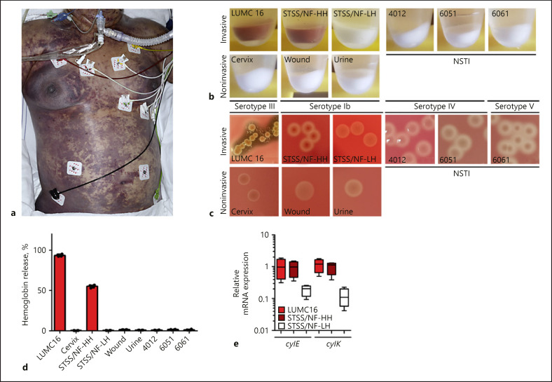
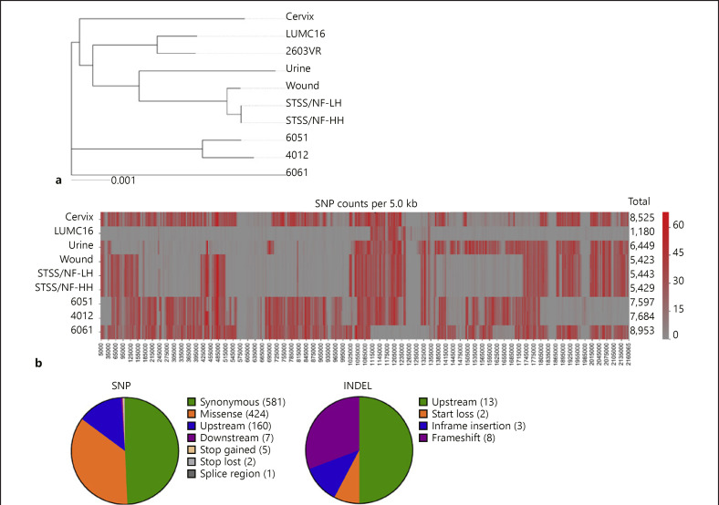
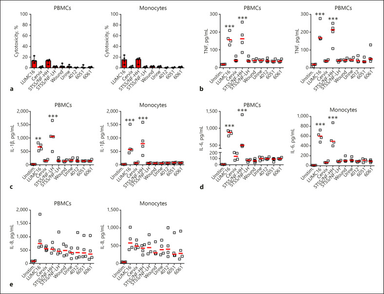
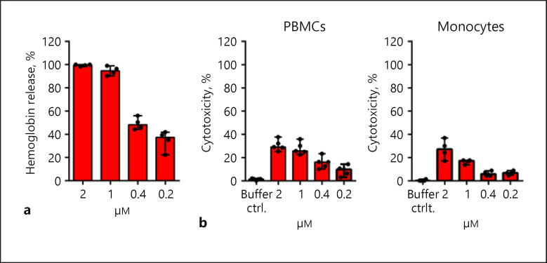
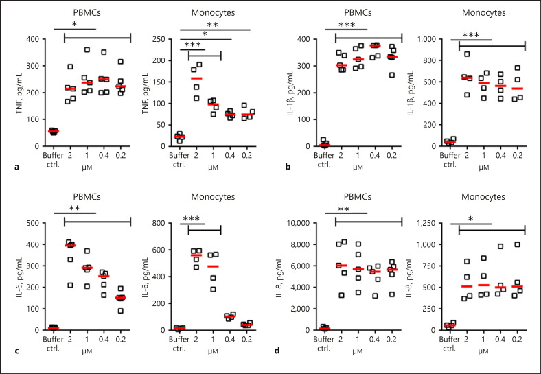
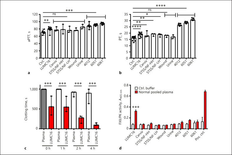
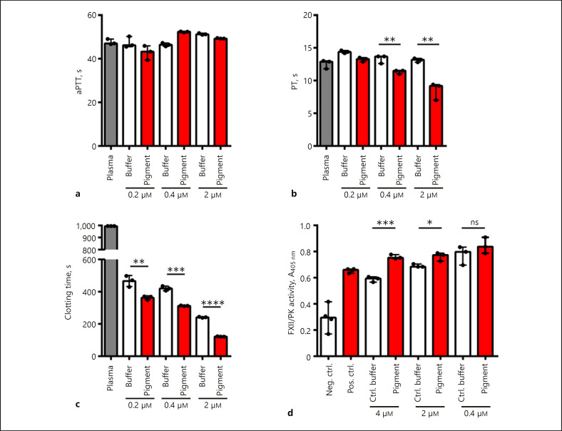
A prominent feature of severe streptococcal infections is the profound inflammatory response that contributes to systemic toxicity. In sepsis the dysregulated host response involves both immunological and nonimmunological pathways. Here, we report a fatal case of an immunocompetent healthy female presenting with toxic shock and purpura fulminans caused by group B streptococcus (GBS; serotype III, CC19). The strain (LUMC16) was pigmented and hyperhemolytic. Stimulation of human primary cells with hyperhemolytic LUMC16 and STSS/NF-HH strains and pigment toxin resulted in a release of proinflammatory mediators, including tumor necrosis factor, interleukin (IL)-1β, and IL-6. In addition, LUMC16 induced blood clotting and showed factor XII activity on its surface, which was linked to the presence of the pigment. The expression of pigment was not linked to a mutation within the CovR/S region. In conclusion, our study shows that the hemolytic lipid toxin contributes to the ability of GBS to cause systemic hyperinflammation and interferes with the coagulation system.
Keywords: Coagulation; Group B streptococcus; Hemolysis; Inflammation; Pigment; Streptococcus agalactiae.
© 2019 The Author(s) Published by S. Karger AG, Basel.
Conflict of interest statement
The authors have no conflicts of interest to declare.
Figures
Similar articles
-
A streptococcal lipid toxin induces membrane permeabilization and pyroptosis leading to fetal injury.EMBO Mol Med. 2015 Apr;7(4):488-505. doi: 10.15252/emmm.201404883. EMBO Mol Med. 2015. PMID: 25750210 Free PMC article.
-
Genetic Basis Underlying the Hyperhemolytic Phenotype of Streptococcus agalactiae Strain CNCTC10/84.J Bacteriol. 2020 Nov 4;202(23):e00504-20. doi: 10.1128/JB.00504-20. Print 2020 Nov 4. J Bacteriol. 2020. PMID: 32958630 Free PMC article.
-
Phenotypic and molecular characterization of hyperpigmented group B Streptococci.Int J Med Microbiol. 2014 Jul;304(5-6):717-24. doi: 10.1016/j.ijmm.2014.05.003. Epub 2014 May 16. Int J Med Microbiol. 2014. PMID: 24933304
-
SIRS and group-B streptococcal sepsis in newborns: pathogenesis and perspectives in adjunctive therapy.Semin Fetal Neonatal Med. 2006 Oct;11(5):333-42. doi: 10.1016/j.siny.2006.03.003. Epub 2006 May 11. Semin Fetal Neonatal Med. 2006. PMID: 16690364 Review.
-
Streptococcal toxins: role in pathogenesis and disease.Cell Microbiol. 2015 Dec;17(12):1721-41. doi: 10.1111/cmi.12531. Epub 2015 Nov 17. Cell Microbiol. 2015. PMID: 26433203 Review.
Cited by
-
Some Like It Hot.J Innate Immun. 2021;13(6):321-322. doi: 10.1159/000520270. Epub 2021 Oct 26. J Innate Immun. 2021. PMID: 34724673 Free PMC article. No abstract available.
-
Single-Molecule Identification of the Isomers of a Lipidic Antibody Activator.J Phys Chem Lett. 2024 Jul 11;15(27):6935-6942. doi: 10.1021/acs.jpclett.4c00164. Epub 2024 Jun 27. J Phys Chem Lett. 2024. PMID: 38935930 Free PMC article.
-
Group B Streptococcal Hemolytic Pigment Impairs Platelet Function in a Two-Step Process.Cells. 2022 May 13;11(10):1637. doi: 10.3390/cells11101637. Cells. 2022. PMID: 35626674 Free PMC article.
-
Virulence, phenotype and genotype characteristics of invasive group B Streptococcus isolates obtained from Swedish pregnant women and neonates.Ann Clin Microbiol Antimicrob. 2022 Oct 13;21(1):43. doi: 10.1186/s12941-022-00534-2. Ann Clin Microbiol Antimicrob. 2022. PMID: 36229877 Free PMC article.
-
An opportunistic pathogen under stress: how Group B Streptococcus responds to cytotoxic reactive species and conditions of metal ion imbalance to survive.FEMS Microbiol Rev. 2024 May 8;48(3):fuae009. doi: 10.1093/femsre/fuae009. FEMS Microbiol Rev. 2024. PMID: 38678005 Free PMC article. Review.
References
-
- Ikebe T, Tominaga K, Shima T, Okuno R, Kubota H, Ogata K, et al. Working Group for Beta-haemolytic Streptococci in Japan Increased prevalence of group A streptococcus isolates in streptococcal toxic shock syndrome cases in Japan from 2010 to 2012. Epidemiol Infect. 2015 Mar;143((4)):864–72. - PMC - PubMed
-
- Ikebe T, Chiba K, Shima T, Masuda C, Okuno R, Ohya H, et al. Working group for beta-hemolytic streptococci in Japan Evaluation of streptococcal toxic shock-like syndrome caused by group B streptococcus in adults in Japan between 2009 and 2013. J Infect Chemother. 2015 Mar;21((3)):207–11. - PubMed
-
- Llewelyn M, Cohen J. Superantigens: microbial agents that corrupt immunity. Lancet Infect Dis. 2002 Mar;2((3)):156–62. - PubMed
-
- Fraser JD, Proft T. The bacterial superantigen and superantigen-like proteins. Immunol Rev. 2008 Oct;225((1)):226–43. - PubMed
-
- Chang B, Ikebe T, Wada A, Ogata K, Tomita M, Katsukawa C, et al. Working Group for Streptococci in Japan Surveillance of group B streptococcal toxic shock-like syndrome in nonpregnant adults and characterization of the strains in Japan. Jpn J Infect Dis. 2006 Jun;59((3)):182–5. - PubMed
Publication types
MeSH terms
Substances
LinkOut - more resources
Full Text Sources
Other Literature Sources
Medical
