. 2020 Feb;9(2):e969.
doi: 10.1002/mbo3.969. Epub 2019 Nov 19.

Lysosome assembly and disassembly changes endocytosis rate through the Leishmania cell cycle

Affiliations

Lysosome assembly and disassembly changes endocytosis rate through the Leishmania cell cycle

Ziyin Wang et al. Microbiologyopen. 2020 Feb.

Abstract

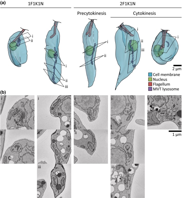
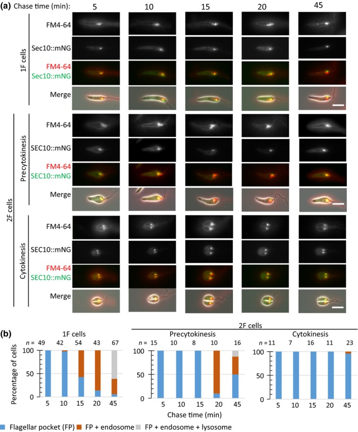
The Leishmania lysosome has an atypical structure, consisting of an elongated vesicle-filled tubule running along the anterior-posterior axis of the cell, which is termed the multivesicular tubule (MVT) lysosome. Alongside, the MVT lysosome is one or more microtubules, the lysosomal microtubule(s). Previous work indicated there were cell cycle-related changes in MVT lysosome organization; however, these only provided snapshots and did not connect the changes in the lysosomal microtubule(s) or lysosomal function. Using mNeonGreen tagged cysteine peptidase A and SPEF1 as markers of the MVT lysosome and lysosomal microtubule(s), we examined the dynamics of these structures through the cell cycle. Both the MVT lysosome and lysosomal microtubule(s) elongated at the beginning of the cell cycle before plateauing and then disassembling in late G2 before cytokinesis. Moreover, the endocytic rate in cells where the MVT lysosome and lysosomal microtubule(s) had disassembled was extremely low. The dynamic nature of the MVT lysosome and lysosomal microtubule(s) parallels that of the Trypanosoma cruzi cytostome/cytopharynx, which also has a similar membrane tubule structure with associated microtubules. As the cytostome/cytopharynx is an ancestral feature of the kinetoplastids, this suggests that the Leishmania MVT lysosome and lysosomal microtubule(s) are a reduced cytostome/cytopharynx-like feature.

Keywords: Leishmania; cell cycle; endocytosis; lysosome.


Conflict of interest statement

The authors declare no conflict of interest.

Figures

Figure 1
Figure 1
Localization and morphological changes in CPA::mNG and mNG::SPEF1 throughout the cell cycle. Images of CPA::mNG (a) or mNG::SPEF1 (d) localization during the cell cycle in the Leishmania promastigote. Micrographs of major cell cycle stages, cells were ordered based on the number of kinetoplasts (K), nuclei (N), and flagella (F). Nuclear and kinetoplast DNA were labeled with Hoechst 33342. The scale bar represents 5 µm. Scatter plot of cell body length or new flagellum length (measured from cell tip to flagellum tip) against CPA::mNG (B) or mNG::SPEF1 (e) length in 1F or 2F stage, respectively. Each dot represents one cell, n = 303 for 1F and 101 for 2F CPA::mNG, n = 297 for 1F and 98 for 2F mNG::SPEF1. Bar charts of CPA::mNG (c) or mNG::SPEF1 (f) signal categories in 1F and 2F cells. (g) Micrographs of CPA:: mNG (green) mCh::SPEF1 (red) and colocalization during the cell cycle in the Leishmania promastigote. MtQ—microtubule quartet; LMt—lysosomal microtubule(s)
Figure 2
Figure 2
Three‐dimensional structure of the MVT lysosome throughout the cell cycle. (a) Three‐dimensional reconstructions of the nuclei (green), flagella (red), and MVT lysosomes (purple) of Leishmania promastigotes from serial block‐face scanning electron microscopy images. Major cell cycle stages similar to those in Figure 1 are shown. From left to right: Early cell cycle 1F1K1N, midcell cycle 1F1K1N, precytokinesis 2F1K1N, early cytokinesis 2F1K1N, and late cytokinesis 2F1K1N. (b) SBFSEM images illustrating example features of the MVT lysosome of the cells shown in (a). The images labeled i, ii, or iii correspond to the sections with the matching label in (a) and the MVT lysosome in each section is outlined
Figure 3
Figure 3
Distribution of the CPA::mNG labeled vesicles in cells undergoing cytokinesis. (a) Example of a cell undergoing cytokinesis divided into four quadrants depending on whether they were in the posterior or anterior portion of the cell which would inherit either the old or the new flagellum. (b) Bar chart showing the number (none, few, and many) of vesicles present in each quadrant. n = 19
Figure 4
Figure 4
FM4‐64 pulse‐chase in the CPA::mNG cell line. (a) FM4‐64 pulse‐chase assay with promastigotes expressing CPA::mNG. Promastigotes were chilled on ice for 20 min and then pulsed with FM4‐64 for 1 min before imaging every 5 min over a 30 min time course. Three major categories of FM4‐64 localization were observed: flagellar pocket; flagellar pocket and endosome; and flagellar pocket, endosome, and lysosome. The scale bar represents 5 µm. (b) Proportion of each category observed at each time point for cells for 1F, 2F, and cells in cytokinesis. Numbers counted for each time point are indicated above the columns. The uptake assays were done independently three times, and results from a representative experiment are shown
Figure 5
Figure 5
FM4‐64 pulse‐chase in the mNG::SPEF1 cell line. (a) FM4‐64 pulse‐chase assay with promastigotes expressing mNG::SPEF1. Promastigotes were chilled on ice for 20 min and then pulsed with FM4‐64 for 1 min before imaging every 5 min over a 30 min time course. Three major categories of FM4‐64 localization were observed: flagellar pocket; flagellar pocket and endosome; and flagellar pocket, endosome, and lysosome. The scale bar represents 5 µm. (b) Proportion of each category observed at each time point for cells for 1F, 2F, and cells in cytokinesis. Numbers counted for each time point are indicated above the columns. The uptake assays were done independently three times, and results from a representative experiment are shown
Figure A1
Figure A1
Localization of CPA::mNG and mCh::SPEF1 in axenic amastigotes. Images of CPA::mNG (green) and mCh::SPEF1 (red) colocalization in axenic amastigotes. The scale bar represents 5 µm. White asterisk in the mCh::SPEF1 channel indicates the anterior end of the cell. mCh::SPEF1 localized to two structures a bright spot close to the flagellar pocket and a fainter curved line extending toward the posterior end of the cell. Two types of CPA::mNG localization pattern were observed, either a curved line extending from near the flagellar pocket to the posterior end of the cell or a series of variable sized points that followed a line through the cell. These large spots correlate with the megasomes previously observed by TEM (Waller & McConville, 2002). Both types of CPA::mNG localization pattern run alongside the mCh::SPEF1 signal, indicating a close association of these proteins in the amastigote form
Figure A2
Figure A2
Interpretation of cell structure from scanning block‐face electron microscopy (relating to Figure 2). (a) Three‐dimensional reconstructions of the major membrane‐bound cytoplasmic structures, endoplasmic reticulum (orange), mitochondrion (blue), and MVT lysosome (purple), in an example 1K1N Leishmania promastigote from serial block‐face scanning electron microscopy images. The MVT lysosome is a third extended membrane‐bound network in addition to the endoplasmic reticulum and mitochondrion. (b) SBFSEM image illustrating identifiable structures in the cell. A combination of appearance (electron density) and 3D shape allow organelle identification. Acidocalcisomes and lipid droplets are near‐spherical organelles with high and low electron density, respectively. Glycosomes are more elongated with intermediate electron density. The endoplasmic reticulum, nuclear envelope, mitochondrion, and MVT lysosome form extended tubes/networks. The endoplasmic reticulum and nuclear envelope have a narrow lumen and the two membranes are not well resolved, instead appearing as a single highly electron dense line. The nucleus is identifiable from the chromatin contents. The mitochondrion lumen is well resolved with cristae occasionally visible. The double membrane is not well resolved, again appearing as a single highly electron dense line. The MVT lysosome has a wider lumen than the endoplasmic reticulum and does not have lamellar regions. The MVT lysosome is bounded by a single membrane which appears less electron dense than the mitochondrion or endoplasmic reticulum membranes
Figure A3
Figure A3
Localization of proteins identified in L. mexicana with identity to known regulators of lysosome function (relating to Table A1). Images of cell lines expressing identified proteins tagged at either the N‐ or C‐terminus with mNG in Leishmania promastigotes. Nuclear and kinetoplast DNA were labeled with Hoechst 33342. The name of the human protein is on the right with the corresponding L. mexicana gene ID in white text in the mNG channel image. Beneath each set of images is the description of the protein localization. The proteins in bold are those cell lines which have a proportion of cells in which a lysosome localization was observed
Figure A4
Figure A4
FM4‐64 pulse‐chase in the SEC10::mNG cell line. (a) FM4‐64 pulse‐chase assay with promastigotes expressing SEC10::mNG. Promastigotes were chilled on ice for 20 min and then pulsed with FM4‐64 for 1 min before imaging over a 45‐min time course. Three major categories of FM4‐64 localization were observed: flagellar pocket; flagellar pocket and endosome; and flagellar pocket, endosome, and lysosome. The scale bar represents 5 µm. (b) Proportion of each category observed at each time point for cells for 1F, 2F, and cells in cytokinesis. Numbers counted for each time point are indicated above the columns. The uptake assays were done independently three times, and results from a representative experiment are shown

Similar articles

Cited by

References

    1. Alcantara, C. L. , Vidal, J. C. , de Souza, W. , & Cunha‐e‐Silva, N. L. (2014). The three‐dimensional structure of the cytostome‐cytopharynx complex of Trypanosoma cruzi epimastigotes. Journal of Cell Science, 127, 2227–2237. - PubMed
    1. Alcantara, C. L. , Vidal, J. C. , de Souza, W. , & Cunha‐E‐Silva, N. L. (2017). The cytostome–cytopharynx complex of Trypanosoma cruzi epimastigotes disassembles during cell division. Journal of Cell Science, 130, 164–176. - PubMed
    1. Besteiro, S. , Williams, R. A. M. , Coombs, G. H. , & Mottram, J. C. (2007). Protein turnover and differentiation in Leishmania. International Journal for Parasitology, 37, 1063–1075. 10.1016/j.ijpara.2007.03.008 - DOI - PMC - PubMed
    1. Cunha‐e‐Silva, N. , Sant’Anna, C. , Pereira, M. G. , Porto‐Carreiro, I. , Jeovanio, A. L. , & de Souza, W. (2006). Reservosomes: Multipurpose organelles? Parasitology Research, 99, 325–327. 10.1007/s00436-006-0190-3 - DOI - PubMed
    1. Dean, S. , Sunter, J. , Wheeler, R. J. , Hodkinson, I. , Gluenz, E. , & Gull, K. (2015). A toolkit enabling efficient, scalable and reproducible gene tagging in Trypanosomatids. Open Biology, 5, 140197 10.1098/rsob.140197 - DOI - PMC - PubMed

Publication types