Review
. 2019 Nov 17;19(22):5019.
doi: 10.3390/s19225019.

Sensors for Positron Emission Tomography Applications

Affiliations
Review

Sensors for Positron Emission Tomography Applications

Wei Jiang et al. Sensors (Basel). .

Abstract

Positron emission tomography (PET) imaging is an essential tool in clinical applications for the diagnosis of diseases due to its ability to acquire functional images to help differentiate between metabolic and biological activities at the molecular level. One key limiting factor in the development of efficient and accurate PET systems is the sensor technology in the PET detector. There are generally four types of sensor technologies employed: photomultiplier tubes (PMTs), avalanche photodiodes (APDs), silicon photomultipliers (SiPMs), and cadmium zinc telluride (CZT) detectors. PMTs were widely used for PET applications in the early days due to their excellent performance metrics of high gain, low noise, and fast timing. However, the fragility and bulkiness of the PMT glass tubes, high operating voltage, and sensitivity to magnetic fields ultimately limit this technology for future cost-effective and multi-modal systems. As a result, solid-state photodetectors like the APD, SiPM, and CZT detectors, and their applications for PET systems, have attracted lots of research interest, especially owing to the continual advancements in the semiconductor fabrication process. In this review, we study and discuss the operating principles, key performance parameters, and PET applications for each type of sensor technology with an emphasis on SiPM and CZT detectors-the two most promising types of sensors for future PET systems. We also present the sensor technologies used in commercially available state-of-the-art PET systems. Finally, the strengths and weaknesses of these four types of sensors are compared and the research challenges of SiPM and CZT detectors are discussed and summarized.

Keywords: avalanche photodiode (APD); cadmium zinc telluride (CZT); digital silicon photomultiplier (dSiPM); photomultiplier tubes (PMT); positron emission tomography (PET); silicon photomultiplier (SiPM); single-photon avalanche diode (SPAD).


Conflict of interest statement

The authors declare no conflict of interest.

Figures

Figure 1
Figure 1
The basic principle of a positron emission tomography (PET) system: A PET detector ring detects a pair of gamma photons with an energy of 511 keV (red arrows) which results from the annihilation of an electron with a positron emitted by the radiotracer (FDG).
Figure 2
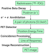
Figure 2
Detection flow of a positron emission tomography (PET) system.
Figure 3
Figure 3
Concept of time-of-flight positron emission tomography (ToF PET): (a) Illustration of a detector ring detecting pairs of gamma photons from the annihilation events with (green) and without (red) the ToF technique; (b) The probability distribution of the annihilation position along the line of response (LoR) in ToF PET; (c) The equal probability of the annihilation position along the LoR in non-ToF PET.
Figure 4
Figure 4
Positron emission tomography (PET) detector: (a) Structure of a PET detector; (b) Detection flow chart of a PET detector.
Figure 5
Figure 5
Principle of the photomultiplier tube (PMT): (a) Simplified conventional structure; (b) Simplified high-voltage biasing network.
Figure 6
Figure 6
Illustration of a microchannel plate photomultiplier tube (MCP-PMT) (the microchannels are usually made of highly resistive materials (e.g., lead glass cladding) with the inner wall coated by high secondary emission materials such as MgO and Al2O3).
Figure 7
Figure 7
Structure of a position-sensitive photomultiplier tube (PS-PMT) with microchannel plates.
Figure 8
Figure 8
Resistive network for a 3 × 3 anode array (each blue dot represents one signal anode).
Figure 9
Figure 9
The impact ionization process. Here, αn1 is the average distance between each electron multiplication event, while Vtotal is the applied reverse bias plus the built-in potential.
Figure 10
Figure 10
Principle of single-photon avalanche diode (SPAD) operation: (a) Avalanche breakdown process in a reverse biased p-n junction; (b) I-V characteristic representation of SPAD operation.
Figure 11
Figure 11
Cross-sectional view of p-n junctions in the standard TSMC 180 nm complementary metal-oxide (CMOS) technology: (a) Three diodes at the n+/p-well, p-well/deep n-well (DNW), and DNW/p-substrate junctions; (b) Two diodes at the p+/n-well and DNW/p-substrate junctions.
Figure 12
Figure 12
Simplified schematic for passive quench and reset (PQR) and the equivalent circuit simulation model.
Figure 13
Figure 13
Simplified schematic for an active quench-reset (AQR) circuit.
Figure 14
Figure 14
Readout electronics of a PET detector based on: (a) Analog SiPM and (b) Digital SiPM.
Figure 15
Figure 15
Illustration of a positron emission tomography (PET) detector: (a) pixelated scintillator (2 × 4 array) mounted on a photosensor; (b) monolithic scintillator mounted on a photosensor.
Figure 16
Figure 16
Planar illustration of a cadmium zinc telluride (CZT) detector. A negative high voltage is applied on the cathode and the anode is grounded. The e–h pairs are generated by absorbing the energy from the gamma rays.
Figure 17
Figure 17
Illustration of electrode patterns of a cadmium zinc telluride (CZT) detector: (a) pixelated pattern; (b) cross-strip pattern.

References

    1. Giger M.L. Machine Learning in Medical Imaging. J. Am. Coll. Radiol. 2018;15:512–520. doi: 10.1016/j.jacr.2017.12.028. - DOI - PubMed
    1. Rohren E.M., Turkington T.G., Coleman R.E. Clinical Applications of PET in Oncology. Radiology. 2004;231:305–332. doi: 10.1148/radiol.2312021185. - DOI - PubMed
    1. Kadir A., Almkvist O., Forsberg A., Wall A., Engler H., Långström B., Nordberg A. Dynamic Changes in PET Amyloid and FDG Imaging at Different Stages of Alzheimer’s Disease. Neurobiol. Aging. 2012;33:198.e1–198.e14. doi: 10.1016/j.neurobiolaging.2010.06.015. - DOI - PubMed
    1. Keng F.Y. Clinical Applications of Positron Emission Tomography in Cardiology: A Review. Ann. Acad. Med. Singap. 2004;33:175–182. - PubMed
    1. Nader M.A., Czoty P.W. Brain Imaging in Nonhuman Primates: Insights into Drug Addiction. ILAR J. 2008;49:89–102. doi: 10.1093/ilar.49.1.89. - DOI - PubMed