Review
. 2019 Oct;54(5):443-465.
doi: 10.1080/10409238.2019.1691497. Epub 2019 Nov 20.

Structural and functional modularity of the U2 snRNP in pre-mRNA splicing

Affiliations
Review

Structural and functional modularity of the U2 snRNP in pre-mRNA splicing

Clarisse van der Feltz et al. Crit Rev Biochem Mol Biol. 2019 Oct.

Abstract

The U2 small nuclear ribonucleoprotein (snRNP) is an essential component of the spliceosome, the cellular machine responsible for removing introns from precursor mRNAs (pre-mRNAs) in all eukaryotes. U2 is an extraordinarily dynamic splicing factor and the most frequently mutated in cancers. Cryo-electron microscopy (cryo-EM) has transformed our structural and functional understanding of the role of U2 in splicing. In this review, we synthesize these and other data with respect to a view of U2 as an assembly of interconnected functional modules. These modules are organized by the U2 small nuclear RNA (snRNA) for roles in spliceosome assembly, intron substrate recognition, and protein scaffolding. We describe new discoveries regarding the structure of U2 components and how the snRNP undergoes numerous conformational and compositional changes during splicing. We specifically highlight large scale movements of U2 modules as the spliceosome creates and rearranges its active site. U2 serves as a compelling example for how cellular machines can exploit the modular organization and structural plasticity of an RNP.

Keywords: Pre-mRNA; RNA; SF3B1; snRNP; spliceosome; splicing.


Conflict of interest statement

Disclosure statement

The authors have no potential conflicts of interest to report.

Figures

Figure 1.
Figure 1.
Overview of the stages of pre-mRNA splicing. The spliceosome catalyzes splicing of the pre-mRNA by recognition of the 5ʹ SS, BS, and the 3ʹ SS. Many steps are required for excision of the intron and concomitant ligation of the 5ʹ (white box) and 3ʹ (black box) exons. The five U snRNPs and the NTC are labeled. BBP and Mud2 are splicing factors that recognize the BS in the E complex. The spliceosome complexes shown are the best characterized, and their respective names (E, A, etc…) are indicated in bold italics. DExD/H-box ATPases necessary for splicing are named in italics below the corresponding stage during which they function (Sub2, Prp5, etc…). A color version of this figure is available online.
Figure 2.
Figure 2.
Sequences and secondary structures of the conserved modules of the yeast (A) and human (B) U2 snRNAs. The modules consist of the Stem I (pink), branch point recognition sequence (BPRS, green), Stem II (blue), and Sm binding site/3ʹ stem loop (tan) regions. Also depicted are constitutively incorporated pseudouridines (Ψ) and positions of 2ʹ O-methylation (bold purple). An alternate structure for the yeast stem I and BPRS module called the branch point interacting stem loop (BSL) is shown in the inset. Human U2 snRNA includes a m6Am post-transcriptional modification at position 30. A color version of this figure is available online.
Figure 3.
Figure 3.
Organization of the protein components of the U2 snRNP in the spliceosome B complex (PDB ID:5NRL). The BPRS, Stem II, and Sm binding site/3ʹ stem loop modules of U2 snRNA (green) scaffold an arrangement of snRNP proteins that anchors the SF3b subcomplex near the BPRS. The region of Hsh155 which interacts with Prp5, Prp2 and the RES complex is noted. A schematic representation of this organization is shown in the inset. A color version of this figure is available online.
Figure 4.
Figure 4.
Pairing between the U2 stem I module and the U6 snRNA in the spliceosome. (A) 2D schematic of U2/U6 helices Ia (light blue), Ib (navy) and II (purple) in the spliceosome prior to 5ʹ SS cleavage. The U2 bases that participate in base pairing are bolded. Pink nucleotides represent those found in U2 stem I (Figure 2). The internal stem loop of U6 coordinates magnesium ions essential for catalysis. U6’s catalytic triplex is boxed and interactions with triplex bases are shown by dashed lines. The site of intron lariat formation is denoted by a gold star. (B) Cryo-EM structure of U2/U6 helices Ia, Ib, and II in the yeast Bact spliceosome. These RNA helices are encased by a number of proteins including Prp8 and a number of components of the NTC (Cef1, Clf1, Cwc15, Prp45, and Syf2; PDB ID: 5GM6). A color version of this figure is available online.
Figure 5.
Figure 5.
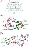
Structure of the U2/BS duplex and its RNA interactions during splicing. (A) Schematic of the U2/BS RNA duplex. U2 snRNA (green) pairs to the branch site (black) resulting in expulsion of the branch site adenosine from the duplex. (B) Positioning of the 2’ OH group of the branch point adenosine into the spliceosome active site for 5ʹ SS cleavage involves hydrogen bonding (black dashes) between the adenosine and a conserved uridine in the BS sequence (PDB ID:5LJ5). (C) The branch point adenosine participates in 3ʹ SS recognition during exon ligation by Hoogsteen pairing with the conserved adenosine of the 3ʹ SS (black dashes, PDB ID:6EXN). In panels (B) and (C), U2 snRNA position 35 was modeled as a uridine based on the cryo-EM data although U2 contains a highly conserved pseudouridine at this position. A color version of this figure is available online.
Figure 6.
Figure 6.
Structure of the human U2 protein SF3B1. (A) Domain organization of human SF3B1. HEAT repeats 4–7 (green) is hot spot for mutations associated a variety of cancers. HEAT repeats 15 and 16 (black) form a binding pocket for the bulged branch site adenosine in the U2/BS RNA duplex. (B) Open (gray, PDB ID: 5IFE) and closed (pink, PDB ID: 6AHD) structures of SF3B1. The U2 snRNA BPRS (green) and intron (black) are shown as ribbons. Frequently mutated positions in SF3B1 are shown as green spheres (E622, R625, H662, K666, K700, and G742). (C) Structure of SF3B1 bound to the splicing inhibitor pladienolide B (red, PDB ID: 6EXN). (D) Chemical structure of pladienolide B. A color version of this figure is available online.
Figure 7.
Figure 7.
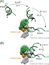
Movements of the U2/BS duplex during splicing. (A) Movements of the duplex during B to Bact to C complex transitions. Movements are shown in relation to the active site and U6 catalytic triplex (orange). The U2 snRNA (green) in U2/U6 helices is shown space-filled and the U2 BPRS region shown as a ribbon. Base pairing between the BPRS and the intron (black; 5ʹ end, blue; 3ʹ end, red) varies between complexes (PDB IDs: 5NRL, 5GM6, and 5LJ3). (B) Movements occurring during the C to C* transition (PDB IDs: 5LJ3 and 5MQ0). Distances shown are measured from the U2 U47 backbone between structures. A color version of this figure is available online.
Figure 8.
Figure 8.
U2 Stem II dynamics during splicing. (A) Stem IIa (blue) is a mutually exclusive structure to stem IIc (blue/red). Stem IIb (lavender) can be accommodated by both stem IIa and IIb pairing. In the spliceosome B complex, the stem loop of IIa interacts with Cus1 and Prp9 and the stem loop of IIb interacts with Hsh49 (PDB ID: 5NRL). In the spliceosome C complex, stem IIc has formed and interacts with U2’s Sm ring. Also in this complex, stem loop IIb interacts with Ecm2 and Cwc2 (PDB ID: 5LJ3). (B) View of stem II’s movement between the spliceosome C and C* complexes. As the active site of the spliceosome is remodeled, stem loop IIb undocks from Ecm2 and Cwc2 and moves ~50 Å (PDB ID: 5LJ3 and 5MQ0). A color version of this figure is available online.
Figure 9.
Figure 9.
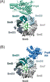
Interactions between the U2 Sm ring and splicing factors in spliceosomes. The Sm binding site in U2 snRNA is shown in green. (A) Interaction between the SmD1 and SmD2 proteins and the SF3a protein Prp9 in B complex (PDB ID: 5NRL). (B) Interaction between SmD1 and SmD2 and the Prp8 RNase H (RH) domain in B* complex (PDB ID: 6J6Q). A color version of this figure is available online.
Figure 10.
Figure 10.
Long-distance movements of the U2 BPRS, stem II, and 3ʹ modules during splicing. (A) Yeast U2 snRNA (nt 1–105) movement as the spliceosome progresses from pre-B to ILS complex formation in two orientations (PDB IDs: 5NRL, 5GM6, 6J6H, 6J6Q, 5LJ5, 5MQ0, 6EXN and 5Y88). For B* complex where multiple structures have been solved, B*a is PDB ID: 6J6H and B*b is PDB ID:6J6Q. Complexes were aligned to U2/U6 helix II (pre-B and B) or the U6 snRNA (Bact to ILS). The U6 snRNA from C complex is shown in grey for reference. (B) Motions of the U2 snRNP in context of the entire spliceosome. The U2 snRNP makes large movements accompanying U2 snRNA. Shown is a view of the spliceosome in the same orientation as the right panel in part (A). U2 snRNA, U2 snRNP subcomplexes, and the branch site region of the intron (black) are highlighted. U2/U6 helix II is boxed in red. The remaining spliceosome components in each complex are shown in grey. A color version of this figure is available online.

References

    1. Abovich N, Liao XLC, Rosbash M. 1994. The yeast Mud2 protein - An interaction with Prp11 defines a bridge between commitment complexes and U2 snRNP addition. Genes Dev. 8(7):843–854. - PubMed
    1. Achsel T, Brahms H, Kastner B, Bachi A, Wilm M, Luhrmann R. 1999. A doughnut-shaped heteromer of human Sm-like proteins binds to the 3 ‘-end of U6 snRNA, thereby facilitating U4/U6 duplex formation in vitro. EMBO J. 18(20):5789–5802. - PMC - PubMed
    1. Agrawal AA, Yu L, Smith PG, Buonamici S. 2018. Targeting splicing abnormalities in cancer. Current opinion in genetics & development. 48:67–74. - PubMed
    1. Alsafadi S, Houy A, Battistella A, Popova T, Wassef M, Henry E, Tirode F, Constantinou A, Piperno-Neumann S, Roman-Roman S et al. 2016. Cancer-associated SF3B1 mutations affect alternative splicing by promoting alternative branchpoint usage. Nature communications. 7:10615. - PMC - PubMed
    1. Arenas JE, Abelson JN. 1997. Prp43: An RNA helicase-like factor involved in spliceosome disassembly. Proc Natl Acad Sci U S A. 94(22):11798–11802. - PMC - PubMed

Publication types

LinkOut - more resources