. 2019 Nov 19;19(1):318.
doi: 10.1186/s12906-019-2718-y.

Weipiling ameliorates gastric precancerous lesions in Atp4a-/- mice

Affiliations

Weipiling ameliorates gastric precancerous lesions in Atp4a-/- mice

Wei Liu et al. BMC Complement Altern Med. .

Retraction in

Abstract

Background: Altered cellular metabolism is considered to be one of the hallmarks of cancer (Coller, Am J Pathol 184:4-17, 2014; Kim and Bae, Curr Opin Hematol 25:52-59, 2018). However, few studies have investigated the role of metabolism in the development of gastric precancerous lesions (GPLs). Weipiling (WPL), a traditional Chinese medicine formula for treatment of GPLs. In this study, we evaluated the amelioration of GPLs by WPL and investigated the possible role of WPL in regulating glucose metabolism.

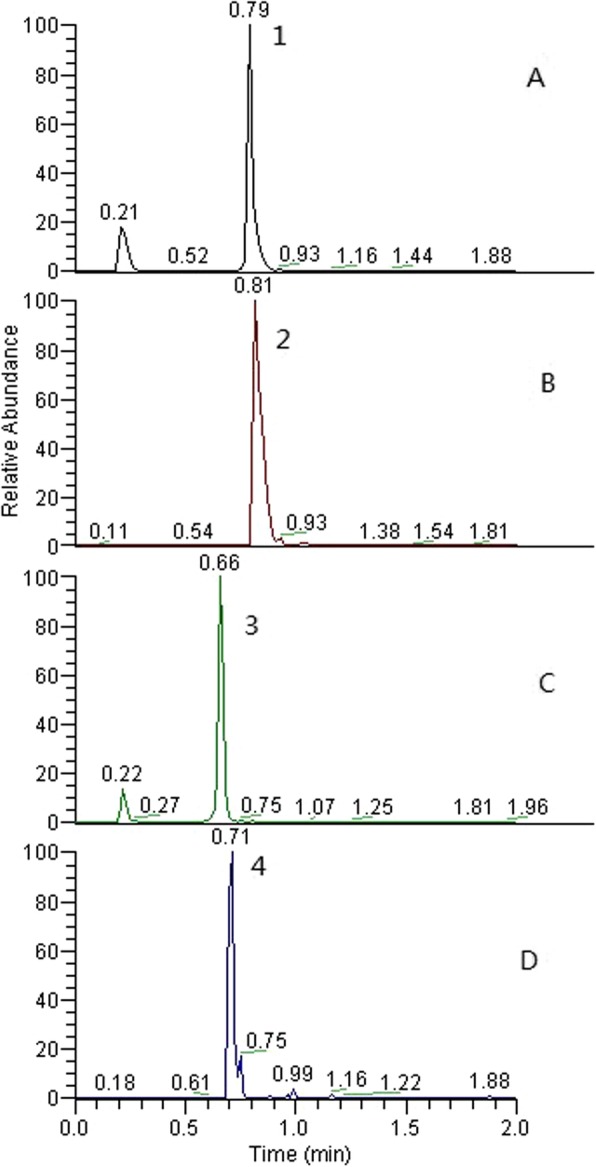
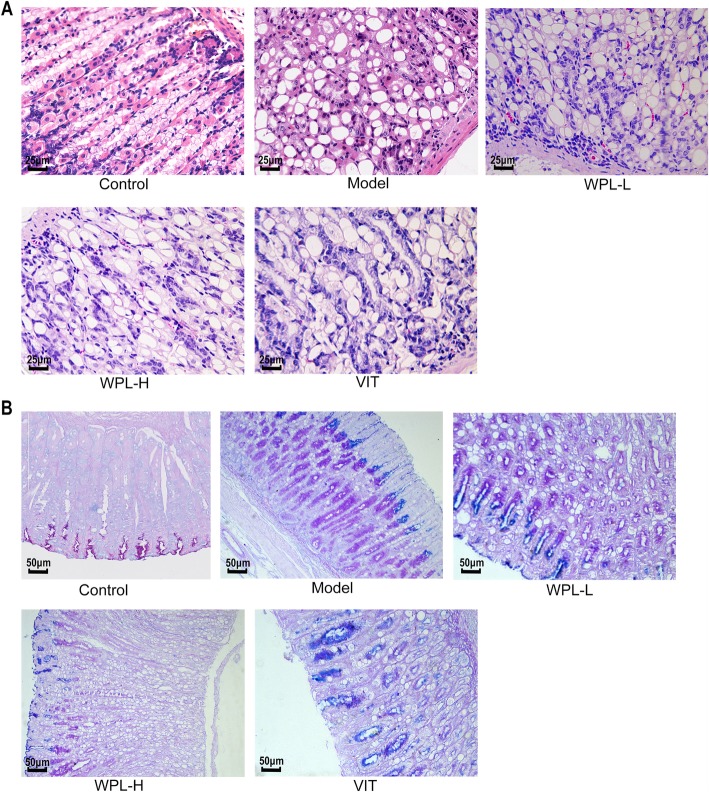
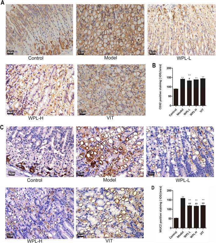
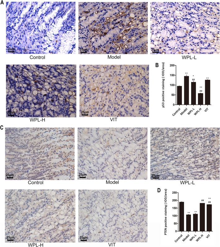
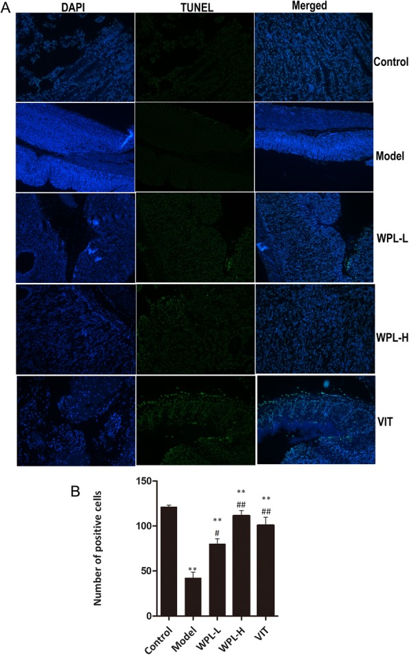
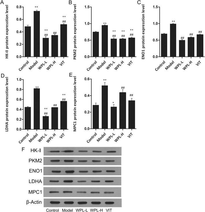
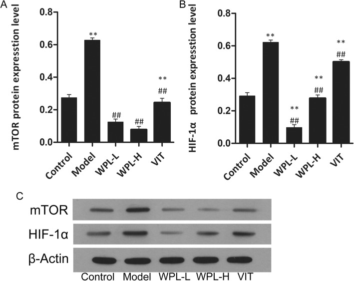
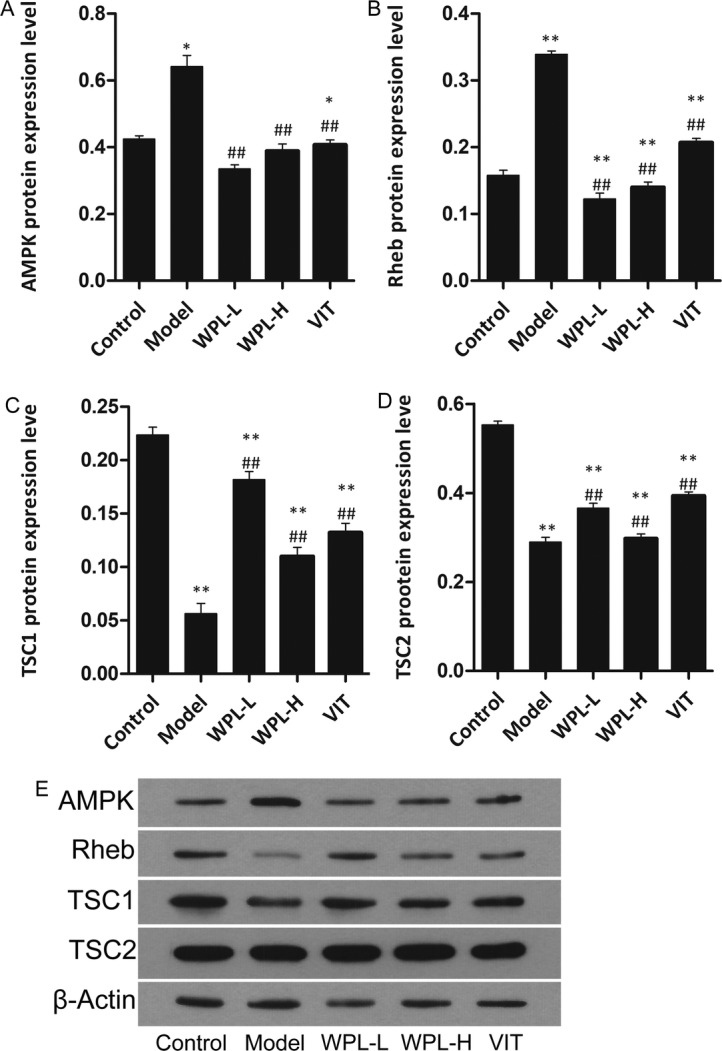
Methods: Firstly, the major components of WPL are chemically characterized by HPLC analytical method. In this study, we chose the Atp4a-/- mouse model (Spicer etal., J Biol Chem 275:21555-21565, 2000) for GPL analysis. Different doses of WPL were administered orally to mice for 10 weeks. Next, the pathological changes of gastric mucosa were assessed by the H&E staining and AB-PAS staining. In addition, TUNEL staining was used to evaluate apoptosis, and we further used immunohistochemically labelled CDX2, MUC2, ki-67, PTEN, and p53 proteins to assess the characteristic changes of gastric mucosa in precancerous lesions. The levels of such transporters as HK-II, PKM2, ENO1, MPC1, and LDHA were determined by Western blot analysis. Finally, we assessed the expression of mTOR, HIF-1α, AMPK, Rheb, TSC1 and TSC2 protein in the gastric mucosa of Atp4a-/-mice.

Results: In this work, we evaluated the protective effect of WPL on gastric mucosa in mice with precancerous lesions. The aberrant apoptosis in gastric mucosa of gastric pre-cancerous lesions was controlled by WPL (P<0.05). Furthermore, WPL suppressed the expression of CDX2, MUC2, ki-67, PTEN and p53, as the levels of these proteins decreased significantly compared with the model group (P<0.05). In parallel, WPL significantly suppressed the expression of transporters, such as HK-II, PKM2, ENO1, MPC1 and LDHA (P<0.05). In addition, mTOR, HIF-1a, AMPK, Rheb, TSC1 and TSC2 protein levels in gastric mucosa of Atp4a-/- mice in the high- and low-dose WPL groups were significantly lower than those in the model group (P<0.05), while the expression of TSC1 and TSC2 protein was significantly higher (P<0.05).

Conclusions: Conclusively, WPL could ameliorate GPLs in Atp4a-/- mice by inhibiting the expression of transporters and suppressing the aberrant activation of mTOR/HIF-1α.

Keywords: Atp4a−/−mice; Gastric precancerous lesions; Glycolysis; Traditional Chinese medicine; Weipiling; mTOR/HIF-1α pathways.


Conflict of interest statement

The authors declare that they have no competing interests.

Figures

Fig. 1
Fig. 1
HPLC chromatogram of WPL text samples (b, d) and reference samples (a, c). Note: Peak 1 is isoflavone glucoside, and Peak 2 is notoginsenoside R1
Fig. 2
Fig. 2
Histological evaluation of gastric epithelial dysplasia and intestinal metaplasia (H&E staining, 400×). a The model gastric epithelium displayed dysplasia pathology characterized by glandular architectural abnormalities. After WPL intervention, these dysplasia pathological alterations, especially irregularities of glandular structure, were regressed to varying degrees. n = 6 in each group. b Histological evaluation of gastric intestinal metaplasia (AB-PAS staining, 200×). Neutral mucins present in normal mucosa were stained red. Sialomucins expressed only in intestinal-type metaplasia (IM) were stained blue. Images of the model gastric epithelium depicted prominent IM lesions, which were dramatically reduced after WPL administration. n = 5 in each group
Fig. 3
Fig. 3
Evaluation of CDX2 and MUC2 in gastric mucosa. a Representative immunohistochemical staining of CDX2 in the mouse gastric mucosa. b Analysis of gastric mucosa total protein of CDX2. c Representative immunohistochemical staining of MUC2 in the mouse gastric mucosa. d Analysis of gastric mucosa total protein of MUC2. Data are expressed as the mean ± SD. ** indicates P < 0.01 compared with the control group. # indicates P < 0.05 and ## indicates P < 0.01 compared with the model group (IHC, 400×). n = 5 in each group
Fig. 4
Fig. 4
Evaluation of ki-67 in gastric mucosa. a Representative immunohistochemical staining of ki-67 in the mouse gastric mucosa. b Analysis of gastric mucosa total protein. Data are expressed as the mean ± SD. ** indicates P < 0.01 compared with the control group. ## indicates P < 0.01 compared with the model group (IHC, 400×). n = 5 in each group
Fig. 5
Fig. 5
Evaluation of p53 and PTEN in gastric mucosa. a Representative immunohistochemical staining of p53 in the mouse gastric mucosa. b Analysis of gastric mucosa total protein of p53. c Representative immunohistochemical staining of PTEN in the mouse gastric mucosa. d Analysis of gastric mucosa total protein of PTEN. Data are expressed as the mean ± SD. ** indicates P < 0.01 compared with the control group. ## indicates P < 0.01 compared with the model group (IHC, 400×). n = 5 in each group
Fig. 6
Fig. 6
Effects of WPL on apoptosis. a The images of TUNEL positive cells were captured by a fluorescence microscope (× 200). left panel: DAPI staining; middle panel: TUNEL assay, right panel: merged image showing the results of DAPI staining and the TUNEL assay. b Quantitative result of TUNEL assay was analyzed. Data are expressed as the mean ± SD. ** indicates P < 0.01 compared with the control group. indicates P < 0.05 and indicates P < 0.01 compared with the model group. n = 5 in each group
Fig. 7
Fig. 7
Expression of key enzymes for glucose metabolism in gastric tissue treated with WPL (a-e) Western blotting was used for the analysis. (f) Expression of proteins in the mouse gastric. Data are expressed as the mean ± SD. * indicates P < 0.05 and ** indicates P < 0.01 compared with the control group. # indicates P < 0.05 and ## indicates P < 0.01 compared with the model group. n = 5 in each group
Fig. 8
Fig. 8
Expression of mTOR and HIF-1α protein. a-b Western blotting was used for the analysis. c Expression of proteins in the mouse gastric. Data are expressed as the mean ± SD. ** indicates P < 0.01 compared with the control group. ## indicates P < 0.01 compared with the model group. n = 5 in each group
Fig. 9
Fig. 9
Effect of WPL on AMPK pathway protein levels in gastric epithelial cells. a-d Western blotting was used for the analysis. e Expression of proteins in the mouse gastric. Data are expressed as the mean ± SD. * indicates P < 0.05, and** indicates P < 0.01, compared with the control group. ## indicates P < 0.01 compared with the model group. n = 5 in each group

References

    1. World Health Organization. GLOBOCAN 2012: Estimated cancer incidence, mortality and prevalence worldwide. 2012, http:// globocan. iarc. fr/Pages/fact_sheets_cancer. aspx (accessed 20 June 2016).
    1. Siegel R, Naishadham D, Jemal A. Cancer statistics, 2012. CA Cancer J Clin. 2012;62:10–29. doi: 10.3322/caac.20138. - DOI - PubMed
    1. Correa P. Human gastric carcinogenesis: a multistep and multifactorial process–first American Cancer Society award lecture on cancer epidemiology and prevention. Cancer Res. 1992;52:6735–6740. - PubMed
    1. Sun SB, Chen ZT, Zheng D, Huang ML, Xu D, Zhang H, Wang P, Wu J. Clinical pathology and recent follow-up study on gastric intraepithelial neoplasia and gastric mucosal lesions. Hepatogastroenterology. 2013;60(127):1597–1601. - PubMed
    1. de Vries AC, van Grieken NC, Looman CW, Casparie MK, de Vries E, Meijer GA, Kuipers EJ. Gastric cancer risk in patients with premalignant gastric lesions: a nationwide cohort study in the Netherlands. Gastroenterology. 2008;134(4):945–952. doi: 10.1053/j.gastro.2008.01.071. - DOI - PubMed

Publication types

MeSH terms

Substances