. 2019 Nov 19;14(1):42.
doi: 10.1186/s13024-019-0341-5.

Wild-type Cu/Zn-superoxide dismutase is misfolded in cerebrospinal fluid of sporadic amyotrophic lateral sclerosis

Affiliations

Wild-type Cu/Zn-superoxide dismutase is misfolded in cerebrospinal fluid of sporadic amyotrophic lateral sclerosis

Eiichi Tokuda et al. Mol Neurodegener. .

Abstract

Background: A subset of familial forms of amyotrophic lateral sclerosis (ALS) are caused by mutations in the gene coding Cu/Zn-superoxide dismutase (SOD1). Mutant SOD1 proteins are susceptible to misfolding and abnormally accumulated in spinal cord, which is most severely affected in ALS. It, however, remains quite controversial whether misfolding of wild-type SOD1 is involved in more prevalent sporadic ALS (sALS) cases without SOD1 mutations.

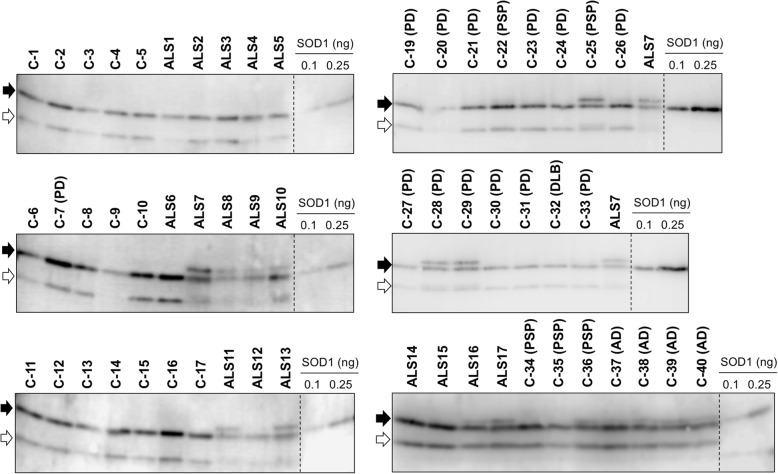
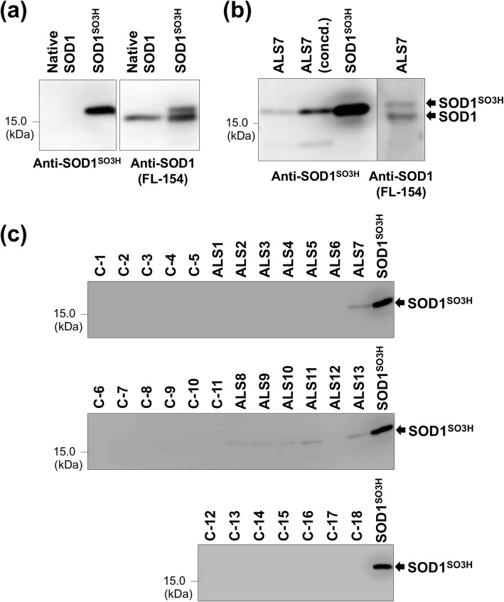
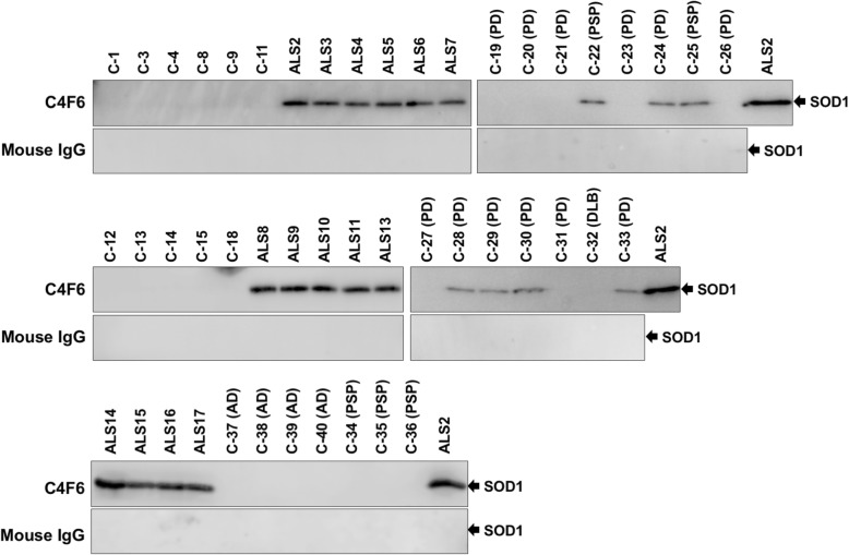
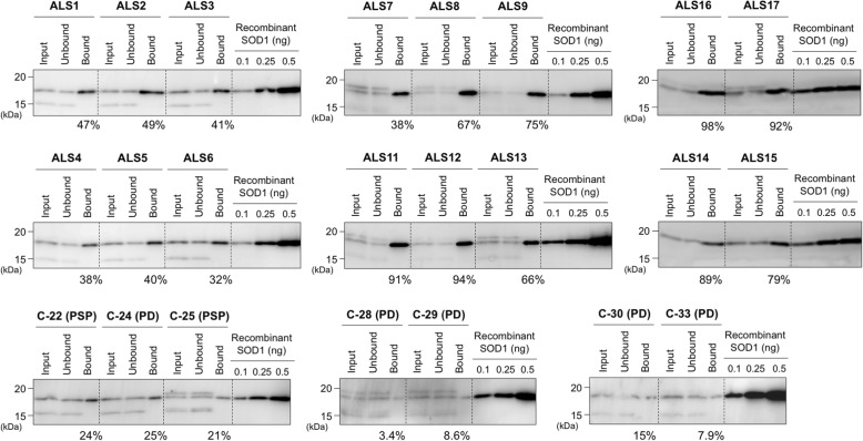
Methods: Cerebrospinal fluid (CSF) from patients including sALS as well as several other neurodegenerative diseases and non-neurodegenerative diseases was examined with an immunoprecipitation assay and a sandwich ELISA using antibodies specifically recognizing misfolded SOD1.

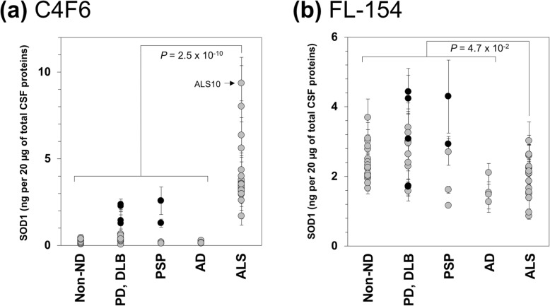
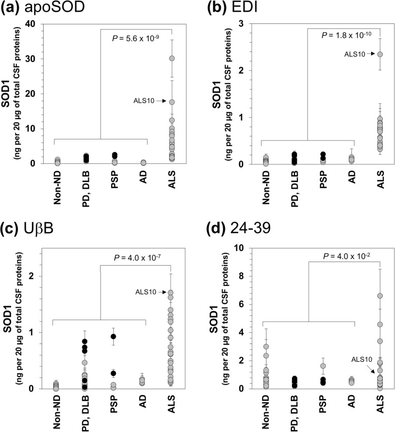
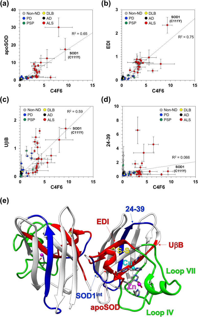
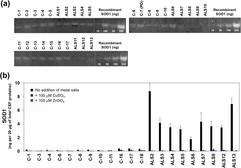
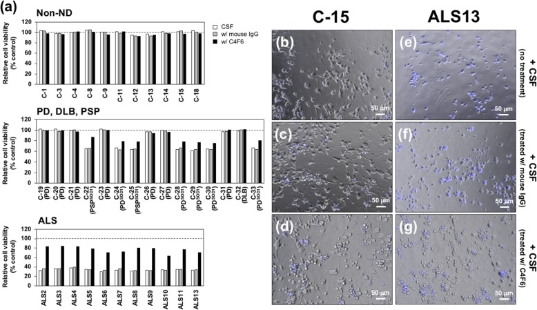
Results: We found that wild-type SOD1 was misfolded in CSF from all sALS cases examined in this study. The misfolded SOD1 was also detected in CSF from a subset of Parkinson's disease and progressive supranuclear palsy, albeit with smaller amounts than those in sALS. Furthermore, the CSF samples containing the misfolded SOD1 exhibited significant toxicity toward motor neuron-like NSC-34 cells, which was ameliorated by removal of the misfolded wild-type SOD1 with immunoprecipitation.

Conclusions: Taken together, we propose that misfolding of wild-type SOD1 in CSF is a common pathological process of ALS cases regardless of SOD1 mutations.

Keywords: Amyotrophic lateral sclerosis (ALS); Cerebrospinal fluid (CSF); Cu/Zn-superoxide dismutase (SOD1); Protein misfolding.


Conflict of interest statement

The authors declare that they have no competing interests.

Figures

Fig. 1
Fig. 1
Sulfonylation of wild-type SOD1 in CSF from some neurodegenerative disease cases. CSF samples (4 μg of total proteins) as well as recombinant human SOD1 proteins (0.1 and 0.25 ng) were analyzed by Western blotting with an anti-SOD1 antibody FL-154. The blots on the membranes were first reacted with FL-154 (0.2 μg/mL) in PBS-T with 1% (w/v) skim milk and then probed with anti-rabbit secondary antibody in PBS-T with 0.5% (w/v) skim milk (1:2000 dilution). A major band corresponding to full-length SOD1 is indicated with a filled arrow, while a band with an open arrow shows a truncated form of SOD1
Fig. 2
Fig. 2
Detection of SOD1SO3H in CSF of ALS cases using anti-SOD1SO3H antibody. (a) Native SOD1 (Sigma, #S9636: 20 μU) and a recombinant sample containing SOD1SO3H (2.5 ng) were analyzed by Western blotting side-by-side with (left) anti-SOD1SO3H and (right) FL-154 antibodies. The recombinant sample containing SOD1SO3H produced doublet bands in the Western blot with FL-154 (right), but only the upper band was recognized with anti-SOD1SO3H antibody (left). Also, native SOD1 without any oxidation was detected as a single band in the Western blot with FL-154 (right) but was not recognized with anti-SOD1SO3H antibody (left). These results confirm the retarded electrophoretic mobility of SOD1 upon sulfonylation of Cys and also support the specificity of our anti-SOD1SO3H antibody toward SOD1SO3H. (b) The CSF sample from ALS7 (4 μg of total proteins; ALS7 concd. Sample contained 10 μg of total proteins) was analyzed by Western blotting side-by-side with (left) anti-SOD1SO3H and (right) FL-154 antibodies. Doublet SOD1-positive bands were observed in the Western blot probed with FL-154 (right), among which only the upper band was recognized with anti-SOD1SO3H antibody and also exhibited the same electrophoretic mobility with that of recombinant SOD1SO3H (20 ng). (c) CSF samples (4 μg of total proteins) and recombinant SOD1SO3H (10 ng) were analyzed by Western blotting with anti-SOD1SO3H antibodies. In all panels, the blots on the membranes were reacted with either FL-154 (0.2 μg/mL) in PBS-T with 1% (w/v) skim milk or anti-SOD1SO3H (0.06 μg/mL) in PBS-T with 3% (w/v) BSA, and then probed with anti-rabbit secondary antibody in PBS-T with 0.5% (w/v) skim milk (1:2000 dilution)
Fig. 3
Fig. 3
CSF of ALS cases contains the species recognized by misfolded SOD1-specific antibody C4F6. The CSF samples (20 μg of total proteins, 100 μL of volume) from Non-ND, PD/DLB, PSP, AD, and ALS cases were analyzed by a sandwich ELISA with (a) C4F6 and (b) FL-154 as capture antibodies, and Pan-SOD1 was used as a detection antibody. As described in the Methods, absorbance values at 490 nm measured in the ELISA were substituted into equations fitted to standard curves (Additional file 3: Figure S2), and apparent amounts of SOD1 were thereby estimated and plotted as SOD1 (ng/20 μg of total CSF proteins). The data on the PD and PSP cases containing the C4F6-reactive SOD1 (also see Fig. 4) were shown in black filled circles. Three independent experiments were performed to estimate error bars (standard deviation). The statistical differences were analyzed with Mann-Whitney U-test, and P values are shown
Fig. 4
Fig. 4
Immunoprecipitation experiments confirm the presence of C4F6-reactive SOD1 in CSF of all ALS cases as well as some of PD and PSP cases. The C4F6-crosslinked or mouse IgG-crosslinked magnetic beads were first incubated with CSF (20 μL) at 4 °C for 24 h, and the SOD1 proteins bound to those magnetic beads were then eluted with 10 μL of 100 mM citrate buffer at pH 3.1. The eluates (10 μL) were analyzed by Western blotting with FL-154 antibody. Due to the limited availability of the CSF samples, we could not examine several cases including C-2, C-5, C-6, C-7, C-10, C-16, C-17, ALS1, ALS12, ALS18, ALS19, ALS20, and ALS21
Fig. 5
Fig. 5
Most of SOD1 in CSF from ALS were recognized by misfolded SOD1-specific antibody C4F6. C4F6-crosslinked magnetic beads were first incubated with CSF (40 μL) at 4 °C for 24 h, and the solutions were collected as the “unbound” fraction. The remaining magnetic beads were then treated with 10 μL of 100 mM citrate buffer at pH 3.1 in order to elute SOD1 proteins captured by the magnetic beads (the “bound” fraction). Ten microliter of CSF (input), the unbound, and the bound fractions were analyzed by Western blotting with FL-154 antibody. The percentage of C4F6-reactive SOD1 in the CSF samples was roughly estimated by measuring the band intensity of SOD1 in the unbound and bound fractions and indicated at the bottom of each gel. Details were described in the Methods
Fig. 6
Fig. 6
CSF of ALS cases contains the SOD1 species recognized with several misfolded SOD1-specific antibodies. CSF samples (20 μg of total proteins, 100 μL of volume) were analyzed by sandwich ELISA with (a) apoSOD, (b) EDI, (c) UβB, or (d) 24–39 as a capture antibody. For all cases, Pan-SOD1 antibody was used as a detection antibody. As described in the Methods, absorbance values at 490 nm measured in the ELISA were substituted into equations fitted to standard curves (Additional file 3: Figure S2), and apparent amounts of SOD1 were thereby estimated and plotted as SOD1 (ng/20 μg of total CSF proteins). The data on PD and PSP cases with C4F6-reactive SOD1 were shown as black filled circles. Three independent experiments were performed to estimate error bars (standard deviation). The statistical differences were analyzed with Mann-Whitney U-test, and P values are shown in each panel
Fig. 7
Fig. 7
Correlation in apparent amounts of SOD1 was detected with a distinct set of misfolded SOD1-specific antibodies. Apparent amounts of SOD1 in CSF detected with (a) apoSOD, (b) EDI, (c) UβB, and (d) 24–39 (Fig. 6) were plotted against those of C4F6 (Fig. 3a). The data are represented as follows: gray circles for non-ND; blue circles for PD; green circles for PSP; a yellow circle for DLB; black circles for AD; red circles for ALS. The data of the PDSOD1 and PSPSOD1 are shown as triangles. In each panel, linear least-squares fitting to the data was performed, and the best fitting line is indicated (broken line) with a value of R2. (e) A map of epitopes recognized by the antibodies for the misfolded SOD1 is shown on a crystal structure of a native, enzymatically active form of the SOD1 homodimer (PDB ID: 2C9V). A copper ion (Cu, cyan), a zinc ion (Zn, pink), and a conserved disulfide bond (S-S, yellow) are shown. The epitopes of EDI, UβB, and apoSOD are colored red, while those of 24–39 and SOD1int are colored blue. Loops IV and VII are shown green
Fig. 8
Fig. 8
Misfolded SOD1 in CSF of ALS decreases the affinity toward metal ions. (a) The CSF samples (20 μg of total proteins) were approximately 4-fold concentrated with a centrifugal filter device and then examined for the SOD1 activities with the in-gel assay. Recombinant wild-type SOD1 protein in the holo state was analyzed as a positive control. (b) The CSF samples (20 μg of total proteins, 100 μL of total volume) were first incubated with either (blue bars) 100 μM CuSO4 or (red bars) 100 μM ZnSO4 at 4 °C overnight and then analyzed by sandwich ELISA with C4F6 and Pan-SOD1 as capture and detection antibodies, respectively. As a control, the CSF samples incubated at 4 °C overnight without addition of any metal ions (black bars) were also examined. As described in the Methods, absorbance values at 490 nm measured in the ELISA were substituted into equations fitted to standard curves (Additional file 3: Figure S2), and apparent amounts of SOD1 were thereby estimated and plotted as SOD1 (ng/20 μg of total CSF proteins). The experiments were performed in triplicate to estimate error bars (standard deviation)
Fig. 9
Fig. 9
Toxicity of CSF samples from sALS toward motor neuronal cell line comes from misfolded SOD1. (a) Differentiated NSC-34 cells were exposed to (white bars) the CSF samples, (gray bars) the CSF samples preabsorbed with normal mouse IgG, and (black bars) the CSF samples preabsorbed with mouse monoclonal C4F6 antibody for 48 h, and the cell viability was assayed with Cell Counting Kit-8. Each of the CSF samples was tested in duplicate, and the data are shown as the averaged cell viability relative to that of the negative control, in which PBS instead of CSF samples was exposed to the cells. (b-g) Representative images of the differentiated NSC-34 cells exposed to the CSF samples from (b-d) non-ND (C-15) and (e-g) sALS (ALS13) are shown. The cells were incubated with (b, e) the CSF samples, (c, f) the CSF samples preabsorbed with mouse IgG, and (d, g) the CSF samples preabsorbed with mouse monoclonal C4F6 antibody. The dying cells were stained with DAPI (shown in blue)

References

    1. Andersen PM, Al-Chalabi A. Clinical genetics of amyotrophic lateral sclerosis: what do we really know? Nat Rev Neurol. 2011;7:603–615. doi: 10.1038/nrneurol.2011.150. - DOI - PubMed
    1. Robberecht W, Philips T. The changing scene of amyotrophic lateral sclerosis. Nat Rev Neurosci. 2013;14:248–264. doi: 10.1038/nrn3430. - DOI - PubMed
    1. Rosen DR, Siddique T, Patterson D, Figlewicz DA, Sapp P, Hentati A, et al. Mutations in Cu/Zn superoxide dismutase gene are associated with familial amyotrophic lateral sclerosis. Nature. 1993;362:59–62. - PubMed
    1. Chio A, Traynor BJ, Lombardo F, Fimognari M, Calvo A, Ghiglione P, et al. Prevalence of SOD1 mutations in the Italian ALS population. Neurology. 2008;70:533–537. doi: 10.1212/01.wnl.0000299187.90432.3f. - DOI - PubMed
    1. Kwon MJ, Baek W, Ki CS, Kim HY, Koh SH, Kim JW, et al. Screening of the SOD1, FUS, TARDBP, ANG, and OPTN mutations in Korean patients with familial and sporadic ALS. Neurobiol Aging. 2012;33:1017 e1017–1017 e1023. doi: 10.1016/j.neurobiolaging.2011.12.003. - DOI - PubMed

Publication types