

Optic disc drusen in children: morphologic features using EDI-OCT
- PMID: 31745329
- PMCID: PMC7608464
- DOI: 10.1038/s41433-019-0694-6
Optic disc drusen in children: morphologic features using EDI-OCT
Abstract
Aims: This study aimed to investigate morphologic features of optic disc drusen (ODD) and peripapillary hyperreflective ovoid mass-like structures (PHOMS) in children, using enhanced depth imaging optical coherence tomography (EDI-OCT). It also assessed if the presence of these features were associated with decreased peripapillary retinal nerve fibre layer (RNFL) thickness.
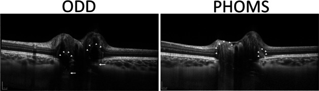
Methods: Retrospective observational study of children with ODD. All subjects underwent complete ophthalmic examination and multimodal imaging. ODD were identified on EDI-OCT as circumscribed hyporeflective spheroidal elements located in front of lamina cribrosa, fully or partially surrounded by a hyperreflective border. PHOMS were identified as hyperreflective ovoid structures located in the peripapillary circumference. Both associations between ODD and RNFL loss and PHOMS and RNFL loss were tested using chi-squared test.
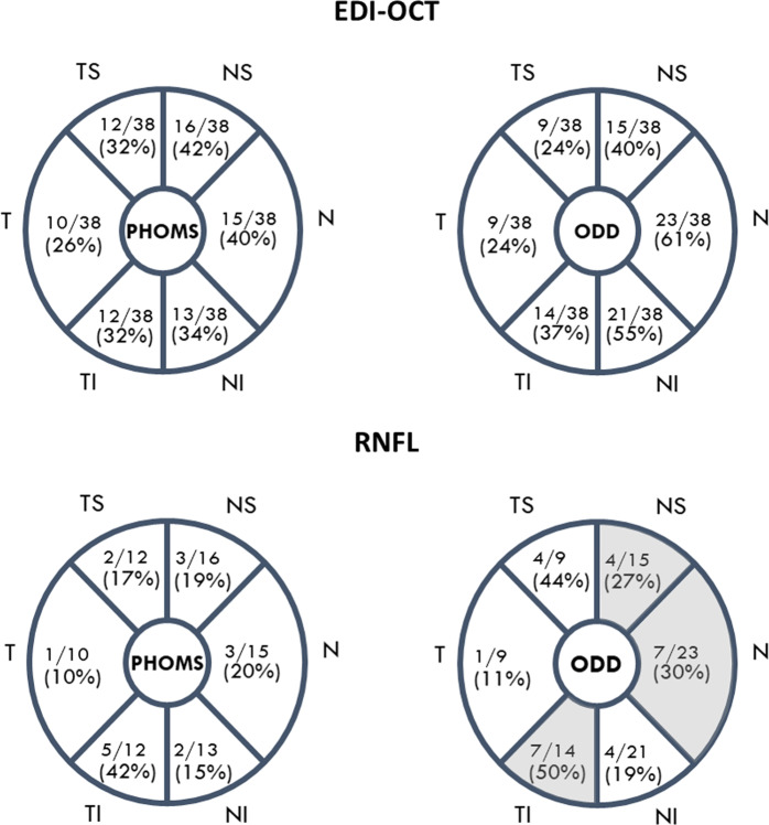
Results: In total, 38 eyes of 20 children were analysed. PHOMS were present in 90% of patients. ODD and PHOMS were predominantly found in the nasal, superonasal and inferonasal sectors. A significant positive association was found between ODD and decreased RNFL thickness in the nasal (p = 0.02), superonasal (p = 0.05) and inferotemporal (p = 0.04) sectors. There was no significant association found with the presence of PHOMS.
Conclusion: EDI-OCT allowed morphological analysis of ODD and PHOMS in children. Drusen were found to be distinct from PHOMS both in their appearance and impact on the RNFL. ODD are hyporeflective and appear on the ONH above the lamina cribrosa and were associated with decreased thickness of the RNFL. On the contrary, PHOMS are hyperreflective structures located around the ONH and were not associated with RNFL loss.
Conflict of interest statement
The authors declare that they have no conflict of interest.
Figures
References
-
- Malmqvist L, Li XQ, Eckmann CL, Skovgaard AM, Olsen EM, Larsen M, et al. Optic disc drusen in children: the copenhagen child cohort 2000 eye study. J Neuro-Ophthalmol. 2017;0:1–7. - PubMed
-
- Kiegler HR. Comparison of functional findings with results of standardized echography of the optic nerve in optic disk drusen. Wien Klin Wochenschr. 1999;107:651–3. - PubMed
-
- Hamann S, Malmqvist L, Costello F. Optic disc drusen: understanding an old problem from a new perspective. Acta Ophthalmol. 2018;1:1–12. - PubMed
Publication types
MeSH terms
LinkOut - more resources
Full Text Sources
