. 2020 Jan 10;48(1):486-499.
doi: 10.1093/nar/gkz1098.

Phylogenetic debugging of a complete human biosynthetic pathway transplanted into yeast

Affiliations

Phylogenetic debugging of a complete human biosynthetic pathway transplanted into yeast

Neta Agmon et al. Nucleic Acids Res. .

Abstract

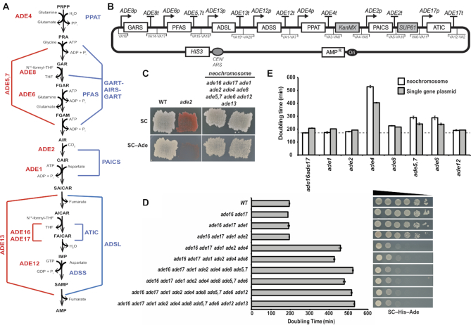
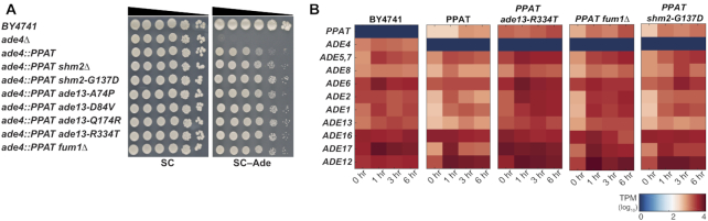
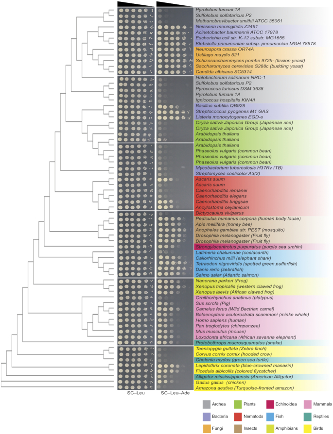
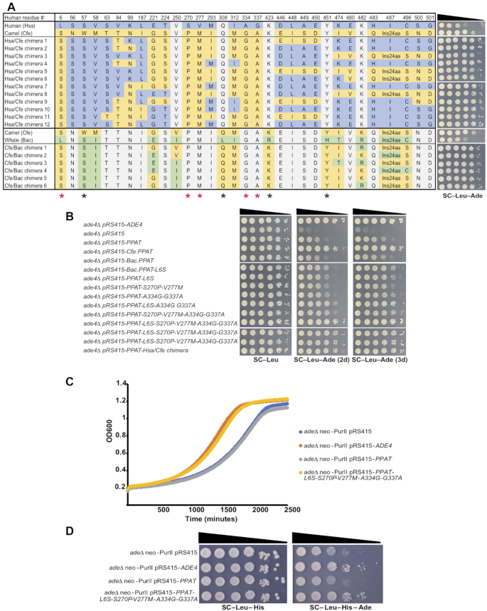
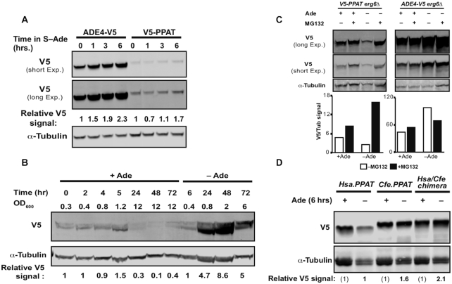
Cross-species pathway transplantation enables insight into a biological process not possible through traditional approaches. We replaced the enzymes catalyzing the entire Saccharomyces cerevisiae adenine de novo biosynthesis pathway with the human pathway. While the 'humanized' yeast grew in the absence of adenine, it did so poorly. Dissection of the phenotype revealed that PPAT, the human ortholog of ADE4, showed only partial function whereas all other genes complemented fully. Suppressor analysis revealed other pathways that play a role in adenine de-novo pathway regulation. Phylogenetic analysis pointed to adaptations of enzyme regulation to endogenous metabolite level 'setpoints' in diverse organisms. Using DNA shuffling, we isolated specific amino acids combinations that stabilize the human protein in yeast. Thus, using adenine de novo biosynthesis as a proof of concept, we suggest that the engineering methods used in this study as well as the debugging strategies can be utilized to transplant metabolic pathway from any origin into yeast.


Figures

Figure 1.
Figure 1.
Yeast and human adenine de novo pathway and humanized strain construction. (A) Schematic representation of the adenine de-novo biosynthesis pathway. Red, yeast genes; blue human. (B) Complete design of the human Purine de-novo Neochromosome for expression in yeast cells. Using yeast golden-gate assembly (18,22) we cloned each human gene (synthesized codon optimized for expression in S. cerevisiae) with its yeast ortholog's promoter and terminator and appropriate adaptors for VEGAS assembly (VA1-VA20) (22) of the neochromosome (asterisk indicate previously unpublished VEGAS adaptors, for details see methods section). (C) Comparing growth on media with and without adenine shows complementation in the humanized strain deleted for all yeast genes and expressing all human genes from the neochromosome. Comparison is to a ade2Δ strain that cannot grow on media without adenine and a WT (wild type; BY4741) that can grow on both. (D) Graphic representation of doubling time in medium without adenine of strains with increasing number of native adenine de novo genes deleted carrying the humanized neochromosome. Grow assay indicates a dramatic increase in doubling time following ADE4 deletion. Spot assay on the left shows a similar trend to the growth assay, showing a dramatic growth defect following deletion of ADE4. (E) Graphic representation of doubling time to verify incomplete complementation of ade4 by its human ortholog PPAT by testing single gene deletions and comparing neochromosome complementation and single gene plasmids in medium without adenine. Dotted line represents doubling time of a wild-type strain grown in the same medium.
Figure 2.
Figure 2.
PPAT suppressor analysis. (A) Reconstitution of mutations found in suppressor strain genomes. All reconstructed suppressors alleviate the PPAT phenotype. Comparing mutants to deletion strain indicates that in all suppressors the phenotype is probably due to loss of function mutations. (B) Heat-map showing ADE gene transcripts in the yeast strains tested (BY4741, ade4::PPAT, ade4::PPAT ade13-R334T, ade4::PPAT fum1Δ and ade4::PPAT shm2-G137D) grown in SC and moved to SC–Ade. Sampled were collected after 1, 3 and 6 h for RNA preparation. RNA-seq analysis was performed on all samples and results are presented in a heat-map (scale represents log10, transcripts per million [tpm]).
Figure 3.
Figure 3.
Phylogenetic analysis of PPATs. Phylogenetic tree generate by multiple sequence alignment CLUSTAL-OMEGA (44) on the EMPL-EBI site (49,50) aligned with a spot assay showing the growth of ade4Δ strains expressing PPATs from different organisms and their ability to complement ade4 deletion.
Figure 4.
Figure 4.
Phylogenetic analysis reveals distinct residues that affect PPATs function in yeast. (A) Left – Table showing the variable residues between human (Hsa), camel (Cfe) and whale (Bac) PPATs in the parental strain and in the DNA shuffling chimera products. DNA shuffling experiment was done in two separated reaction: Hsa + Cfe and Cfe + Bac. Colors represent the residues specific to each organism: Human (blue), camel (Yellow) and whale (green). Residues which are identical to both shuffled parents are marked in gray. Asterisks on the bottom indicate residues common to all chimeras, red asterisks are those absent from the human sequence. Right – Spot assay of ade4Δ strains carrying plasmids expressing the products of DNA shuffling between human (Hsa) and camel (Cfe) PPATs and camel and whale (Bac) on SC–Leu/SC–Leu–Ade. All 18 candidates sequenced show 5 invariable common amino acids changes L6S, S270P, V227M, A334G and G337A (relative to the human reference sequence). (B) Growth assay of ade4Δ strains carrying plasmids expressing different version of PPATs on SC–Leu/SC–Leu–Ade. We have reconstructed the five substitutions that were invariant in all DNA shuffling products into the human PPAT sequence in different combinations. Results shows that several of the combinations show increase complementation. However, the five substitutions variants (pRS415-PPAT-L6S-S270M-V227M-A334G-G337A) shows similar complementation as the Has/Cfe chimera. (C, D) Graphic representation of the growth in medium without adenine and spot assay of fully humanized yeast cells supplemented with either an empty vector (pRS415), vector expressing yeast ADE4 gene (pRS415-ADE4), vector expressing human PPAT (pRS415-PPAT) or vector expressing human PPAT with the five substitution variant (pRS415-PPAT-L6S-S270M-V227M A334G G337A). Indicating that PPAT carrying the 5 substitutions complement as well as yeast ADE4.
Figure 5.
Figure 5.
Ppat protein level is sensitive to presence of adenine in the medium. (A) Immunoblot of cells expressing either Ade4 tagged with V5 or Ppat tagged with V5 integrated into the native ADE4 locus and transcribed from its native promoter. Both Ade4-V5 and V5-Ppat show an increase in protein level during prolonged time in media without adenine. Relative V5 signal is calculated V5/α-Tubulin divided by the signal in time 0 h. (B) Immunoblot of cells expressing Ppat tagged with V5 in medium with or without adenine. Relative V5 signal is calculated V5/α-Tubulin similar to (a). Accumulation of significantly more Ppat in media without adenine. Results were repeated in two biological replicates. (C) Immunoblot of cells expressing either Ppat or Ade4 tagged with V5 6 h in medium with or without adenine and with or without 50 μM MG132. Ade4-V5 expressing cells show similar levels of protein in the presence and absence of MG132, while V5-PPAT expressing cells show an increase in protein level in the presence of MG132 without adenine. The graph indicates the signal for V5 divided by the α-tubulin signal. (D) Immunoblot of cells expressing either Human Ppat (Hsa.PPAT), Camel PPAT (Cfe.PPAT) or Human/Camel chimera PPAT (Hsa/Cfe.PPAT) tagged with V5 expressed from a CEN/ARS plasmid regulated by ADE4 native promoter and terminator. Cells were grown for 6 h in either SC–Leu or SC–Ade–Leu medium to maintain the expression plasmid. Samples were collected and total protein was prepared. Relative V5 signal is calculated as V5/α-Tubulin divided by the signal in Hsa.PPAT sample grown in SC–Ade media. Camel PPAT and chimera PPAT variants show higher levels of protein in adenine depleted medium.

References

    1. Zhang N., Osborn M., Gitsham P., Yen K., Miller J.R., Oliver S.G.. Using yeast to place human genes in functional categories. Gene. 2003; 303:121–129. - PubMed
    1. Kruger W.D., Cox D.R.. A yeast system for expression of human cystathionine beta-synthase: structural and functional conservation of the human and yeast genes. Proc. Natl. Acad. Sci. U.S.A. 1994; 91:6614–6618. - PMC - PubMed
    1. Marini N.J., Gin J., Ziegle J., Keho K.H., Ginzinger D., Gilbert D.A., Rine J.. The prevalence of folate-remedial MTHFR enzyme variants in humans. Proc. Natl. Acad. Sci. U.S.A. 2008; 105:8055–8060. - PMC - PubMed
    1. Lee M.G., Nurse P.. Complementation used to clone a human homologue of the fission yeast cell cycle control gene cdc2. Nature. 1987; 327:31–35. - PubMed
    1. Brachmann R.K., Vidal M., Boeke J.D.. Dominant-negative p53 mutations selected in yeast hit cancer hot spots. Proc. Natl. Acad. Sci. U.S.A. 1996; 93:4091–4095. - PMC - PubMed

Publication types

MeSH terms