. 2020 Jan;21(1):69-76.
doi: 10.3892/mmr.2019.10803. Epub 2019 Nov 6.

Silencing of RBP‑JK promotes the differentiation of bone marrow mesenchymal stem cells into vascular endothelial cells

Affiliations

Silencing of RBP‑JK promotes the differentiation of bone marrow mesenchymal stem cells into vascular endothelial cells

Zhengliang Luo et al. Mol Med Rep. 2020 Jan.

Abstract

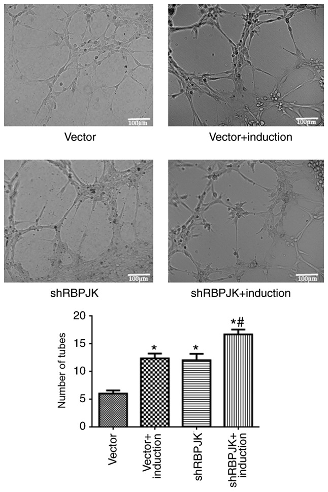
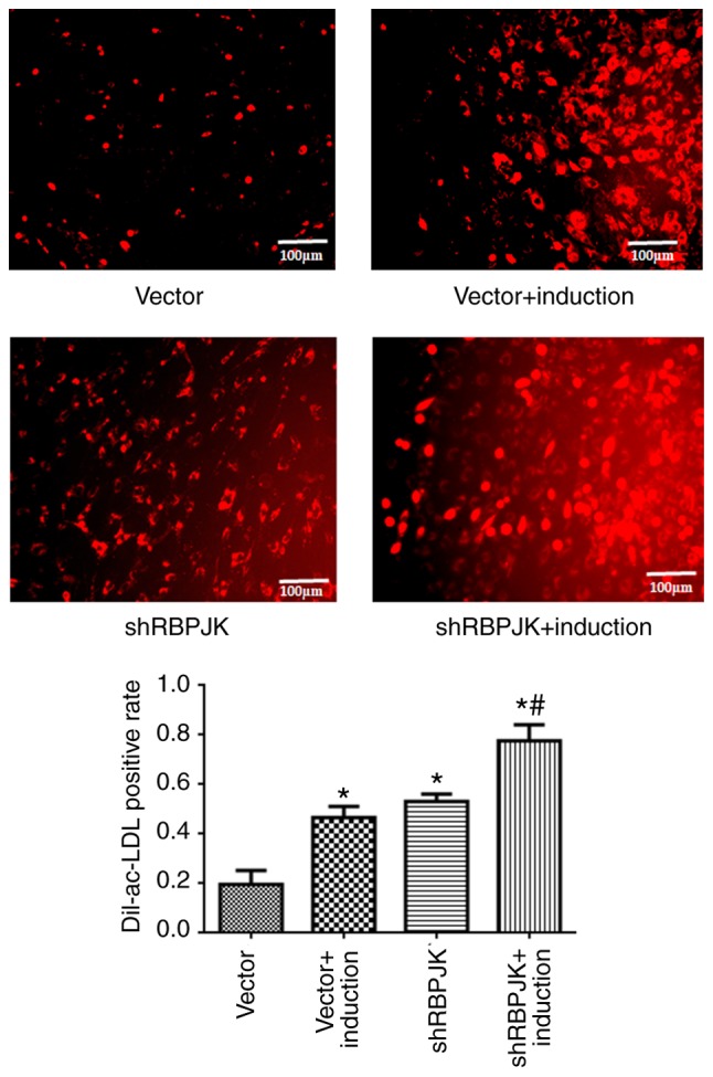
Bone marrow mesenchymal stem cells (BM‑MSCs) are important for postnatal angiogenesis and are suitable for use in construction of blood vessels by tissue engineering. The present study aimed to investigate the influence of recombination signal binding protein for immunoglobulin kappa J region (RBP‑JK) on the differentiation of BM‑MSCs into vascular endothelial cells, and to assess the underlying mechanisms. BM‑MSCs were isolated and identified by flow cytometry. Lentiviral vectors encoding RBP‑JK shRNA (shRBPJK) were constructed to knockdown RBP‑JK expression and endothelial differentiation of BM‑MSCs was induced. The experimental groups were treated with: empty lentiviral vector (vector group), growth factors (bFGF and VEGF; induced group), shRBPJK (shRBPJK group), and growth factors + shRBPJK (induced + shRBPJK group). The expression of endothelial markers, vascular endothelial growth factor receptor 2 (Flk‑1), and von Willebrand factor (vWF) were detected by immunofluorescence. Additionally, in vitro blood vessel formation and phagocytosis were assessed using acetylated LDL, Dil complex and the underlying molecular mechanisms evaluated by western blotting. BM‑MSCs were separated and transduced with shRBPJK to reduce RBP‑JK expression. Compared with the vector group, the expression of the endothelial cell markers, Flk‑1 and vWF, in vitro tubule formation, and phagocytosis ability increased, while the expression levels of p‑AKT/AKT and p‑NF‑κB/NF‑κB were significantly decreased (P<0.05) in the induced, shRBPJK, and induced + shRBPJK groups. Compared with the induced group, the expression of Flk‑1 and vWF, the number of tubules, and phagocytosis were higher in the induced + shRBPJK group, while the expression levels of p‑AKT/AKT and p‑NF‑κB/NF‑κB were lower (P<0.05). Collectively, the present data indicated that silencing of RBP‑JK promotes the differentiation of MSCs into vascular endothelial cells, and this process is likely regulated by AKT/NF‑κB signaling.

Keywords: rBP-JK; bone marrow mesenchymal stem cells; endothelial cells.


Figures

Figure 1.
Figure 1.
Identification of BM-MSCs. The majority of cells were CD90+ CD105+ CD34. BM-MSCs, bone marrow mesenchymal stem cells.
Figure 2.
Figure 2.
Verification of the interference effect of shRBPJK. (A) RBP-JK mRNA levels. (B) RBP-JK protein levels. shRBPJK2 had the optimal influence on the expression of RBP-JK. Upper panel: Representative blots. Lower panel: quantification data. *P<0.05, compared with the control group. RBP-JK, recombination signal binding protein for immunoglobulin kappa J region; shRBPJK, RBP-JK shRNA.
Figure 3.
Figure 3.
The expression of endothelial cell surface markers. (A) Representative images of Flk-1 and vWF expression. Left panel: Flk-1 expression. Right panel: vWF expression. (B) Quantification data for Flk-1 and vWF expression. *P<0.05, compared with the vector control group; #P<0.05, compared with the induced group. Scale bars, 100 µm. Flk-1, vascular endothelial growth factor receptor 2; vWF, von Willebrand factor; shRBPJK, RBP-JK shRNA.
Figure 4.
Figure 4.
Results of in vitro angiogenesis assay. Upper panel: representative images. Lower panel: Quantification data. *P<0.05, compared with the vector control group; #P<0.05, compared with the induced group. Scale bars, 100 µm. shRBPJK, RBP-JK shRNA.
Figure 5.
Figure 5.
Dil-ac-LDL phagocytosis assay. Upper panel: representative images. Lower panel: Quantification data. *P<0.05, compared with the vector control group; #P<0.05, compared with the induced group. Scale bars, 100 µm. shRBPJK, RBP-JK shRNA.
Figure 6.
Figure 6.
Expression of AKT, p-AKT, NF-κB, and p-NF-κB. (A) Representative blots. (B) Quantification data for p-AKT/AKT. (C) Quantification data for p-NF-κB/ NF-κB. *P<0.05, compared with the vector control group; #P<0.05, compared with the induced group. shRBPJK, RBP-JK shRNA.

References

    1. Wang W, Yeung KWK. Bone grafts and biomaterials substitutes for bone defect repair: A review. Bioact Mater. 2017;2:224–247. doi: 10.1016/j.bioactmat.2017.05.007. - DOI - PMC - PubMed
    1. Henkel J, Woodruff MA, Epari DR, Steck R, Glatt V, Dickinson IC, Choong PF, Schuetz MA, Hutmacher DW. Bone regeneration based on tissue engineering conceptions-A 21st century perspective. Bone Res. 2013;1:216–248. doi: 10.4248/BR201303002. - DOI - PMC - PubMed
    1. Socie G, Selimi F, Sedel L, Frija J, Devergie A, Esperou Bourdeau H, Ribaud P, Gluckman E. Avascular necrosis of bone after allogeneic bone marrow transplantation: Clinical findings, incidence and risk factors. Br J Haematol. 1994;86:624–628. doi: 10.1111/j.1365-2141.1994.tb04795.x. - DOI - PubMed
    1. Bing W, Pang X, Qu Q, Bai X, Yang W, Bi Y, Bi X. Simvastatin improves the homing of BMSCs via the PI3K/AKT/miR-9 pathway. J Cell Mol Med. 2016;20:949–961. doi: 10.1111/jcmm.12795. - DOI - PMC - PubMed
    1. Zheng H, Zou W, Shen J, Xu L, Wang S, Fu YX, Fan W. Opposite effects of coinjection and distant injection of mesenchymal stem cells on breast tumor cell growth. Stem Cells Transl Med. 2016;5:1216–1228. doi: 10.5966/sctm.2015-0300. - DOI - PMC - PubMed

MeSH terms

Substances