. 2020 Jan;21(1):161-172.
doi: 10.3892/mmr.2019.10815. Epub 2019 Nov 12.

Roxatidine inhibits fibrosis by inhibiting NF‑κB and MAPK signaling in macrophages sensing breast implant surface materials

Affiliations

Roxatidine inhibits fibrosis by inhibiting NF‑κB and MAPK signaling in macrophages sensing breast implant surface materials

Litong Ji et al. Mol Med Rep. 2020 Jan.

Abstract

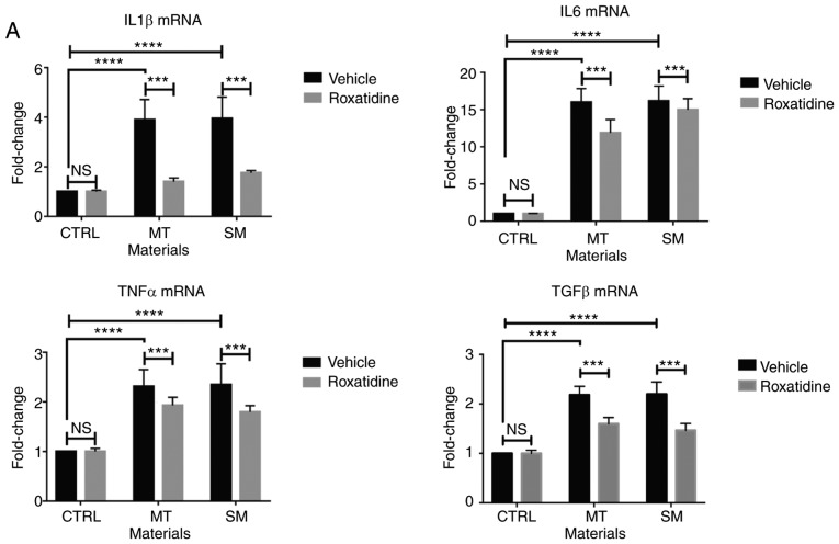
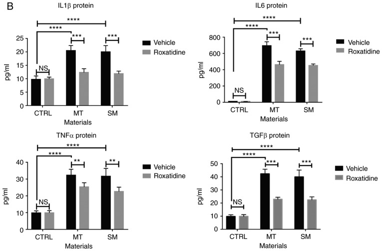
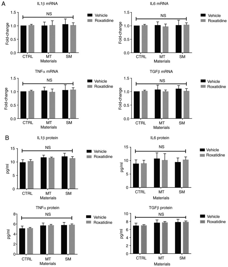
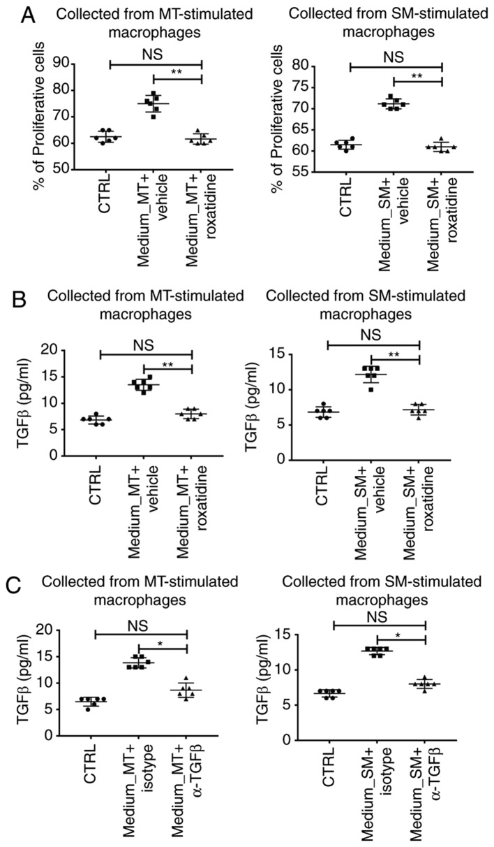
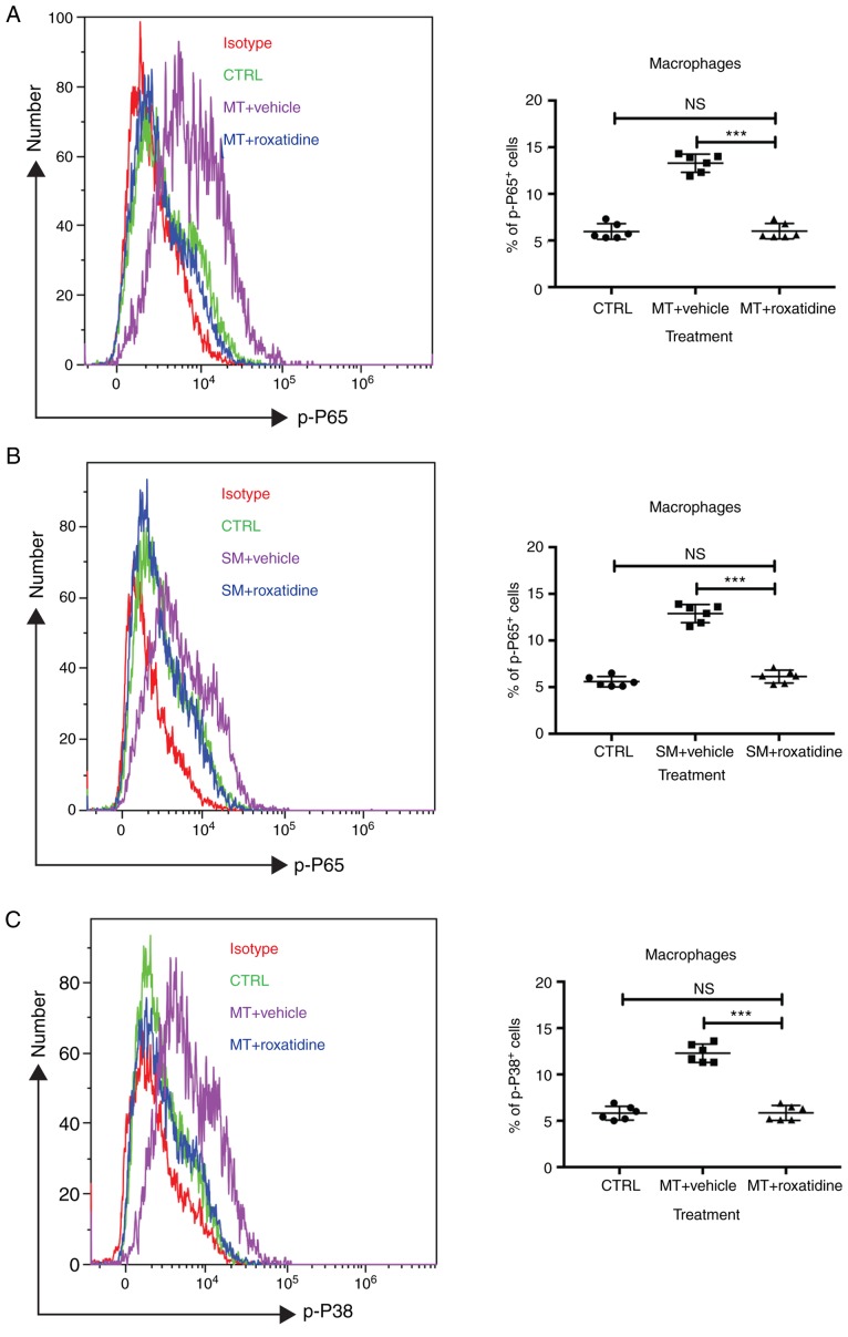
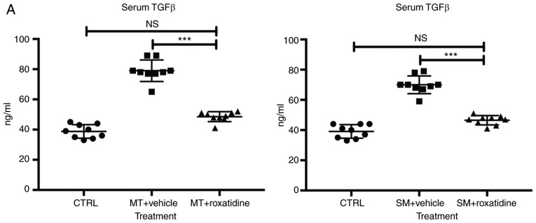
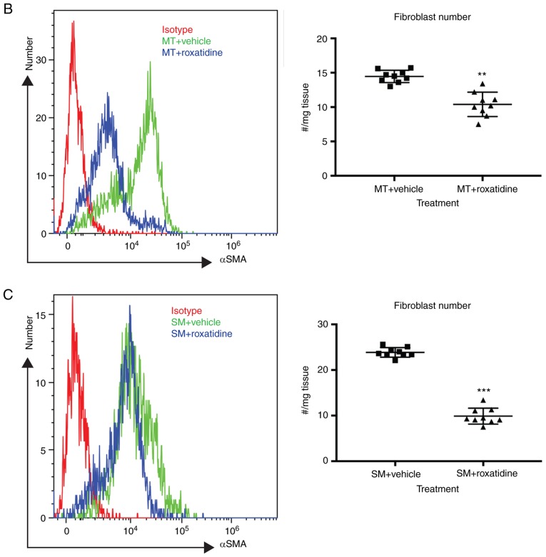
Capsular contracture is an important complication after silicone mammary implant surgery. Fibroblasts and macrophages play critical roles in the pathogenesis of capsular contracture, making these two cell types therapeutic targets. It has been reported that inhibiting histamine receptors results attenuates fibrosis, but the role of roxatidine (a histamine receptor 2 inhibitor) in preventing fibrosis caused by breast implant materials remains unknown. The aim of the present study was to assess the hypothesis that roxatidine might have a prophylactic effect in capsular contracture induced by implant material. Inflammation induced by breast implant materials was mimicked by co‑culturing macrophages or fibroblasts with these materials in vitro. Capsular contracture was modeled in mice by planting breast implant materials in a subcutaneous pocket. Roxatidine was added in the culture medium or administered to mice bearing breast implant materials. By co‑culturing macrophages or fibroblasts with common breast implant materials (micro‑textured or smooth breast implants), the present study demonstrated that macrophages respond to these materials by producing pro‑inflammatory cytokines, a process that was abolished by addition of roxatidine to the culture medium. Although fibroblasts did not respond to implant surface materials in the same way as macrophages, the conditioned media of macrophages induced proliferation of fibroblasts. Mechanistically, administration of roxatidine inhibited activation of NF‑κB and p38/mitogen‑activated protein kinase (MAPK) signaling in macrophages. Furthermore, treatment with roxatidine in implant‑bearing mice reduced serum concentrations of transforming growth factor‑β and the abundance of fibroblasts around the implant. The present study concluded that roxatidine plays an important role in preventing fibrosis by inhibiting activation of NF‑κB and p38/MAPK signaling in macrophages.

Keywords: mammary implant; capsular contracture; macrophage; fibroblast; roxatidine.


Figures

Figure 1.
Figure 1.
Pretreatment of roxatidine in the culture medium of macrophages stimulated with silicone surface materials reduced the production of proinflammatory cytokines. Cells were pretreated with roxatidine (25 µM) or vehicle for 1 h at 37°C before adding implant materials. At the end of culture (24 h after co-culture with implant materials at 37°C), cells were collected for another 24 h culture in serum-free medium at 37°C for subsequent RT-qPCR and ELISA. (A) Administration of roxatidine reduced proinflammatory cytokine production by macrophages stimulated with breast implant materials at the mRNA level. (B) Roxatidine decreased secretion of proinflammatory cytokines by stimulated macrophages. n=6 wells/group, from one of triplicated experiments. n.s., not significant, **P<0.01, ***P<0.001 and ****P<0.0001, as indicated. Data were analyzed using one-way ANOVA followed by a Bonferroni post hoc test. RT-qPCR, reverse transcription-quantitative PCR; IL, interleukin; TNF, tumor necrosis factor; TGF, transforming growth factor; CTRL, control; MT, micro-textured; SM, smooth.
Figure 1.
Figure 1.
Pretreatment of roxatidine in the culture medium of macrophages stimulated with silicone surface materials reduced the production of proinflammatory cytokines. Cells were pretreated with roxatidine (25 µM) or vehicle for 1 h at 37°C before adding implant materials. At the end of culture (24 h after co-culture with implant materials at 37°C), cells were collected for another 24 h culture in serum-free medium at 37°C for subsequent RT-qPCR and ELISA. (A) Administration of roxatidine reduced proinflammatory cytokine production by macrophages stimulated with breast implant materials at the mRNA level. (B) Roxatidine decreased secretion of proinflammatory cytokines by stimulated macrophages. n=6 wells/group, from one of triplicated experiments. n.s., not significant, **P<0.01, ***P<0.001 and ****P<0.0001, as indicated. Data were analyzed using one-way ANOVA followed by a Bonferroni post hoc test. RT-qPCR, reverse transcription-quantitative PCR; IL, interleukin; TNF, tumor necrosis factor; TGF, transforming growth factor; CTRL, control; MT, micro-textured; SM, smooth.
Figure 2.
Figure 2.
Pretreatment of roxatidine in the culture medium of fibroblasts stimulated with silicone surface materials did not affect production of proinflammatory cytokines. Cells were pretreated with roxatidine (25 µM) or vehicle for 1 h at 37°C before adding implant materials. At the end of culture (24 h after co-culture with implant materials at 37°C), cells were collected for another 24 h culture in serum-free medium at 37°C for RT-qPCR and ELISA. (A) mRNA levels of proinflammatory cytokines. (B) Protein levels of proinflammatory cytokines. n=6 wells/group, from one of triplicated experiments. n.s., not significant, by one-way ANOVA among all groups. Data were analyzed by one-way ANOVA. RT-qPCR, reverse transcription-quantitative PCR; IL, interleukin; TNF, tumor necrosis factor; TGF, transforming growth factor; CTRL, control; MT, micro-textured; SM, smooth.
Figure 3.
Figure 3.
Addition of roxatidine to the culture medium of fibroblasts stimulated with silicone surface materials did not affect proliferation, as measured by Ki67 expression using flow cytometry. Cells were pretreated with roxatidine (25 µM) or vehicle for 1 h at 37°C before adding implant materials. Fresh complete media was replaced every 24 h for up to 5 days. The primary gate of cells was set for viable cells according to the plots of forward scatter side scatter. (A) Representative histogram curves of Ki67 staining in fibroblasts stimulated with MT in the presence or absence of roxatidine at day 5. (B) Dynamic change in the percentage of Ki67-positive fibroblasts stimulated with MT in the presence or absence of roxatidine at day 5. (C) Representative histogram curves of Ki67 staining in fibroblasts stimulated with SM in the presence or absence of roxatidine at day 5. (D) Dynamic change in the percentage of Ki67-positive fibroblasts stimulated with SM in the presence or absence of roxatidine at day 5. n=6 wells per group, from one of triplicated experiments. n.s., not significant among all groups. Data were analyzed by two-way ANOVA. MT, micro-textured; SM, smooth; CTRL, control.
Figure 4.
Figure 4.
Conditioned media collected from stimulated macrophages increased L929 cell proliferation and TGFβ production. Cell-free conditioned media, collected from the co-culture system of macrophages and silicone material implants with or without pretreatment with roxatidine, were added to L929 culture for 24 h at 37°C, and then the medium was switched to the serum-free medium for another 24 h at 37°C. By the end of L929 culture, cells and media were harvested for analyses. (A) Presence of roxatidine during macrophage culture inhibited macrophage-conditioned media-induced enhanced proliferation in L929 cells. (B) The presence of roxatidine during macrophage culture decreased TGFβ production by L929 cultured in macrophage-conditioned media. (C) Presence of TGFβ neutralizing antibodies during fibroblast culture suppressed production of TGFβ by L929 cultured in macrophage-conditioned media. Left panels, media collected from MT-stimulated macrophages. Right panels, media collected from SM-stimulated macrophages. n=6 wells/group, from one of triplicated experiments. n.s., no significance; *P<0.05 and **P<0.01, as indicated. Data were analyzed by one-way ANOVA followed by the Bonferroni post hoc test. TGF, transforming growth factor; MT, micro-textured; SM, smooth; CTRL, control.
Figure 5.
Figure 5.
Addition of roxatidine to the culture medium of RAW 264.7 macrophages inhibited the activation of the NF-κB and MAPK signaling pathways. RAW 264.7 macrophages were cultured in serum-free media at 37°C. Then, 24 h after cells were seeded, roxatidine (25 µM) was added to the media 1 h prior to stimulation with silicone surface materials. Cells were collected for analysis of phosphorylation of NF-κB subunit p65 and p38 MAPK by flow cytometry 15 minutes after adding silicone surface materials to culture media. Administration of roxatidine suppressed p65 activation stimulated by (A) MT and (B) SM, as well as p38 phosphorylation stimulated by (C) MT. p38 phosphorylation stimulated by (D) SM. n=6 wells/group, from one of triplicated experiments. n.s., not significant and ***P<0.001, as indicated. Data were analyzed by one-way ANOVA followed by the Bonferroni post hoc test. MAPK, mitogen-activated protein kinase; MT, micro-textured; SM, smooth; CTRL, control.
Figure 5.
Figure 5.
Addition of roxatidine to the culture medium of RAW 264.7 macrophages inhibited the activation of the NF-κB and MAPK signaling pathways. RAW 264.7 macrophages were cultured in serum-free media at 37°C. Then, 24 h after cells were seeded, roxatidine (25 µM) was added to the media 1 h prior to stimulation with silicone surface materials. Cells were collected for analysis of phosphorylation of NF-κB subunit p65 and p38 MAPK by flow cytometry 15 minutes after adding silicone surface materials to culture media. Administration of roxatidine suppressed p65 activation stimulated by (A) MT and (B) SM, as well as p38 phosphorylation stimulated by (C) MT. p38 phosphorylation stimulated by (D) SM. n=6 wells/group, from one of triplicated experiments. n.s., not significant and ***P<0.001, as indicated. Data were analyzed by one-way ANOVA followed by the Bonferroni post hoc test. MAPK, mitogen-activated protein kinase; MT, micro-textured; SM, smooth; CTRL, control.
Figure 6.
Figure 6.
Treatment with roxatidine in implant-bearing mice reduced the severity of fibrosis. Implant materials (5 mm in diameter) were surgically seeded in a subcutaneous fat pocket in mice as described. Mice were treated daily by oral gavage with roxatidine (15.62 mg/kg) or PBS (control) for 14 days. At day 90 post-surgery, serum samples and fibrosis around implant materials were collected. (A) Administration of roxatidine reduced levels of TGFβ in the serum. n=9 mice/group, from one of duplicated experiments. n.s., not significant and ***P<0.001, as indicated. Data were analyzed by one-way ANOVA followed by the Bonferroni post hoc test. Administration of roxatidine reduced the number of fibroblasts in surrounding tissue of (B) MT and (C) SM in mice. Fibroblasts were defined as αSMA-positive cells by flow cytometry. n=9 mice/group, from one of duplicated experiments. **P<0.01 and ***P<0.001. Data were analyzed by the two-tailed Student's t-test. TGF, transforming growth factor; MT, micro-textured; SM, smooth; SMA, smooth muscle actin; CTRL, control.

Similar articles

Cited by

References

    1. Vegas MR, Martin del Yerro JL. Stiffness, compliance, resilience, and creep deformation: Understanding implant-soft tissue dynamics in the augmented breast: Fundamentals based on materials science. Aesthetic Plast Surg. 2013;37:922–930. doi: 10.1007/s00266-013-0197-y. - DOI - PubMed
    1. Johnson M. Breast implants: History, safety, and imaging. Radiol Technol. 2013;84:439M–520M. - PubMed
    1. Cassileth L, Kohanzadeh S, Amersi F. One-stage immediate breast reconstruction with implants: A new option for immediate reconstruction. Ann Plast Surg. 2012;69:134–138. doi: 10.1097/SAP.0b013e3182250c60. - DOI - PubMed
    1. Cappellano G, Ploner C, Lobenwein S, Sopper S, Hoertnagl P, Mayerl C, Wick N, Pierer G, Wick G, Wolfram D. Immunophenotypic characterization of human T cells after in vitro exposure to different silicone breast implant surfaces. PLoS One. 2018;13:e0192108. doi: 10.1371/journal.pone.0192108. - DOI - PMC - PubMed
    1. Headon H, Kasem A, Mokbel K. Capsular contracture after breast augmentation: An update for clinical practice. Arch Plast Surg. 2015;42:532–543. doi: 10.5999/aps.2015.42.5.532. - DOI - PMC - PubMed