

Radiomics approach to distinguish between well differentiated liposarcomas and lipomas on MRI
- PMID: 31747074
- PMCID: PMC6899528
- DOI: 10.1002/bjs.11410
Radiomics approach to distinguish between well differentiated liposarcomas and lipomas on MRI
Abstract
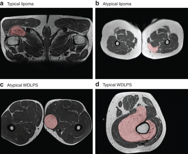
Background: Well differentiated liposarcoma (WDLPS) can be difficult to distinguish from lipoma. Currently, this distinction is made by testing for MDM2 amplification, which requires a biopsy. The aim of this study was to develop a noninvasive method to predict MDM2 amplification status using radiomics features derived from MRI.
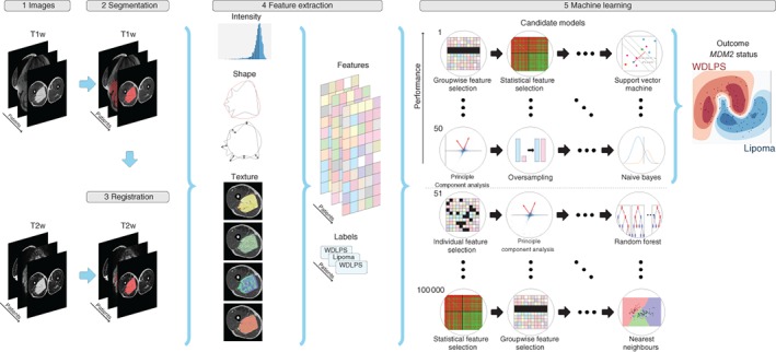
Methods: Patients with an MDM2-negative lipoma or MDM2-positive WDLPS and a pretreatment T1-weighted MRI scan who were referred to Erasmus MC between 2009 and 2018 were included. When available, other MRI sequences were included in the radiomics analysis. Features describing intensity, shape and texture were extracted from the tumour region. Classification was performed using various machine learning approaches. Evaluation was performed through a 100 times random-split cross-validation. The performance of the models was compared with the performance of three expert radiologists.
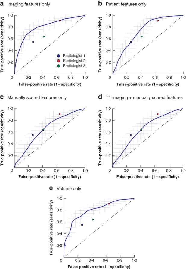
Results: The data set included 116 tumours (58 patients with lipoma, 58 with WDLPS) and originated from 41 different MRI scanners, resulting in wide heterogeneity in imaging hardware and acquisition protocols. The radiomics model based on T1 imaging features alone resulted in a mean area under the curve (AUC) of 0·83, sensitivity of 0·68 and specificity of 0·84. Adding the T2-weighted imaging features in an explorative analysis improved the model to a mean AUC of 0·89, sensitivity of 0·74 and specificity of 0·88. The three radiologists scored an AUC of 0·74 and 0·72 and 0·61 respectively; a sensitivity of 0·74, 0·91 and 0·64; and a specificity of 0·55, 0·36 and 0·59.
Conclusion: Radiomics is a promising, non-invasive method for differentiating between WDLPS and lipoma, outperforming the scores of the radiologists. Further optimization and validation is needed before introduction into clinical practice.
Antecedentes: Es difícil distinguir los liposarcomas bien diferenciados (well-differentiated liposarcomas, WDLPS) de los lipomas. En la actualidad, esta distinción se realiza mediante la prueba de amplificación del gen MDM2 por biopsia. El objetivo de este estudio fue predecir de forma no invasiva el estado de amplificación del gen MDM2 para diferenciar los lipomas de los WDLPS utilizando características radiómicas a partir de la resonancia magnética. MÉTODOS: Se incluyeron los pacientes remitidos al instituto Erasmus MC entre 2009-2018 por un lipoma MDM2 negativo o WDLPS MDM2 positivo y las resonancias magnéticas potenciadas en T1 correspondientes antes del tratamiento. Cuando estaban disponibles, se incluyeron otras secuencias de MRI en el análisis radiómico. Se describieron la intensidad, forma y textura de la región tumoral. Para la clasificación se utilizaron varios modelos de aprendizaje automático (machine learning). La evaluación se realizó mediante una validación cruzada aleatoria 100x. Se comparó el rendimiento de los modelos con la clasificación realizada por tres radiólogos expertos.
Resultados: Se incluyeron 116 pacientes (58 lipomas, 58 WDLPS) y 41 aparatos de MRI, con una gran heterogeneidad en las técnicas y protocolos para la adquisición de imágenes. El modelo radiómico basado únicamente en las características de las imagen en T1 dio como resultado una AUC media de 0,83, con una sensibilidad de 0,68 y una especificidad de 0,84. Un análisis adicional incorporando las imágenes ponderadas en T2 mejoró el modelo con una AUC media de 0,89, una sensibilidad de 0,74 y una especificidad de 0,88. Los tres radiólogos obtuvieron una AUC de 0,74/0,72/0,61, una sensibilidad de 0,74/0,91/0,64 y una especificidad de 0,55/0,36/0,59, respectivamente. CONCLUSIÓN: La radiómica es un método prometedor y no invasivo para diferenciar entre WDLPS y lipomas, superando la valoración de los radiólogos. Sin embargo, se necesita la optimización y validación de esta técnica antes de su introducción en la práctica clínica diaria.
© 2019 The Authors. BJS published by John Wiley & Sons Ltd on behalf of BJS Society Ltd.
Figures
References
-
- Fletcher CDM, Bridge JA, Hogendoorn PCW, Mertens F; World Health Organization, International Agency for Research on Cancer . WHO Classification of Tumours of Soft Tissue and Bone. IARC Press: Lyon, 2013.
-
- Brisson M, Kashima T, Delaney D, Tirabosco R, Clarke A, Cro S et al MRI characteristics of lipoma and atypical lipomatous tumor/well‐differentiated liposarcoma: retrospective comparison with histology and MDM2 gene amplification. Skeletal Radiol 2013; 42: 635–647. - PubMed
-
- Kransdorf MJ, Bancroft LW, Peterson JJ, Murphey MD, Foster WC, Temple HT. Imaging of fatty tumors: distinction of lipoma and well‐differentiated liposarcoma. Radiology 2002; 224: 99–104. - PubMed
-
- Gupta P, Potti TA, Wuertzer SD, Lenchik L, Pacholke DA. Spectrum of fat‐containing soft‐tissue masses at MR imaging: the common, the uncommon, the characteristic, and the sometimes confusing. Radiographics 2016; 36: 753–766. - PubMed
-
- Drevelegas A, Pilavaki M, Chourmouzi D. Lipomatous tumors of soft tissue: MR appearance with histological correlation. Eur J Radiol 2004; 50: 257–267. - PubMed
Publication types
MeSH terms
LinkOut - more resources
Full Text Sources
Medical
Research Materials
