. 2020 Jan 1;123(1):22-33.
doi: 10.1152/jn.00532.2019. Epub 2019 Nov 20.

Alterations in sleep, sleep spindle, and EEG power in mGluR5 knockout mice

Affiliations

Alterations in sleep, sleep spindle, and EEG power in mGluR5 knockout mice

David D Aguilar et al. J Neurophysiol. .

Abstract

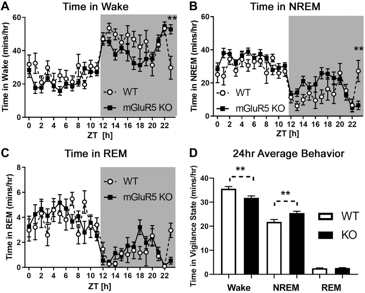
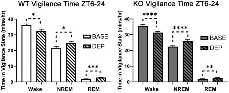
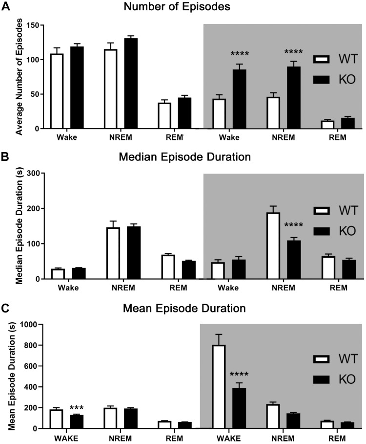
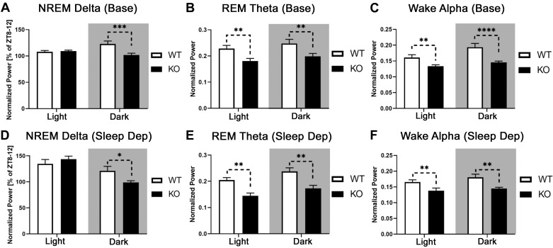
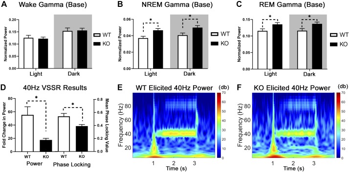
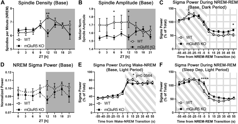
The type 5 metabotropic glutamate receptor (mGluR5) represents a novel therapeutic target for schizophrenia and other disorders. Schizophrenia is associated with progressive abnormalities in cortical oscillatory processes including reduced spindles (8-15 Hz) during sleep and increased delta (0.5-4 Hz)- and gamma-band activity (30-80 Hz) during wakefulness. mGluR5 knockout (KO) mice demonstrate many schizophrenia-like behaviors, including abnormal sleep. To examine the effects of mGluR5 on the maintenance of the neocortical circuitry responsible for such neural oscillations, we analyzed sleep/wake electroencephalographic (EEG) activity of mGluR5 KO mice at baseline, after 6 h of sleep deprivation, and during a visual method of cortical entrainment (visual steady state response). We hypothesized mGluR5-KO mice would exhibit translationally relevant abnormalities in sleep and neural oscillations that mimic schizophrenia. Power spectral and spindle density analyses were performed across 24-h EEG recordings in mGluR5-KO mice and wild-type (WT) controls. Novel findings in mGluR5 KO mice include deficits in sleep spindle density, wake alpha power, and 40-Hz visual task-evoked gamma power and phase locking. Sigma power (10-15 Hz), an approximation of spindle activity, was also reduced during non-rapid eye movement sleep transitions. Our observations on abnormal sleep/wake are generally in agreement with previous reports, although we did not replicate changes in rapid eye movement sleep. The timing of these phenotypes may suggest an impaired circadian process in mGluR5 KO mice. In conclusion, EEG phenotypes in mGluR5 KO mice resemble deficits observed in patients with schizophrenia. These findings implicate mGluR5-mediated pathways in several translationally relevant phenotypes associated with schizophrenia, and suggest that agents targeting this receptor may have harmful consequences on sleep health and daily patterns of EEG power.NEW & NOTEWORTHY Metabotropic glutamate receptor type 5 (mGluR5) knockout (KO) mice show several translationally relevant abnormalities in neural oscillatory activity associated with schizophrenia. These include deficits in sleep spindle density, sigma and alpha power, and 40-Hz task-evoked gamma power. The timing of these phenotypes suggests an impaired circadian process in these mice. Previously reported rapid eye movement sleep deficits in this model were not observed. These findings suggest mGluR5-enhancing drugs may improve sleep stability and sleep spindle density, which could impact memory and cognition.

Keywords: N-methyl-d-aspartate receptors; metabotropic glutamate receptor 5; polysomnography; sigma; visual evoked potentials.


Conflict of interest statement

No conflicts of interest, financial or otherwise, are declared by the authors.

Figures

Fig. 1.
Fig. 1.
Metabotropic glutamate receptor type 5 (mGluR5) knockout (KO) mice (n = 12) had decreased time in wake (A) and increased time in non-rapid eye movement (NREM) sleep (B) across 24 h compared with wild-type (WT; n = 8) controls (D; 2-way RM ANOVA; main effects, P < 0.01). No differences in rapid eye movement (REM) sleep were observed (C). In contrast to the 24-h average trends represented in D, KO mice had more wake and less NREM activity during Zeitgeber time (ZT) 23–24 (A and B). In A and B: **Significant Holm–Sidak post hoc test (P < 0.01) following a 2-way RM ANOVA significant interaction (P < 0.01). In D: **Significant main effect of strain (P < 0.01, 2-way RM ANOVA). EEG data from mice were sleep scored using 10-s epochs. Values are means (error bars are SE). Shaded area represents the dark cycle/lights-off period (ZT 12–24, or 7:00 PM–7:00 AM).
Fig. 2.
Fig. 2.
Sleep deprivation from Zeitgeber time (ZT) 0–6 decreased wake and increased non-rapid eye movement (NREM) and rapid eye movement (REM) sleep in both wild-type (WT; n = 8; left) and metabotropic glutamate receptor type 5 knockout (KO; n = 10; right) animals. This baseline recording was taken 24 h before the automated sleep deprivation. *P < 0.05; **P < 0.01; ***P < 0.001; ****P < 0.0001, significant main effects of sleep deprivation for WT and KO (2-way RM ANOVA). BASE, baseline; DEP, sleep deprivation.
Fig. 3.
Fig. 3.
Metabotropic glutamate receptor type 5 (mGluR5) knockout (KO) mice displayed more fragmented sleep and wake activity in the dark (active) period. Light [Zeitgeber time (ZT) 0–12, or 7:00 AM–7:00 PM) and dark periods (ZT 12–24, or 7:00 PM–7:00 AM; shaded areas) were analyzed separately. mGluR5 KO mice (n = 12) showed a greatly increased number of non-rapid eye movement (NREM) sleep and wake episodes compared with wild-type (WT) controls (n = 8) in the dark period (A). This pattern persisted after 6 h of sleep deprivation (not shown). Next, the median duration of episodes was calculated for each mouse and then averaged within each strain. Analysis of median episode duration revealed shorter NREM episodes in mGluR5 KO mice than in WT during the dark cycle in baseline (B) and post-sleep-deprived conditions (not shown). Mean episode duration analysis revealed significantly shorter wake episodes in KO mice than in WT across both light and dark cycles at baseline (C) and across the dark cycle after sleep deprivation (not shown). ***P < 0.001, ****0.0001, significant difference in the Holm–Sidak post hoc test following a significant interaction (P < 0.05) in a 2-way RM ANOVA. REM, rapid eye movement.
Fig. 4.
Fig. 4.
Metabotropic glutamate receptor type 5 (mGluR5) knockout (KO) mice demonstrated decreased delta (0.5–4 Hz), theta (7–9 Hz), and alpha power (8–13 Hz). Normalized non-rapid eye movement (NREM) delta power was analyzed as a percentage of individual mean delta power from the last 4 h of the light period [Zeitgeber time (ZT) 8–12, as seen in Ahnaou et al. (2015b) and Holst et al. (2017)]. This represents a block of time when delta power is normally lowest. For other frequency bands, each epoch was normalized to the sum power (0–100 Hz) of all epochs within a behavioral state across the baseline day ZT 0–24. mGluR5 KO mice (n = 12) had lower normalized NREM delta power than wild-type (WT) controls (n = 8) during the dark cycle (A). A similar relationship was observed in mice after sleep deprivation (D; WT, n = 8; KO, n = 10). Rapid eye movement (REM) theta power was decreased across light and dark cycles in mGluR5 KO mice at baseline (B) and after sleep deprivation (E). Compared with WT animals, mGluR5 KO mice demonstrated a decrease in wake alpha power across light and dark cycles during baseline (C) and after sleep deprivation (F). In A, C, and D: *P < 0.05; **P < 0.01; ***P < 0.001; ****P < 0.0001, significant Holm–Sidak following a strain × light cycle interaction in a 2-way RM ANOVA. In B, E, and F: **P < 0.01, significant main effect of strain in a 2-way RM ANOVA. In the recovery sleep condition (D–F), the hours of active sleep deprivation (ZT 0–6) have been omitted from the light cycle. Sleep Dep, sleep deprivation.
Fig. 5.
Fig. 5.
Elevated baseline gamma and reduced 40-Hz task-evoked gamma are present in metabotropic glutamate receptor type 5 (mGluR5) knockout (KO) mice. Baseline gamma activity was unchanged in our KO animals during normal waking conditions (A) but enhanced during non-rapid eye movement (NREM; B) and rapid eye movement (REM: C) sleep compared with wild-type (WT) mice (n = 8 WT, n = 12 KO; 2-way RM ANOVA, main effect of strain, *P < 0.05). We utilized the visual steady-state response (VSSR) to measure task-evoked gamma power in 9 WT (7 males, 2 females) and 6 KO animals (3 males, 3 females). This is a visual analog to the auditory steady-state response (ASSR), where a 2-s train of light flashes was presented to a mouse in complete darkness at a range of frequencies (10–60 Hz in 10-Hz steps) while frontal cortical EEG response was measured. The evoked response (elicited power of stimulus period/average power of baseline period) resembles the ASSR with a distribution peaking around 30 Hz. KO mice have a smaller fold increase in power (D, left) and reduced phase-locking value (D, right) than WT mice at 40 Hz, which mimics the ASSR in patients with schizophrenia (Welch’s t test, *P < 0.05). Grand-averaged spectrograms of task-evoked power during a 40-Hz light stimulus are shown for WT (E) and mGluR5 KO animals (F). Colors represent the elicited power in decibels (dB) with blues representing lower power and reds representing higher power. Base, baseline.
Fig. 6.
Fig. 6.
Sleep spindle density and non-rapid eye movement (NREM) sigma power is decreased during transition periods in metabotropic glutamate receptor type 5 (mGluR5) knockout (KO) mice. A custom MATLAB script (Uygun et al. 2019) was used to extract the following NREM sleep spindle data from the EEG trace: spindle density (NREM spindles per minute), median spindle duration, and median normalized spindle amplitude. During baseline activity, mGluR5 KO animals (n = 12) exhibited significantly lower NREM spindle density than wild-type (WT) controls (n = 8) during the first 3 hours of the dark (active) period at Zeitgeber time (ZT) 12–15 (A) and lower spindle amplitude at ZT 6–9 and 12–15 (B). These differences disappeared after sleep deprivation (data not shown). NREM sigma power (10–15 Hz), which is sometimes used to approximate spindle activity, was not significantly different between groups (D). Sigma power was measured during the transitions to and from NREM sleep [C, E, and F; diagonal shaded lines indicate NREM sleep, grid indicates rapid eye movement (REM) sleep]. At baseline, the surge in sigma power is lower in KO than in WT mice 35 s before the NREM-REM transition (C, dark period) and 15 s after the wake-NREM transition (E, light period; Holm–Sidak P = 0.0564). Conversely, after 6 h of sleep deprivation, mGluR5 KO mice (n = 10) had increased sigma power when transitioning from NREM to REM sleep compared with WT controls (n = 8) (F, light period; −5 to +5 s of transition, and main effect P < 0.05). Together, these results suggest abnormal regulation of sigma power during transitions to and from NREM sleep in KO mice. *P < 0.05; **P < 0.01; ****P < 0.0001, significance from multiple t tests with a Holm–Sidak correction alone (A, B, D) or following a significant interaction in a 2-way RM ANOVA (C, E, F). Base, baseline.

References

    1. Ahnaou A, Langlois X, Steckler T, Bartolome-Nebreda JM, Drinkenburg WH. Negative versus positive allosteric modulation of metabotropic glutamate receptors (mGluR5): indices for potential pro-cognitive drug properties based on EEG network oscillations and sleep-wake organization in rats. Psychopharmacology (Berl) 232: 1107–1122, 2015a. doi:10.1007/s00213-014-3746-4. - DOI - PubMed
    1. Ahnaou A, Raeymaekers L, Steckler T, Drinkenbrug WH. Relevance of the metabotropic glutamate receptor (mGluR5) in the regulation of NREM-REM sleep cycle and homeostasis: evidence from mGluR5 (−/−) mice. Behav Brain Res 282: 218–226, 2015b. doi:10.1016/j.bbr.2015.01.009. - DOI - PubMed
    1. Andreasen NC, Arndt S, Swayze V 2nd, Cizadlo T, Flaum M, O’Leary D, Ehrhardt JC, Yuh WT. Thalamic abnormalities in schizophrenia visualized through magnetic resonance image averaging. Science 266: 294–298, 1994. doi:10.1126/science.7939669. - DOI - PubMed
    1. Ango F, Robbe D, Tu JC, Xiao B, Worley PF, Pin JP, Bockaert J, Fagni L. Homer-dependent cell surface expression of metabotropic glutamate receptor type 5 in neurons. Mol Cell Neurosci 20: 323–329, 2002. doi:10.1006/mcne.2002.1100. - DOI - PubMed
    1. Annamalai A, Palmese LB, Chwastiak LA, Srihari VH, Tek C. High rates of obstructive sleep apnea symptoms among patients with schizophrenia. Psychosomatics 56: 59–66, 2015. doi:10.1016/j.psym.2014.02.009. - DOI - PMC - PubMed

Publication types

Substances