. 2019 Dec 26;62(24):11280-11300.
doi: 10.1021/acs.jmedchem.9b01530. Epub 2019 Dec 10.

Structure-Based Discovery of SD-36 as a Potent, Selective, and Efficacious PROTAC Degrader of STAT3 Protein

Structure-Based Discovery of SD-36 as a Potent, Selective, and Efficacious PROTAC Degrader of STAT3 Protein

Haibin Zhou et al. J Med Chem. .

Abstract

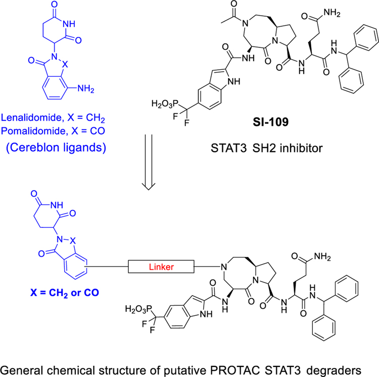
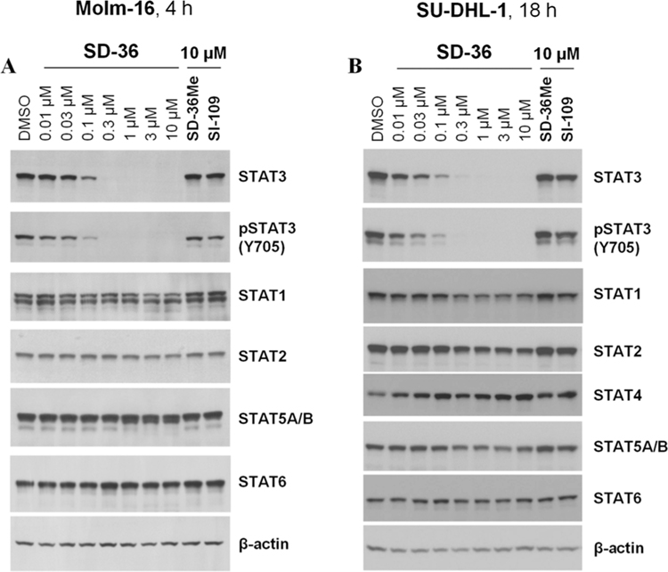
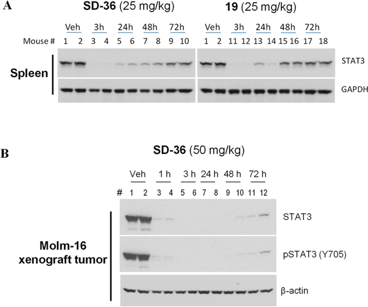
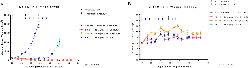
Signal transducer and activator of transcription 3 (STAT3) is a transcription factor and an attractive therapeutic target for cancer and other human diseases. Despite 20 years of persistent research efforts, targeting STAT3 has been very challenging. We report herein the structure-based discovery of potent small-molecule STAT3 degraders based upon the proteolysis targeting chimera (PROTAC) concept. We first designed SI-109 as a potent, small-molecule inhibitor of the STAT3 SH2 domain. Employing ligands for cereblon/cullin 4A E3 ligase and SI-109, we obtained a series of potent PROTAC STAT3 degraders, exemplified by SD-36. SD-36 induces rapid STAT3 degradation at low nanomolar concentrations in cells and fails to degrade other STAT proteins. SD-36 achieves nanomolar cell growth inhibitory activity in leukemia and lymphoma cell lines with high levels of phosphorylated STAT3. A single dose of SD-36 results in complete STAT3 protein degradation in xenograft tumor tissue and normal mouse tissues. SD-36 achieves complete and long-lasting tumor regression in the Molm-16 xenograft tumor model at well-tolerated dose-schedules. SD-36 is a potent, selective, and efficacious STAT3 degrader.


Conflict of interest statement

The authors declare the following competing financial interest(s): The University of Michigan has filed patent applications on SD-36 and its analogues, for which S. Wang, H. Zhou, R Xu, L. Bai, D. McEachem, C.-Y. Yang, and J. Stuckey are co-inventors. These patents have been licensed by Oncopia Therapeutics Inc., of which S. Wang is a co-founder, a paid consultant, and owns stock The University of Michigan also owns stock in Oncopia. This study is supported in part by a research contract from Oncopia.

Figures

Figure 1.
Figure 1.
Design of potent and cell-permeable small molecule inhibitors of the STAT3 SH2 domain: chemical structures and binding affinities of our previously reported compound CJ-887 and newly designed and synthesized inhibitors.
Figure 2.
Figure 2.
Cocrystal structure of SI-109 with STAT3 (PDB code 6NUQ). (A) The FoFc electron density generated from an omit map contoured at 3σ shows placement of atoms of SI-109. (B) Detailed interactions of STAT3 with SI-109. SI-109 (cyan) and side chains of interacting residues of STAT3 with SI-109 are shown in stick form. Hydrogen bonds are depicted as dashed lines.
Figure 3.
Figure 3.
Design of putative STAT3 PROTAC degraders based upon cereblon ligands and our STAT3 SH2 domain inhibitor SI-109.
Figure 4.
Figure 4.
Design of SD-36Me as a control compound.
Figure 5.
Figure 5.
Western blotting analysis of STAT proteins in Molm-16 and SU-DHL-1 cells treated with SD-36 or control compounds SD-36Me and SI-109. (A) Molm-16 cells were treated with SD-36 at 0.01–10 μM or SD-36Me and SI-109 at 10 μM for 4 h. (B) SU-DHL-1 cells were treated with SD-36 at 0.01–10 μM or SD-36Me and SI-109 at 10 μM for 18 h.
Figure 6.
Figure 6.
Pharmacodynamics study of SD-36 and 19 in native mouse spleen tissue in CD-1 mice (A) and SD-36 in the Molm-16 xenograft tumors in SCID mice (B). CD-1 and SCID mice were treated intravenously with a single dose of SD-36 or 19 as indicated. Native spleen tissue or tumor lysates were analyzed by immunoblotting.
Figure 7.
Figure 7.
In vivo antitumor activity of SD-36 in the MoIm-16 xenograft model in mice: (A) tumor growth; (B) percentage of animal body weight change.
Scheme 1.
Scheme 1.. Synthesis of 5-((Di-tert-butoxyphosphoryl)methyl)-1H-indole-2-carboxylic Acid (41)a
aReagents and conditions: (a) NaH, Boc2O, THF, 0 °C to rt; (b) NBS, Bz2O2, CCl4, reflux, 77% over 2 steps; (c) (t-BuO)2PONa 1.0 equiv of NaH 2.5 equiv, THF, then reflux for 3 h; (d) LiOH, MeOH–THF–H2O, 60 °C.
Scheme 2.
Scheme 2.. Synthesis of 3-((Diethoxyphosphoryl)difluoromethyl)-1H-indole-2-carboxylic Acid (46)a
aReagents and conditions: (a) P(OEt)3, 100 °C, 84%; (b) Ti(O-i-Pr)4, BnOH, 100 °C, 83%; (c) NaH, Cbz-Cl, THF, 0 °C to rt, 88%; (d) NFBS, NaHMDS, THF, −78 °C to rt, 95%; (e) H2/Pd–C, THF, 94%.
Scheme 3.
Scheme 3.. Synthesis of Compounds 2–4a
aReagents and conditions: (a) EDC–HCl 2.0 equiv, HOBt 2.0 equiv, EtN(i-Pr)2, DCM, rt, 3 h, 89% yield; (b) CF3CO2H 2 mL, Et3Si–H 0.1 mL, DCM 3 mL, rt, 1 h; (c) (1) protected phosphotyrosine mimetics, EDC–HCl, HOBt, EtN(i-Pr)2, DCM, rt, 3 h; (2) TFA–DCM, rt, 3 h; or TMSI, 0 °C, 1 h.
Scheme 4.
Scheme 4.. Synthesis of Compounds 5 and 7a
aReagents and conditions: (a) (1) HATU, EtN(i-Pr)2, DMF, rt, 1 h; (2) TFA, DCM; (b) 46, HATU, EtN(i-Pr)2, DMF, rt, 1 h; (c) H2, Pd/C, MeOH; (d) (1) HCHO, sodium triacetoxyborohydride, DCE; (2) TFA, DCM; (e) R-NH2, HATU, EtN(i-Pr)2, DMF, rt, 1 h.
Scheme 5.
Scheme 5.. Synthesis of Compounds 8–11a
aReagents and conditions: (a) HATU, EtN(i-Pr)2, DMF, rt, 1 h; (b) TFA, DCM; (c) 51, HATU, EtN(i-Pr)2, DMF, rt, 1 h; (d) (1) H2, Pd/C, MeOH; (2) Ac2O, EtN(i-Pr)2, DCM; (3) TFA, DCM; (e) (1) phosphotyrosine mimetics, HATU, EtN(i-Pr)2, DMF, rt, 1 h; (2) TFA–DCM, rt, 3 h; or TMSI, 0 °C, 1 h.
Scheme 6.
Scheme 6.. Synthesis of SD-36a
aReagents and conditions: (a) (1) TFA, DCM; (2) 46, HATU, DIEA, DMF; (b) H2, Pd/C, MeOH; (c) 68, HATU, DIEA, DMF; (d) TMSI, BSTFA, DCM; (e) Pd(PPh3)Cl2, CuI, DMF/TEA, 80 °C, 1 h.
Scheme 7.
Scheme 7.. Synthesis of Compounds 30 and 31a
aReagents and conditions: (a) (1) 68, HATU, DIEA, DMF; (2) TFA, DCM; (b) (i) R-NH2, HATU, DIEA, DMF; (2) TMSI, BSTFA, DCM.
Scheme 8.
Scheme 8.. Synthesis of Compounds 32–34a
aReagents and conditions: (a) (1) H2, Pd/C, MeOH; (2) 68, HATU, DIEA, DMF; (3) TFA, DCM; (b) (1) phosphotyrosine mimetics, HATU, EtN(i-Pr)2, DMF, rt, 1 h; (2) TFA–DCM, rt, 3 h; or TMSI, 0 °C, 1 h.
Scheme 9.
Scheme 9.. Synthesis of Fluorescent Labeled Compound 74a
aReagents and conditions: (a) (1) HATU, DIEA, DMF; (2) TFA, DCM; (b) (1) HATU, DIEA, DMF; (2) TMSI, BSTFA, DCM.

References

    1. Levy DE; Darnell JE STATs: Transcriptional control and biological impact. Nat. Rev. Mol. Cell Biol. 2002, 3 (9), 651–662. - PubMed
    1. Yu H; Jove R The stats of cancer - new molecular targets come of age. Nat. Rev. Cancer 2004, 4 (2), 97–105. - PubMed
    1. Johnson DE; O’Keefe RA; Grandis JR Targeting the IL-6/JAK/STAT3 signalling axis in cancer. Nat. Rev. Clin. Oncol. 2018, 15 (4); 234–248. - PMC - PubMed
    1. Beebe JD,; Liu JY; Zhang JT Two decades of research in discovery of anticancer drugs targeting STAT3, how close are we? Pharmacol. Ther. 2018, 191, 74–91. - PubMed
    1. Yang J,; Stark GR Roles of unphosphorylated STATs in signaling. Cell Res. 2008, 18 (4), 443–451. - PubMed

Publication types

MeSH terms