. 2019 Nov 19;29(8):2450-2460.e5.
doi: 10.1016/j.celrep.2019.10.066.

The Bromodomain Protein 4 Contributes to the Regulation of Alternative Splicing

Affiliations

The Bromodomain Protein 4 Contributes to the Regulation of Alternative Splicing

Sheetal Uppal et al. Cell Rep. .

Abstract

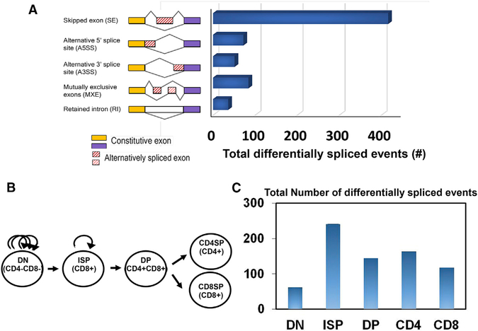
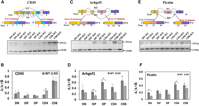
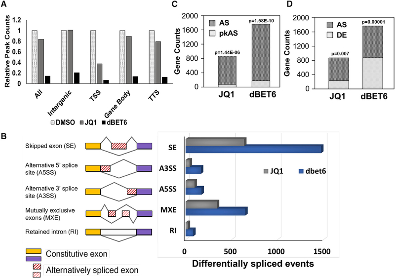
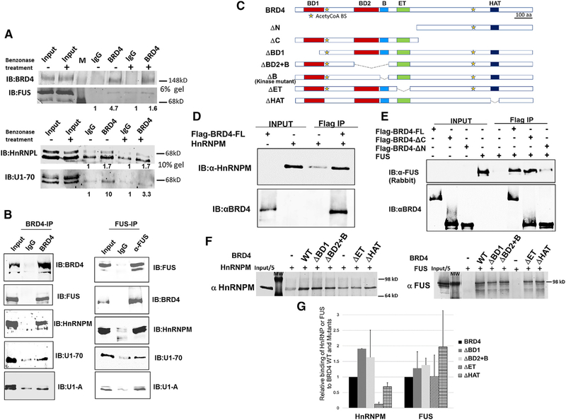
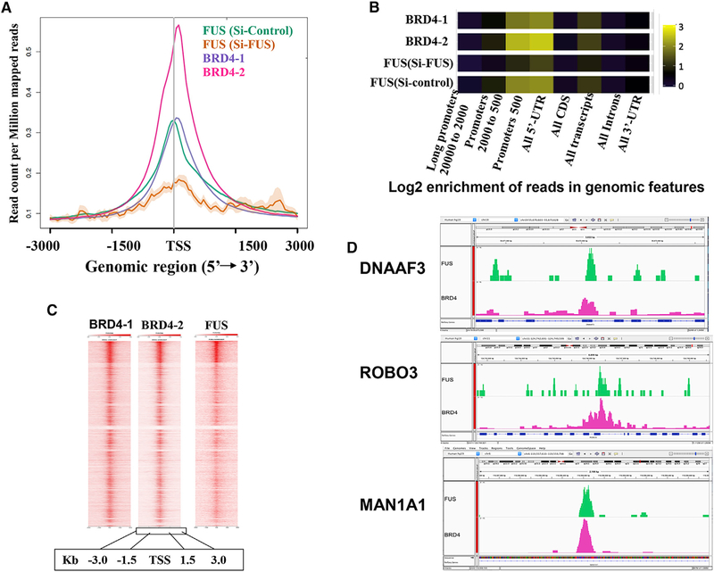
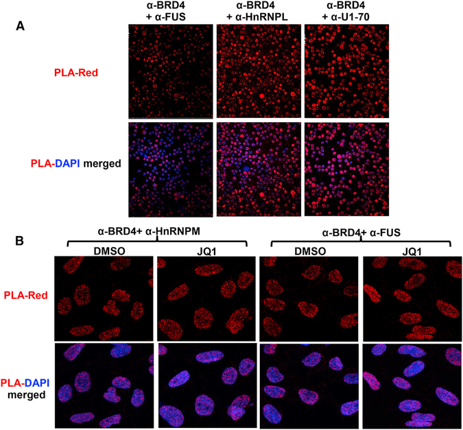
The bromodomain protein 4 (BRD4) is an atypical kinase and histone acetyl transferase (HAT) that binds to acetylated histones and contributes to chromatin remodeling and early transcriptional elongation. During transcription, BRD4 travels with the elongation complex. Since most alternative splicing events take place co-transcriptionally, we asked if BRD4 plays a role in regulating alternative splicing. We report that distinct patterns of alternative splicing are associated with a conditional deletion of BRD4 during thymocyte differentiation in vivo. Similarly, the depletion of BRD4 in T cell acute lymphoblastic leukemia (T-ALL) cells alters patterns of splicing. Most alternatively spliced events affected by BRD4 are exon skipping. Importantly, BRD4 interacts with components of the splicing machinery, as assessed by both immunoprecipitation (IP) and proximity ligation assays (PLAs), and co-localizes on chromatin with the splicing regulator, FUS. We propose that BRD4 contributes to patterns of alternative splicing through its interaction with the splicing machinery during transcription elongation.

Keywords: AML; BET; BRD4; FUS; alternative splicing; thymocyte differentiation.


Conflict of interest statement

DECLARATION OF INTERESTS

The authors declare no competing interests.

Figures

Figure 1.
Figure 1.. BRD4 Regulates Alternative Splicing in Murine Thymocytes
(A) Schematic diagram depicting different types of alternatively spliced events (left). The bar graph (right) shows the distribution of alternatively spliced events among those that are differentially spliced in total thymus in BRD4 knock-out versus wild type (WT) (FDR < 0.05). Comparison of splicing events between WT and BRD4-deficient cells was based on transcripts expressed in both. (B) Developmental stages in thymocyte differentiation, DN (CD4, CD8 DN), ISP (CD8+ ISP), DP (CD4, CD8 DP), CD4, and CD8 single-positive thymocytes are shown. Arched arrows denote level of proliferative activity in DN and ISP thymocytes. (C) Bar graph showing the total number of differentially spliced events in the different thymocyte subpopulations in BRD4 knock-out versus wild-type thymus (FDR < 0.05), derived from RNA-seq analysis. BRD4 was conditionally deleted in DN thymocytes by LCK-Cre (Gegonne et al., 2018). Comparison of splicing events between WT and BRD4-deficient cells was based on transcripts expressed in both. See also Figure S1.
Figure 2.
Figure 2.. Validation of RNA-Seq Data by RT-PCR
RNA from the different thymocyte subpopulations was subjected to RT-PCR for the indicated genes: CD45 (A and B), Arhgef1 (C and D), and Picalm (E and F). (A, C, and E) Upper panels: schematic diagrams depicting partial gene structure of the alternatively spliced genes CD45 (A), Arhgef1 (C), and Picalm (E). Rectangular boxes represent the exons, and the horizontal straight lines connecting the boxes represent the introns; the numbers below the boxes refer to the exon number of the gene, and numbers inside the boxes refer to the length of the exons; the numbers within the terminal exons do not refer to the actual exon length but the length amplifiable by the RT-PCR primers. The arrow heads show the approximate positions of the RT-PCR primers; boxes with hashed lines show the alternative exons; and curved lines connecting the boxes depict the splicing pattern. WT and KO refer to the splicing pattern prevalent in either the wild-type or knock-out thymocytes as determined by RNA-seq analysis. Lower panels: ethidium bromide stained agarose gels showing RT-PCR products derived from total RNA from BRD4 WT and KO thymocytes. (B, D, and F) Bar graphs of the RT-PCR results for CD45 (B), Arhgef1 (D), and Picalm (F). The ratios A/A+B (ratio of included exon transcript/total transcripts) were used as measure of alternative splicing and represent the average of three separate RT-PCR analyses. #, p < 0.05, significant difference between WT subpopulations, relative to WT DN; *p < 0.05, significant difference between WT and KO for the specific subpopulation. See also Figure S2.
Figure 3.
Figure 3.. BET Inhibition (JQ1) or BET Degradation (dBET6) Alter Splicing Patterns in T Cell Acute Lymphoblastic Leukemia (T-ALL) Cells
(A) Effect of JQ1 or dBET6 treatment on the binding of BRD4 across the gene. ALL, TSS+gene body+TTS+intergenic; TSS, transcription start site; Gene body, between TSS and TTS; TTS, transcription termination site; intergenic, all remaining sequences. The peak distribution, in the absence of treatment is as follows: TSS, 1123; gene body, 4065; TTS, 226; intergenic, 1827. (B) Bar graph showing the distribution of alternative splice events among the differentially spliced events in response to JQ1 treatment or dBET6 treatment in T-ALL cells. (C) Bar graph showing the fraction of alternative splice (AS) genes that also have BRD4 associated with them at the TSS (pkAS). The total number of BRD4 peaks detected at the TSS across the genome was 1123. (D) Bar graph showing the fraction of AS genes that are also differentially expressed (DE) in response to JQ1 or dBET6 treatment. p values for (C) and (D) were obtained using a hypergeometric test, which tests the probability that the frequency of AS genes derived from either DE genes (overlap) or genes with BRD4-bound TSS peaks is larger than expected from the population; a low p value suggests the enrichment of AS genes in either DE genes or genes with BRD4 TSS peaks. See also Figures S3 and S4.
Figure 4.
Figure 4.. BRD4 Interacts with Splicing Factors In Vivo
(A) Immunoblot of BRD4 immunoprecipitates from thymocyte nuclear extracts (with and without benzonase treatment) with indicated antibodies to splicing factors FUS, HnRNPL, and U1–70. The immunoprecipitates from a single extract were run on either a 6% gel to visualize BRD4 and Fus or on a 10% gel to visualize HnRNPL and U1–70. The values under the IP lanes indicate the enrichment of anti-BRD4 co-IP, relative to the IgG control. (B, left) Immunoblot of BRD4 immunoprecipitates from HeLa nuclear extracts with indicated antibodies to splicing factors FUS, HnRNPM, U1–70, and U1-A. (B, right) Immunoblot of FUS immunoprecipitates from HeLa nuclear extracts with indicated antibodies to BRD4 and splicing factors HnRNPM, U1–70, and U1-A. (C) Schematic representation of BRD4 and BRD4-deletion mutants. The coordinates of the mouse BRD4 mutations are as follows. WT BRD4, 1402 aa; DN, 722–1402 aa; ΔC, 1–699aa; ΔBD1, 146–1402 aa; ΔBD2+B, 1–349/599–1402 aa; ΔB, 1–502/549–1402 aa; ΔET, 1–600/684–1402 aa; ΔHAT, 1–1156/1198–1402 aa. (D) Immunoblots showing pull-down analysis of recombinant BRD4 with recombinant HnRNPM. rHnRNPM (0.25 μg) was pulled down with rflag-BRD4 (0.5 μg) immobilized on Flag beads. Immunoblots were with anti-HnRNPM (upper) and anti-BRD4 (lower). (E) Immunoblots showing pull-down analysis of recombinant BRD4 with recombinant FUS. rFUS (0.25 μg) was pulled down with rflag-BRD4 (0.5 μg) WT or equimolar amounts of N-terminal or C-terminal BRD4 truncation mutants immobilized on Flag beads. Immunoblots were with anti-FUS (upper) and anti-BRD4 (lower). (F) Binding of HnRNPM (left panel) and FUS (right panel) to BRD4 mutants was assessed in pull-down assays with rBRD4 immobilized on Flag beads and immunoblotting with appropriate antibodies. The results represent the average of two experiments. (G) Retention of FUS and HnRNPM to BRD4 mutants, relative to the WT, was quantified as the fraction of input and normalized to the extent of binding to BRD4 WT. All results are representative of at least two independent experiments. See also Figure S5.
Figure 5.
Figure 5.. BRD4 and FUS Co-localize on the Genome
(A) Metagene profile of BRD4 and FUS CHIP datasets showing colocalization of BRD4 and FUS at the TSS. (B) Log2 enrichment of reads in genomic features along the metagene body. (C) Enrichment heatmap showing co-localization of BRD4 with FUS across the genome. (D) Genome browser views of DNAAF3, ROBO3, and MAN1A1, showing BRD4 and FUS co-localization around the TSS and gene body. See also Figures S6A and S6B.
Figure 6.
Figure 6.. BRD4 Co-localizes with Other Splicing Factors In Situ in Primary Thymocytes and HeLa Cells
(A) Proximity ligation assays (PLAs) were performed on primary thymocytes with anti-BRD4 and the antibodies for the indicated splicing factors. The PLAs are all significantly above the single antibody controls (Figure S6C). (B) PLA was performed using anti-BRD4 and the antibodies for the indicated splicing factors on fixed HeLa cells that had been treated with JQ1 (500 nM)/ DMSO for 6 hr. There is no significant difference (p > 0.05) between the treated and control PLA samples for either HnRNPM or Fus; both PLAs are significantly above single antibody alone controls (Figure S6C). PLA interaction is shown in red; DAPI staining in blue. See also Figures S6C and S6D.

References

    1. Alsarraj J, and Hunter KW (2012). Bromodomain-Containing Protein 4: A Dynamic Regulator of Breast Cancer Metastasis through Modulation of the Extracellular Matrix. Int. J. Breast Cancer 2012, 670632. - PMC - PubMed
    1. Anders S, Pyl PT, and Huber W (2015). HTSeq–a Python framework to work with high-throughput sequencing data. Bioinformatics 31, 166–169. - PMC - PubMed
    1. Baranello L, Wojtowicz D, Cui K, Devaiah BN, Chung HJ, Chan-Salis KY, Guha R, Wilson K, Zhang X, Zhang H, et al. (2016). RNA Polymerase II Regulates Topoisomerase 1 Activity to Favor Efficient Transcription. Cell 165, 357–371. - PMC - PubMed
    1. Berkovits BD, Wang L, Guarnieri P, and Wolgemuth DJ (2012). The testis-specific double bromodomain-containing protein BRDT forms a complex with multiple spliceosome components and is required for mRNA splicing and 3′-UTR truncation in round spermatids. Nucleic Acids Res. 40, 7162–7175. - PMC - PubMed
    1. Bisgrove DA, Mahmoudi T, Henklein P, and Verdin E (2007). Conserved P-TEFb-interacting domain of BRD4 inhibits HIV transcription. Proc. Natl. Acad. Sci. USA 104, 13690–13695. - PMC - PubMed

Publication types

MeSH terms

LinkOut - more resources