. 2019 Nov 20;10(1):332.
doi: 10.1186/s13287-019-1432-5.

Extracellular vesicles derived from human Wharton's jelly mesenchymal stem cells protect hippocampal neurons from oxidative stress and synapse damage induced by amyloid-β oligomers

Affiliations

Extracellular vesicles derived from human Wharton's jelly mesenchymal stem cells protect hippocampal neurons from oxidative stress and synapse damage induced by amyloid-β oligomers

Victor Bodart-Santos et al. Stem Cell Res Ther. .

Abstract

Background: Mesenchymal stem cells (MSCs) have been explored as promising tools for treatment of several neurological and neurodegenerative diseases. MSCs release abundant extracellular vesicles (EVs) containing a variety of biomolecules, including mRNAs, miRNAs, and proteins. We hypothesized that EVs derived from human Wharton's jelly would act as mediators of the communication between hMSCs and neurons and could protect hippocampal neurons from damage induced by Alzheimer's disease-linked amyloid beta oligomers (AβOs).

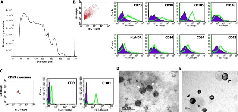
Methods: We isolated and characterized EVs released by human Wharton's jelly mesenchymal stem cells (hMSC-EVs). The neuroprotective action of hMSC-EVs was investigated in primary hippocampal cultures exposed to AβOs.

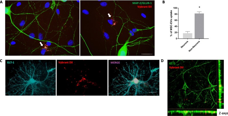
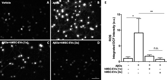
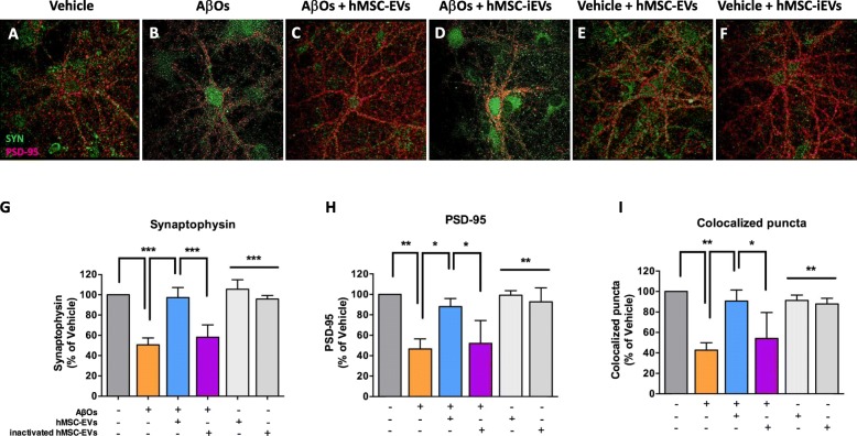
Results: hMSC-EVs were internalized by hippocampal cells in culture, and this was enhanced in the presence of AβOs in the medium. hMSC-EVs protected hippocampal neurons from oxidative stress and synapse damage induced by AβOs. Neuroprotection by hMSC-EVs was mediated by catalase and was abolished in the presence of the catalase inhibitor, aminotriazole.

Conclusions: hMSC-EVs protected hippocampal neurons from damage induced by AβOs, and this was related to the transfer of enzymatically active catalase contained in EVs. Results suggest that hMSC-EVs should be further explored as a cell-free therapeutic approach to prevent neuronal damage in Alzheimer's disease.


Conflict of interest statement

The authors declare that they have no competing interests.

Figures

Fig. 1
Fig. 1
Characterization of hMSC-EVs. a Nanoparticle tracking analysis of hMSC-EVs indicates a mixed population of exosomes and microvesicles, comprising particles with diameters ranging between 30 and 750 nm and an average particle density of 1.7 ± 0.7 × 104 particles/cell (n = 3 EV preparations from independent cord donors). b hMSC-EVs were positive for MSC markers CD73, CD90, CD105 and negative for HLA-DR, CD14, CD34, CD45, and CD146. c CD63-positive exosomes isolated from the total population of hMSC-EVs by CD63-conjugated capture beads are positive for exosome-associated tetraspanins, CD9 and CD81. d, e Representative TEM images of hMSC-EVs. White arrows indicate exosomes; black arrows indicate microvesicles
Fig. 2
Fig. 2
AβOs promote uptake of hMSC-EVs by hippocampal cells. Double-labeled hMSC-EVs (Vybrant DiI shown in red; SYTO RNA shown in green) were incubated for 24 h with hippocampal cultures after a previous 24-h exposure of cultures to vehicle (b) or 500 nM AβOs (c). Control hippocampal cultures in the absence of fluorescent hMSC-EVs showed no labeling (a). Scale bar, 20 μm
Fig. 3
Fig. 3
Uptake of hMSC-EVs is primarily carried out by non-neuronal cells in hippocampal cultures. a Vybrant DiI-labeled hMSC-EVs were incubated for 24 h with hippocampal cultures following previous incubation of cultures for 24 h with 500 nM AβOs. White arrows indicate representative uptake of hMSC-EVs by MAP 2/GluA1-positive cells. b Analysis of co-localization of Vibrant DiI and MAP 2/GluA1 labeling revealed that uptake of hMSC-EVs was mostly carried out by non-neuronal cells in culture. Quantification was performed on ten images from each of three coverslips in each three independent experiments. c GLT-1-positive astrocytes in hippocampal cultures exhibit robust hMSC-EV uptake. d Representative z-stack maximal intensity projection confocal image demonstrating the presence of Vybrant DiI-labeled hMSC-EVs (red) inside GLT-1-labeled astrocytes (green). Scale bar, 20 μm
Fig. 4
Fig. 4
hMSC-EVs rescue AβO-induced ROS formation in hippocampal cultures. a, b Representative DCF fluorescence images from hippocampal cultures exposed to vehicle or 500 nM AβOs for a total period of 6 h. c, d Hippocampal cultures were treated with two different doses of hMSC-EVs (6.1 × 107 particles, identified as [1x], or 1.82 × 108 particles, identified as [3x]). Scale bar, 50 μm. e Integrated DCF fluorescence. Bars represent means ± SEM from three experiments using independent hippocampal culture. In each experiment, three images from each of three coverslips were acquired and analyzed using NIH ImageJ software. *p < 0.05; **p < 0.0001; one-way ANOVA followed by Tukey’s post hoc test
Fig. 5
Fig. 5
Inactivation of catalase abolishes the capacity of hMSC-EVs to block oxidative stress induced by AβOs in hippocampal cultures. a Catalase activity of hMSC-EVs was determined by high-resolution respirometry by measuring O2 release in response to increasing concentrations of added hydrogen peroxide. Treatment with aminotriazole (AMZ, a catalase inhibitor) fully inactivated catalase activity of hMSC-EVs. b Integrated DCF fluorescence from hippocampal cultures exposed to AbOs (or vehicle) in the presence of hMSC-EVs previously treated (or not) with aminotriazole. Bars represent means ± SEM from three experiments using independent neuronal cultures. In each experiment, three images from each of three coverslips were acquired and analyzed using NIH ImageJ software. Scale bar, 50 μm. *p < 0.05; **p < 0.0001; one-way ANOVA followed by Tukey’s post hoc test. cf Representative DCF fluorescence images of hippocampal cultures
Fig. 6
Fig. 6
Catalase mediates the ability of hMSC-EVs to prevent synapse damage induced by AβOs. a, b Representative images from cultured hippocampal neurons exposed to 500 nM AβOs or vehicle for 24 h and immunolabeled for synaptophysin (SYP, green) and PSD-95 (red). c, d Where indicated, hippocampal cultures were treated with hMSC-EVs or hMSC-iEVs. e, f Representative images from vehicle-exposed cultures treated with hMSC-EVs or hMSC-iEVs. Integrated fluorescence intensities for synaptophysin (g) or PSD-95 (h), and numbers of co-localized synaptophysin/PSD-95 punctae (i). Bars represent means ± SEM from three experiments using independent cultures. Nine to 12 images from each of three coverslips were acquired and analyzed in each independent experiment. *p < 0.05; **p < 0.01; ***p < 0.001; one-way ANOVA followed by Tukey’s post hoc test. Scale bar, 20 μm

References

    1. Anand R, Gill KD, Mahdi AA. Therapeutics of Alzheimer’s disease: past, present and future. Neuropharmacology. 2014;76:Pt A:27–Pt A:50. doi: 10.1016/j.neuropharm.2013.07.004. - DOI - PubMed
    1. Imtiaz B, Tolppanen AM, Kivipelto M, Soininen H. Future directions in Alzheimer’s disease from risk factors to prevention. Biochem Pharmacol. 2014;88(4):661–670. doi: 10.1016/j.bcp.2014.01.003. - DOI - PubMed
    1. Ferreira ST, Klein WL. The Abeta oligomer hypothesis for synapse failure and memory loss in Alzheimer’s disease. Neurobiol Learn Mem. 2011;96(4):529–543. doi: 10.1016/j.nlm.2011.08.003. - DOI - PMC - PubMed
    1. Mucke L, Selkoe DJ. Neurotoxicity of amyloid beta-protein: synaptic and network dysfunction. Cold Spring Harb Perspect Med. 2012;2(7):a006338. doi: 10.1101/cshperspect.a006338. - DOI - PMC - PubMed
    1. Ferreira IL, Ferreiro E, Schmidt J, Cardoso JM, Pereira CM, Carvalho AL, Oliveira CR, Rego AC. Abeta and NMDAR activation cause mitochondrial dysfunction involving ER calcium release. Neurobiol Aging. 2015;36(2):680–692. doi: 10.1016/j.neurobiolaging.2014.09.006. - DOI - PubMed

Publication types

MeSH terms

LinkOut - more resources