Review
. 2019 Dec;42(12):2338-2346.
doi: 10.2337/dc19-0475.

Setting the Stage for Islet Autoimmunity in Type 2 Diabetes: Obesity-Associated Chronic Systemic Inflammation and Endoplasmic Reticulum (ER) Stress

Affiliations
Review

Setting the Stage for Islet Autoimmunity in Type 2 Diabetes: Obesity-Associated Chronic Systemic Inflammation and Endoplasmic Reticulum (ER) Stress

Barbara M Brooks-Worrell et al. Diabetes Care. 2019 Dec.

Abstract

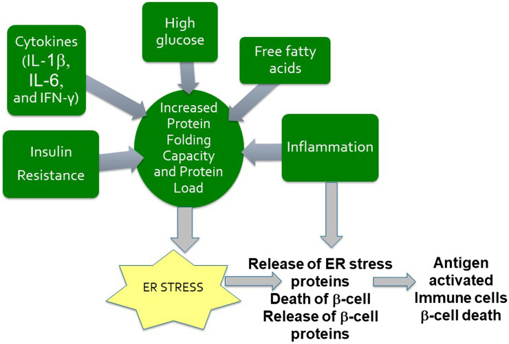
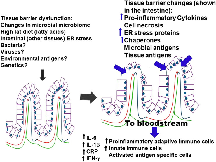
Islet autoimmunity has been identified as a component of both type 1 (T1D) and type 2 (T2D) diabetes, but the pathway through which islet autoimmunity develops in T1D and T2D may be different. Acknowledging the presence of islet autoimmunity in the pathophysiology of T2D, a historically nonautoimmune metabolic disease, would pave the way for important changes in classifications of and therapeutic options for T2D. In order to fully appreciate the importance of islet autoimmunity in T2D, the underlying mechanisms for immune system activation need to be explored. In this review, we focus on the potential origin of immune system activation (innate and adaptive) leading to the development of islet autoimmunity in T2D.


Figures

Figure 1
Figure 1
Obesity-induced changes in adipocytes. In an obese state, the immune composition of the adipose tissue shifts and takes on a proinflammatory (PI) phenotype, whereas in a lean state, the immune system of the adipose tissue primarily has an anti-inflammatory (AI) composition. These changes incorporate cytokines, adipokines, newly expressed proteins on the surface of the adipocytes, and ER stress proteins; apoptosis of adipocytes; and changes toward a PI immune composition. The PI immune cells may recognize the “novel” adipocyte proteins or proteins resulting from adipocyte apoptosis or ER stress. They may include immune cells that were stimulated in the intestine or other tissues and are migrating to the adipose tissue or cells that were stimulated within the adipose tissue. The antigen (Ag)-stimulated PI cells and cytokines may then enter the bloodstream and migrate to other tissues and organs.
Figure 2
Figure 2
ER stress in pancreatic β-cells. Numerous factors such as insulin resistance, high glucose, free fatty acids, cytokines, and incoming inflammation may be responsible for the development of ER stress in pancreatic cells. ER stress may then be responsible for an increase in the number of abnormal posttranslational modifications or in the expression of endogenous ER proteins on the surface of pancreatic cells. This may occur to a level that is sufficient to generate autoimmune responses to these “new” surface proteins, or neoproteins. The “pro-inflammatory” antigen-stimulated immune cells arriving in the pancreas may recognize the “novel” islet proteins or the proteins resulting from β-cell apoptosis or ER stress. Antigen may activate other immune cells within the pancreas, leading to further β-cell destruction or dysfunction.
Figure 3
Figure 3
Intestinal assault and resulting changes. An intestinal assault compromises the intestinal integrity or alters the bacterial microbiome, allowing for external environmental antigens to enter the environment. These changes shift the immune composition of the intestine from an anti-inflammatory phenotype to a proinflammatory phenotype. These changes incorporate novel expression of proteins on the surface of the intestinal epithelial cells (ER stress), apoptosis of intestinal cells, and upregulation of proinflammatory cells (innate and adaptive) and cytokines (IL-6, IL-1β, TNF-α, IFN-γ, CRP). The immune cells and cytokines are released into the bloodstream. The antigen-stimulated proinflammatory immune cells and cytokines arriving in the pancreas or other organs may recognize the “novel” proteins resulting from ER stress to which they were initially stimulated.
Figure 4
Figure 4
ER stress in T1D and T2D pathogenesis. In patients with T1D or T2D, ER stress, tissue barrier dysfunction, or a breakdown in the integrity of the intestines or other organs may be responsible for generating “new” surface proteins, or neo-proteins, leading to antigen activation of innate and adaptive proinflammatory (PI) immune cells and cytokines. These antigen-activated PI immune cells and cytokines may then circulate in the periphery, setting up systemic inflammation. The resulting systemic inflammation creates insulin resistance and allows for the PI cells and cytokines to target other tissues and organs. The arrival of antigen-activated immune cells and circulating PI cytokines may create ER stress in susceptible β-cells of the pancreas and other organs and tissues (e.g., adipose). The development of ER stress in islet β-cells and other tissues may produce altered self-proteins, contributing to the initiation of an autoimmune response. The PI immune cells in the pancreas may recognize the “novel” islet proteins or the proteins resulting from β-cell apoptosis or ER stress. These ER proteins may be the same proteins that are released during ER stress in the intestines or other tissues (adipose tissue), adding to the potential cross-reactivity between various tissues. Alternatively, the ER stress in the β-cell may develop novel proteins that stimulate autoreactive immune cells that are specific to the ER proteins of the pancreas. Differences in genetics, the robustness of the immune responses, antigen presentation, environmental antigens, or microbial antigens may underlie the differences in autoimmune reactivity between T1D and T2D. Immune responses in older individuals may need to be magnified as a result of a less robust immune system by adding inflammation in other tissues/organs in order to shift the immune balance toward autoimmunity. Autoreactive cells and PI cytokines may also migrate to other organs, establishing autoimmunity or cancer.

References

    1. Leslie RD, Palmer J, Schloot NC, Lernmark A. Diabetes at the crossroads: relevance of disease classification to pathophysiology and treatment. Diabetologia 2016;59:13–20 - PubMed
    1. Hosszúfalusi N, Vatay A, Rajczy K, et al. . Similar genetic features and different islet cell autoantibody pattern of latent autoimmune diabetes in adults (LADA) compared with adult-onset type 1 diabetes with rapid progression. Diabetes Care 2003;26:452–457 - PubMed
    1. Sladek R, Rocheleau G, Rung J, et al. . A genome-wide association study identifies novel risk loci for type 2 diabetes. Nature 2007;445:881–885 - PubMed
    1. Balasubramanyam A, Garza G, Rodriguez L, et al. . Accuracy and predictive value of classification schemes for ketosis-prone diabetes. Diabetes Care 2006;29:2575–2579 - PubMed
    1. Bingley PJ, Mahon JL, Gale EAM; European Nicotinamide Diabetes Intervention Trial Group . Insulin resistance and progression to type 1 diabetes in the European Nicotinamide Diabetes Intervention Trial (ENDIT). Diabetes Care 2008;31:146–150 - PubMed

Publication types