. 2020 Jan 6;152(1):e201912451.
doi: 10.1085/jgp.201912451.

Mechanism of modulation of AMPA receptors by TARP-γ8

Affiliations

Mechanism of modulation of AMPA receptors by TARP-γ8

Elisa Carrillo et al. J Gen Physiol. .

Abstract

Fast excitatory synaptic transmission in the mammalian central nervous system is mediated by glutamate-activated α-amino-5-methyl-3-hydroxy-4-isoxazole propionate (AMPA) receptors. In neurons, AMPA receptors coassemble with transmembrane AMPA receptor regulatory proteins (TARPs). Assembly with TARP γ8 alters the biophysical properties of the receptor, producing resensitization currents in the continued presence of glutamate. Using single-channel recordings, we show that under resensitizing conditions, GluA2 AMPA receptors primarily transition to higher conductance levels, similar to activation of the receptors in the presence of cyclothiazide, which stabilizes the open state. To study the conformation associated with these states, we have used single-molecule FRET and show that this high-conductance state exhibits tighter coupling between subunits in the extracellular parts of the receptor. Furthermore, the dwell times for the transition from the tightly coupled state to the decoupled states correlate to longer open durations of the channels, thus correlating conformation and function at the single-molecule level.


Figures

Figure S1.
Figure S1.
Representative single-channel currents in cell-attached mode in the presence of 10 mM glutamate (related to Fig. 1). Each trace is from a different patch and shows variability in different patches.
Figure S2.
Figure S2.
Representative whole-cell recordings of HEK cells expressing WT GluA2, GluA2-D23C, and GluA2-L467C. (A) Each receptor alone. (B) Each receptor in tandem with γ8. (C) Summary data show the percentage resensitization of the constructs in tandem with γ8. Error bars are SEM. Resensitization (%) was quantified as the percentage of the ratio of the steady-state current to the initial peak current response evoked by the application of 10 mM glutamate.
Figure S3.
Figure S3.
Representative smFRET trace obtained with donor excitation and detected in the donor and acceptor emission wavelengths. The trace shows a single photobleaching step for the acceptor (green) and a single photobleaching step for the donor (blue). Only traces showing a single photobleaching step with anticorrelation between the donor and acceptor fluorophores were considered for the smFRET analysis. FRET efficiencies were determined from the donor and acceptor intensities before photobleaching.
Figure 1.
Figure 1.
γ8 induces a GluA2 receptor subconductance landscape similar to that in the presence of CTZ. (A) Single-channel currents recorded from GluA2 WT homomeric receptor alone, as the tandem construct with γ8, and in the presence of 100 µM CTZ during continuous application of 10 mM glutamate. Openings are shown as downward deflections. (B) Amplitude histograms of single-channel events from patches fitted with two Gaussian components. (C) Maps of the transitions between conductance levels.
Figure 2.
Figure 2.
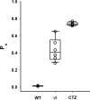
Open probability of GluA2 receptors. Open probability (Po) of GluA2 receptors in the absence of CTZ, in tandem with γ8, and in the presence of CTZ. Error bars are SEM.
Figure 3.
Figure 3.
Dwell-time distribution of GluA2 receptors from single-channel recordings. (A) Open time. (B) Shut time. (C) Burst duration dwell-time distributions for GluA2 receptor alone, in GluA2/γ8, and in GluA2 in the presence of CTZ, with 10 mM glutamate. SQRT, square root.
Figure 4.
Figure 4.
Conformational landscape at the ATD of the GluA2 receptor. (A–E) smFRET histograms for site 23 of the GluA2 receptor under (A) unliganded apo condition, (B) open condition (in the presence of 1 mM glutamate + 100 µM CTZ), (C) desensitized condition (in the presence of 1 mM glutamate), (D) in tandem with γ8 in unliganded apo condition, and (E) in tandem with γ8 in the desensitized condition (in the presence of 1 mM glutamate). Corresponding representative smFRET traces are shown above each histogram. Denoised smFRET histograms are in red, and observed histograms with Gaussian fits are in gray. (F) Fluorophore attachment site 23 at the ATD of GluA2 receptors shown in the side view and top view of full-length GluA2 receptor (PDB accession no. 4U2P; apo).
Figure 5.
Figure 5.
Dwell-time distributions for transitions between the smFRET states at site 23. (I) Comparison of dwell times for transitions in GluA2/γ8 and in the presence of glutamate between (A) the high FRET state 1 to the low FRET state 2 and between (B) the low FRET state 2 to the high FRET state 1. (II) Comparison of dwell times for transitions in GluA2 in the presence of CTZ and glutamate between (A) the high FRET state 1 and the low FRET state 2 and between (B) the low FRET state 2 and the high FRET state 1.
Figure 6.
Figure 6.
Conformational landscape at the LBD of the GluA2 receptor. smFRET histograms for site 467 of the GluA2 receptor under (A) unliganded apo condition, (B) open condition (in the presence of 1 mM glutamate + 100 µM CTZ), (C) desensitized condition (in the presence of 1 mM glutamate), (D) in tandem with γ8 in unliganded apo condition, and (E) in tandem with γ8 in the desensitized condition (in the presence of 1 mM glutamate). Corresponding representative smFRET traces are shown above each histogram. Denoised smFRET histograms are in red, and observed ligand-binding terminal domain of GluA2 receptors is shown in the side view and top view of full-length GluA2 receptor (PDB accession no. 4U2P; apo).EA, apparent FRET efficiency.
Figure 7.
Figure 7.
Dwell time distributions for transitions between the smFRET states at site 467. (I) Comparison of dwell times for transitions in GluA2/γ8 and in the presence of glutamate between (A) the high FRET state 1 and the low FRET state 2 and between (B) the low FRET state 2 and the high FRET state 1. (II) Comparison of dwell times for transitions in GluA2 in the presence of CTZ and glutamate between (A) the high FRET state 1 and the low FRET state 2 and between (B) the low FRET state 2 and the high FRET state 1.

Comment in

  • Resensitizing AMPA receptors.
    Short B. Short B. J Gen Physiol. 2020 Jan 6;152(1):e201912542. doi: 10.1085/jgp.201912542. J Gen Physiol. 2020. PMID: 31825464 Free PMC article.

Similar articles

Cited by

References

    1. Baranovic J., and Plested A.J.R.. 2018. Auxiliary subunits keep AMPA receptors compact during activation and desensitization. eLife. 7:e40548 10.7554/eLife.40548 - DOI - PMC - PubMed
    1. Ben-Yaacov A., Gillor M., Haham T., Parsai A., Qneibi M., and Stern-Bach Y.. 2017. Molecular mechanism of AMPA receptor modulation by TARP/stargazin. Neuron. 93:1126–1137.e4. 10.1016/j.neuron.2017.01.032 - DOI - PubMed
    1. Cais O., Herguedas B., Krol K., Cull-Candy S.G., Farrant M., and Greger I.H.. 2014. Mapping the interaction sites between AMPA receptors and TARPs reveals a role for the receptor N-terminal domain in channel gating. Cell Reports. 9:728–740. 10.1016/j.celrep.2014.09.029 - DOI - PMC - PubMed
    1. Carbone A.L., and Plested A.J.. 2016. Superactivation of AMPA receptors by auxiliary proteins. Nat. Commun. 7:10178 10.1038/ncomms10178 - DOI - PMC - PubMed
    1. Chen S., Zhao Y., Wang Y., Shekhar M., Tajkhorshid E., and Gouaux E.. 2017. Activation and desensitization mechanism of AMPA receptor-TARP complex by cryo-EM. Cell. 170:1234–1246.e14. 10.1016/j.cell.2017.07.045 - DOI - PMC - PubMed

Publication types