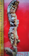
Metastasis of carcinoma of buccal mucosa to small intestine causing ileal perforation
- PMID: 31748358
- PMCID: PMC6887439
- DOI: 10.1136/bcr-2019-231449
Metastasis of carcinoma of buccal mucosa to small intestine causing ileal perforation
Abstract
Oral cancers rarely metastasize to the small intestines. In a previously operated case of squamous cell carcinoma of buccal mucosa without any known preoperative distant metastases, we report a case of solitary ileal perforation 3 months after the surgery. The edge of the ileal perforation was positive for squamous cell carcinoma on histopathology. It is important to remember metastases as a cause of acute abdomen in the prior history of oral malignancies.
Keywords: gastrointestinal surgery; general surgery; head and neck cancer; small intestine cancer.
© BMJ Publishing Group Limited 2019. No commercial re-use. See rights and permissions. Published by BMJ.
Conflict of interest statement
Competing interests: None declared.
Figures


References
-
- Kaneda K, Miyazaki N, Sugimoto T, et al. . A case of perforation of a peritoneal disseminated tumor of the small intestine after total gastrectomy for gastric cancer. The journal of the Japanese Practical Surgeon Society 1994;55:1814–7. 10.3919/ringe1963.55.1814 - DOI
-
- Katuyoshi A, Mikihiro F, Nobuhiro U, et al. . Duodenal metastasis from head and neck cancer with an intestinal obstruction. J Cytol Histol 2014;S14:1–4.
-
- Sharma K. Solitary jejunal metastasis from carcinoma pyriform fossa presenting as intestinal obstruction. Int JO Phono Laryng jan to june 2014;4:36–9. 10.5005/jp-journals-10023-1078 - DOI
Publication types
MeSH terms
LinkOut - more resources
Full Text Sources
Medical
