. 2020 Jan 31;94(4):e01936-19.
doi: 10.1128/JVI.01936-19. Print 2020 Jan 31.

Hepatitis Delta Virus Alters the Autophagy Process To Promote Its Genome Replication

Affiliations

Hepatitis Delta Virus Alters the Autophagy Process To Promote Its Genome Replication

Marwa Khabir et al. J Virol. .

Abstract

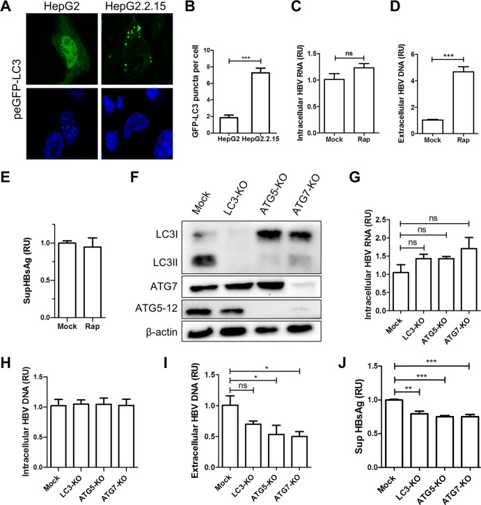
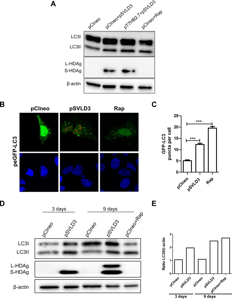
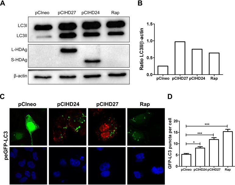
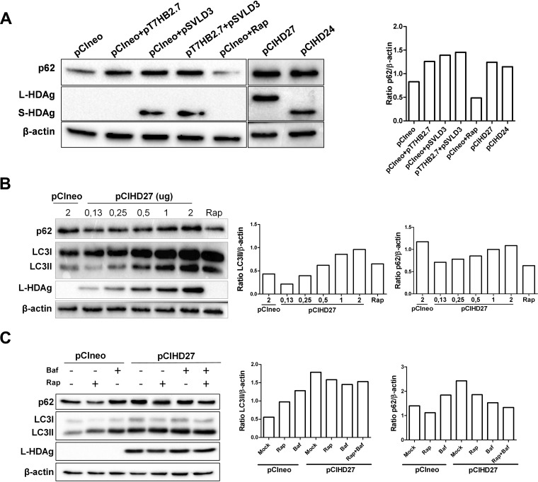
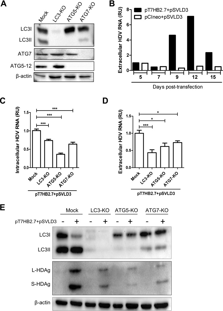
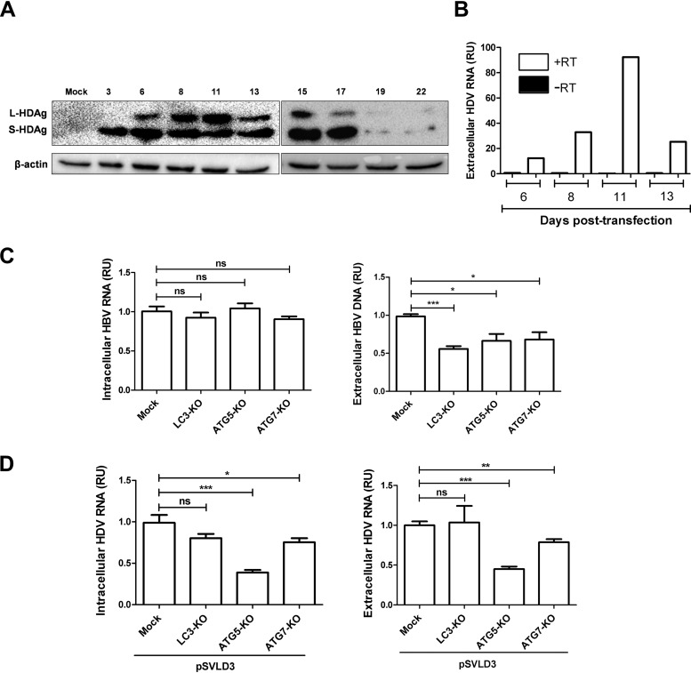
A substantial number of viruses have been demonstrated to subvert autophagy to promote their own replication. Recent publications have reported the proviral effect of autophagy induction on hepatitis B virus (HBV) replication. Hepatitis delta virus (HDV) is a defective virus and an occasional obligate satellite of HBV. However, no previous work has studied the relationship between autophagy and HDV. In this article, we analyze the impact of HBV and HDV replication on autophagy as well as the involvement of the autophagy machinery in the HDV life cycle when produced alone and in combination with HBV. We prove that HBxAg and HBsAg can induce early steps of autophagy but ultimately block flux. It is worth noting that the two isoforms of the HDV protein, the small HDAg (S-HDAg) and large HDAg (L-HDAg) isoforms, can also efficiently promote autophagosome accumulation and disturb autophagic flux. Using CRISPR-Cas9 technology to generate specific knockouts, we demonstrate that the autophagy machinery, specifically the proteins implicated in the elongation step (ATG7, ATG5, and LC3), is important for the release of HBV without affecting the level of intracellular HBV genomes. Surprisingly, the knockout of ATG5 and ATG7 decreased the intracellular HDV RNA level in both Huh7 and HepG2.2.15 cells without an additional effect on HDV secretion. Therefore, we conclude that HBV and HDV have evolved to utilize the autophagy machinery so as to assist at different steps of their life cycle.IMPORTANCE Hepatitis delta virus is a defective RNA virus that requires hepatitis B virus envelope proteins (HBsAg) to fulfill its life cycle. Thus, HDV can only infect individuals at the same time as HBV (coinfection) or superinfect individuals who are already chronic carriers of HBV. The presence of HDV in the liver accelerates the progression of infection to fibrosis and to hepatic cancer. Since current treatments against HBV are ineffective against HDV, it is of paramount importance to study the interaction between HBV, HDV, and host factors. This will help unravel new targets whereby a therapy that is capable of simultaneously impeding both viruses could be developed. In this research paper, we evidence that the autophagy machinery promotes the replication of HBV and HDV at different steps of their life cycle. Notwithstanding their contribution to HBV release, autophagy proteins seem to assist HDV intracellular replication but not its secretion.

Keywords: ATG5; autophagy; chronic infection; hepatitis B virus (HBV); hepatitis delta virus (HDV); viral replication.


Figures

FIG 1
FIG 1
HBsAg and HBxAg expressions induce the accumulation of autophagosomes in Huh7 cells. (A) Huh7 cells were transfected with pCIneo, pCIHBenv(+), or pCIHBenv(−) or treated for 24 h with rapamycin (Rap) (200 nM). Seventy-two hours after transfection, the levels of LC3II, p62, and β-actin were evaluated by Western blotting. (B) Huh7 cells were transfected with pCIneo, pCIHBx-Flag, or pCIHBs-Flag. After 72 h of transfection, the levels of LC3II, p62, and β-actin were evaluated by Western blotting. Moreover, the expressions of HBxAg and HBsAg were verified by Western blotting using an anti-Flag antibody. Cells transfected with pCIneo were treated or not with rapamycin. (C) Huh7 cells were cotransfected with pCIneo, pCIHBx-Flag, or pCIHBs-Flag and peGFP-LC3. As a positive control for autophagy induction, Huh7 cells cotransfected with pCIneo and peGFP-LC3 were treated with rapamycin (100 μM) for 3 h. At 72 h posttransfection, cells were fixed and stained with an anti-Flag antibody (red) or DAPI (blue). (D) The numbers of GFP-LC3 puncta per cell were calculated. (E and F) Huh7 cells were cotransfected with peGFP-LC3 and the plasmid pCIneo, pCIHBx-Flag, or pCIHBs-Flag, expressing the HBV proteins (S-HBsAg or HBxAg) through a cytomegalovirus (CMV) promoter (E), or the plasmid pCIneo, pT7HB2.7x(−) (L-M-S), or pT7HB2.7 (L-M-S-X), expressing the HBV envelope proteins and HBxAg from HBV endogenous promoters (F). The percentage of GFP-LC3-positive cells with punctate LC3 was calculated in transfected cells at 72 h posttransfection. Calculation was based on the count in 100 cells under each condition.
FIG 2
FIG 2
Autophagy machinery is required for HBV maturation/secretion. (A and B) HepG2.2.15 and HepG2 cells were transfected with peGFP-LC3 for 72 h. The nuclei were stained with DAPI (blue). The numbers of GFP-LC3 puncta per cell were calculated (n = 74). (C to E) HepG2.2.15 cells were treated or not with rapamycin (50 nM). After 24 h of treatment, cells were washed and treated for another 24 h. Next, supernatants and cell lysates were harvested. The intracellular HBV RNA levels were measured by RT-qPCR. For monitoring the release of HBV virions, Dane particles from supernatants were immunoprecipitated with anti-preS1 antibody and protein A/G beads, and the viral genomes present in the virions were analyzed by qPCR. The release of HBsAg in the cell supernatants (SupHBsAg) was analyzed by an ELISA. The RT-qPCR, qPCR, and ELISA results are presented as relative units (RU) and compared to values for untreated HepG2.2.15 cells. Error bars show the standard errors of the means from four experiments, measured in triplicate. (F) HepG2.2.15 cells were transduced with lentivirus preparations targeting LC3, ATG5, and ATG7 for 9 days. The expression levels of LC3, ATG5, ATG7, and β-actin were verified by Western blotting. (G) Intracellular HBV RNAs were quantified by RT-qPCR. (H) Intracellular HBV DNAs were quantified by qPCR. (I) The extracellular HBV virions were immunoprecipitated with anti-preS1 antibody and protein A/G beads, and the viral DNA expression levels in the knockout populations were quantified by qPCR. (J) The secretion of HBsAg in the cell culture supernatant was measured by an ELISA. Data were measured in quadruplicate under each condition. RU, relative units; ns, not significant.
FIG 3
FIG 3
Hepatitis delta virus replication induces autophagosome accumulation in Huh7 cells. (A) Huh7 cells were transfected as indicated or treated with rapamycin (200 nM) for 24 h. At 72 h posttransfection, cells were lysed, and the levels of LC3, HDAg, and β-actin were evaluated by Western blotting. (B) Huh7 cells were cotransfected with pCIneo or pSVLD3 and peGFP-LC3. As a positive control for autophagy induction, Huh7 cells cotransfected with pCIneo and peGFP-LC3 were treated with rapamycin (100 μM) for 3 h. After 72 h, cells were fixed and then stained with anti-HDAg antibody from HDV+ human serum (red). The nucleus was stained with DAPI (blue). (C) The number of GFP-LC3 puncta per cell was calculated from confocal images (n = 100). (D) Huh7 cells were transfected with pCIneo or pSVLD3 or treated with rapamycin (200 nM) for 24 h. At 3 and 9 days posttransfection, cells were lysed, and the levels of LC3, HDAg, and β-actin were evaluated by Western blotting. (E) Ratios of LC3II/β-actin.
FIG 4
FIG 4
The two isoforms of HDAg can induce autophagosome accumulation in Huh7 cells. (A) Huh7 cells were transfected with pCIneo or a plasmid coding for L-HDAg (pCIHD27) or S-HDAg (pCIHD24). As a control, Huh7 cells were treated with rapamycin (200 nM) for 24 h. At 72 h posttransfection, cells were lysed, and the levels of LC3, HDAg, and β-actin were evaluated by Western blotting. (B) Ratios of LC3II/β-actin. (C) Confocal images of Huh7 cells cotransfected with pCIneo, pCIHD27, pCIHD24, and peGFP-LC3. The S-HDAg and L-HDAg proteins were labeled using anti-HDAg antibody from HDV+ human serum (red). The nucleus was stained with DAPI (blue). (D) The number of GFP-LC3 puncta per cell was calculated (n = 100).
FIG 5
FIG 5
L-HDAg and S-HDAg expressions impede autophagic flux. (A) Huh7 cells were transfected as indicated or transfected with pCIneo and treated with rapamycin (200 nM) for 24 h. (Left) At 72 h posttransfection, the levels of p62, HDAg, and β-actin were evaluated by Western blotting. (Right) The ratios of p62/β-actin were quantified. (B) Huh7 cells were transfected with pCIneo and treated or not with rapamycin (200 nM for 24 h) or with increasing quantities of pCIHD27. (Left) After 72 h, the levels of expression of p62, LC3, L-HDAg, and β-actin were evaluated by Western blotting. (Right) The ratios of LC3II/β-actin and p62/β-actin were quantified. (C) Huh7 cells were transfected with pCIneo or pCIHD27 and treated or not with rapamycin and/or bafilomycin A1 (Baf), as indicated. (Left) After 72 h, cells were lysed, and the levels of p62, LC3, L-HDAg, and β-actin were evaluated by Western blotting. (Right) The ratios of LC3II/β-actin and p62/β-actin were also quantified.
FIG 6
FIG 6
Autophagy machinery is required for HDV replication in Huh7 cells. (A) Huh7 cells were transduced with lentivirus to produce stable knockout populations for LC3, ATG5, and ATG7 genes. Western blotting was conducted to verify the knockout efficiencies. (B) Huh7 cells were cotransfected with pT7HB2.7 and pSVLD3 to produce HDV virions or with pSVLD3 and pCIneo. The supernatants were harvested at different time points to determinate the kinetics of secretion of HDV virions by RT-qPCR. (C to E) Huh7 cells knocked out for LC3, ATG5, or ATG7 were cotransfected with pT7HB2.7 and pSVLD3. (C and D) At 9 days posttransfection, intracellular (C) and extracellular (D) HDV RNAs were quantified by RT-qPCR. To control DNA plasmid contamination, the supernatants were treated with Benzonase. Error bars show the standard errors of the means from three independent experiments, measured in triplicate. (E) Cell lysates were used for Western blotting to evaluate the LC3, HDAg, and β-actin expression levels.
FIG 7
FIG 7
Autophagy machinery differentially affects the HBV and HDV replication cycle in cells expressing both viruses. (A and B) The kinetics of HDV replication were evaluated in the presence of HBV replication. HepG2.2.15 cells were transfected with pSVLD3, and cells and supernatants were collected at different time points posttransfection. (A) Cell lysates from days 3 to 22 were subjected to Western blotting to evaluate the expression of HDAg and β-actin. (B) HDV RNAs from supernatants collected at 6 to 13 days posttransfection were quantified by RT-qPCR. To control for plasmid DNA contamination, the supernatants were treated with Benzonase, DNase treatment was carried out on columns during RNA extraction, and RT-qPCR was conducted with or without the reverse transcriptase enzyme. (C) HepG2.2.15 cells knocked out for LC3, ATG5, and ATG7 were transfected with pSVLD3. At 11 days posttransfection, cells and supernatants were harvested. (Left) To analyze intracellular HBV RNA, RT-qPCR was conducted. (Right) To measure the release of HBV virions, immunoprecipitated virions were subjected to qPCR. (D) Intracellular (left) and extracellular (right) HDV RNAs were quantified by RT-qPCR. Error bars show the standard errors of the means from three independent experiments, measured in triplicate.

Similar articles

Cited by

References

    1. Klionsky DJ, Emr SD. 2000. Autophagy as a regulated pathway of cellular degradation. Science 290:1717–1721. doi:10.1126/science.290.5497.1717. - DOI - PMC - PubMed
    1. Noda NN, Inagaki F. 2015. Mechanisms of autophagy. Annu Rev Biophys 44:101–122. doi:10.1146/annurev-biophys-060414-034248. - DOI - PubMed
    1. Shpilka T, Mizushima N, Elazar Z. 2012. Ubiquitin-like proteins and autophagy at a glance. J Cell Sci 125:2343–2348. doi:10.1242/jcs.093757. - DOI - PubMed
    1. Mizushima N, Kuma A, Kobayashi Y, Yamamoto A, Matsubae M, Takao T, Natsume T, Ohsumi Y, Yoshimori T. 2003. Mouse Apg16L, a novel WD-repeat protein, targets to the autophagic isolation membrane with the Apg12-Apg5 conjugate. J Cell Sci 116:1679–1688. doi:10.1242/jcs.00381. - DOI - PubMed
    1. Sakoh-Nakatogawa M, Matoba K, Asai E, Kirisako H, Ishii J, Noda NN, Inagaki F, Nakatogawa H, Ohsumi Y. 2013. Atg12-Atg5 conjugate enhances E2 activity of Atg3 by rearranging its catalytic site. Nat Struct Mol Biol 20:433–439. doi:10.1038/nsmb.2527. - DOI - PubMed

Publication types

LinkOut - more resources