. 2020 Feb 21;295(8):2175-2183.
doi: 10.1074/jbc.RA119.009896. Epub 2019 Nov 20.

A proteolytic C-terminal fragment of Nogo-A (reticulon-4A) is released in exosomes and potently inhibits axon regeneration

Affiliations

A proteolytic C-terminal fragment of Nogo-A (reticulon-4A) is released in exosomes and potently inhibits axon regeneration

Yuichi Sekine et al. J Biol Chem. .

Abstract

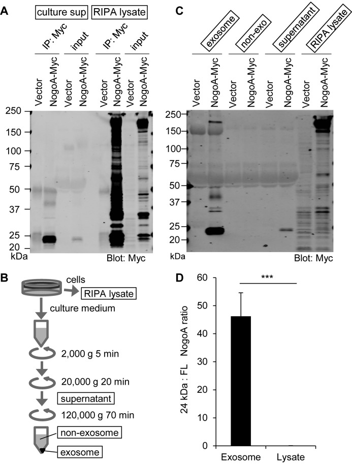
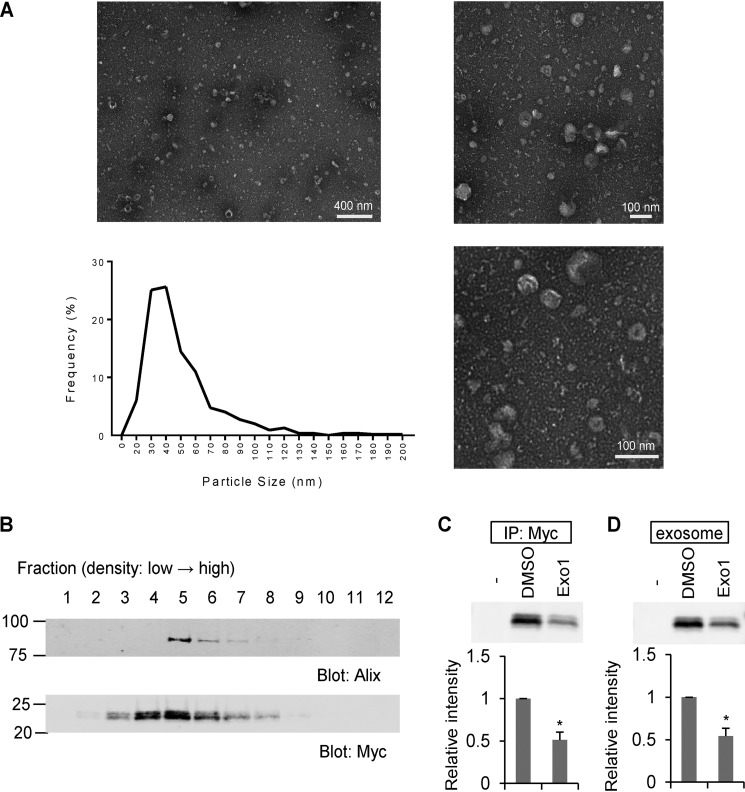
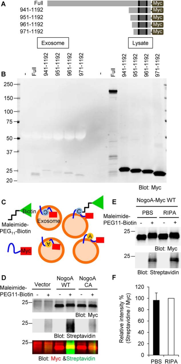
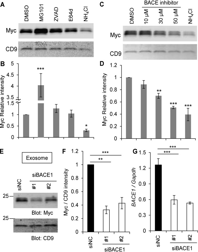
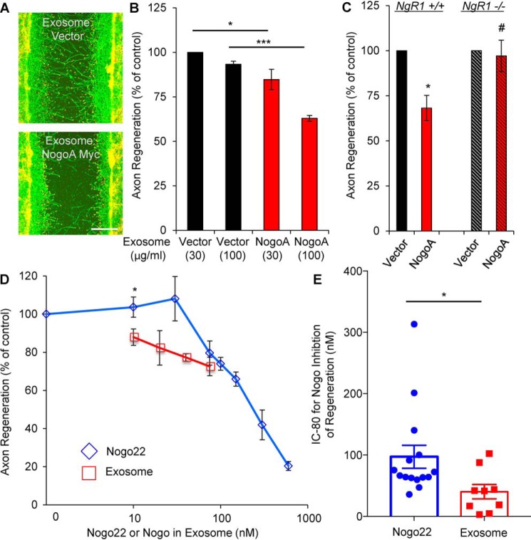
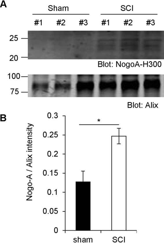
Glial signals are known to inhibit axonal regeneration and functional recovery after mammalian central nervous system trauma, including spinal cord injury. Such signals include membrane-associated proteins of the oligodendrocyte plasma membrane and astrocyte-derived, matrix-associated proteins. Here, using cell lines and primary cortical neuron cultures, recombinant protein expression, immunoprecipitation and immunoblot assays, transmission EM of exosomes, and axon regeneration assays, we explored the secretion and activity of the myelin-associated neurite outgrowth inhibitor Nogo-A and observed exosomal release of a 24-kDa C-terminal Nogo-A fragment from cultured cells. We found that the cleavage site in this 1192-amino-acid-long fragment is located between amino acids 961-971. We also detected a Nogo-66 receptor (NgR1)-interacting Nogo-66 domain on the exosome surface. Enzyme inhibitor treatment and siRNA knockdown revealed that β-secretase 1 (BACE1) is the protease responsible for Nogo-A cleavage. Functionally, exosomes with the Nogo-66 domain on their surface potently inhibited axonal regeneration of mechanically injured cerebral cortex neurons from mice. Production of this fragment was observed in the exosomal fraction from neuronal tissue lysates after spinal cord crush injury of mice. We also noted that, relative to the exosomal marker Alix, a Nogo-immunoreactive, 24-kDa protein is enriched in exosomes 2-fold after injury. We conclude that membrane-associated Nogo-A produced in oligodendrocytes is processed proteolytically by BACE1, is released via exosomes, and is a potent diffusible inhibitor of regenerative growth in NgR1-expressing axons.

Keywords: NgR1; Nogo; Nogo receptor; axon; exosome (vesicle); oligodendrocyte; regeneration; reticulon; spinal cord injury; β-secretase 1 (BACE1).


Conflict of interest statement

The content is solely the responsibility of the authors and does not necessarily represent the official views of the National Institutes of Health. S.M.S. is a cofounder of ReNetX Bio, which seeks to develop NgR1-based therapeutics

Figures

Figure 1.
Figure 1.
The Nogo-A C-terminal region is secreted in culture supernatant as an exosome component. A, culture supernatants (culture sup) collected from vector- and Nogo-A–Myc–transfected HEK293T cells were immunoprecipitated (IP) with anti-Myc antibody and immunoblotted with anti-Myc antibody. Cell lysates were also immunoprecipitated and immunoblotted in the same membrane. B, schematic of exosome fractionation by centrifugation. C, each fraction from vector or Nogo-A–Myc–transfected HEK293T cells was immunoblotted with anti-Myc antibody. D, ratio of 24-kDa Nogo-A fragment to full-length (FL) Nogo-A in exosome and lysate fractions. Mean ± S.E., n = 12 for each group. ***, p < 0.005; Student's two-tailed t test.
Figure 2.
Figure 2.
The Nogo-A C-terminal region is enriched in exosomes. A, negative stain transmission EM images of the exosome fraction derived from HEK293T cell culture medium, with particle diameter distribution represented in a histogram. n = 546 particles. B, culture medium from Nogo-A–Myc–transfected HEK293T cells was separated by sucrose density centrifugation. Gradient fractions from the top were immunoblotted with anti-Alix and anti-Myc antibodies. C and D, HEK293T cells were transfected with vector (−) or Nogo-A–Myc. 24 h after transfection, media were changed to DMSO or Exo1 containing medium and cultured for another 12 h. Then the culture supernatants were immunoprecipitated (IP) with anti-Myc antibody or exosome-fractionated. Mean ± S.E., n = 3 independent experiments. *, p < 0.05; Student's two-tailed t test.
Figure 3.
Figure 3.
C-terminal cleavage site in Nogo-A. A, a series of Nogo-A–Myc C-terminally truncated constructs were generated. B, HEK293T cells were transfected with full-length Nogo-A–Myc or a series of C-terminal constructs of Nogo-A–Myc. 36 h after transfection, culture supernatants were collected, and exosomes were fractionated. Cell lysates were also collected. Samples were then immunoblotted with anti-Myc antibody. C, schematics of maleimide–PEG11–biotin labeling. A cysteine residue of Nogo-A exposed on the surface of exosomes can be labeled with nonpermeable maleimide–PEG11–biotin reagent. Cysteine is shown as a gray circle. Alanine substituted from cysteine is shown as a yellow circle. D, exosomes from vector-, Nogo-A WT–, or Nogo-A C1101A (NogoA CA)–transfected HEK293T cells were incubated with maleimide–PEG11–biotin. After the maleimide reaction, exosomes were lysed with RIPA buffer and immunoprecipitated with anti-Myc antibody. Immunoprecipitates were washed three times and blotted with anti-Myc antibody and Alexa Fluor 488 streptavidin. E, Nogo-A–Myc-exosomes resuspended in PBS or RIPA buffer were incubated with maleimide–PEG11–biotin. After the maleimide reaction, exosomes were resuspended in RIPA buffer and immunoprecipitated with anti-Myc antibody. Immunoprecipitates were washed three times and blotted with anti-Myc antibody and Alexa Fluor 488 streptavidin. F, streptavidin intensity was divided by Myc intensity under both PBS and RIPA conditions and expressed as normalized value as RIPA sample of 100%. Mean ± S.E., n = 9 independent experiments. p = 0.70, Student's two-tailed t test.
Figure 4.
Figure 4.
The Nogo-66 loop region on the surface of exosomes. A and B, HEK293T cells were transfected with Nogo-A–Myc. 24 h after transfection, media were changed. DMSO, MG101 (20 μm), Z-VAD-fmk (20 μm), E64d (20 μm), and NH4Cl (20 mm) were added and cultured for further 12 h. Then culture supernatants were collected, and the exosome fraction was immunoblotted with anti-Myc and anti-CD9 antibodies (A). The graph shows Myc intensity divided by CD9 intensity compared to DMSO control (B). Mean ± S.E., n = 5–8 independent experiments. *, p < 0.05; ***, p < 0.005; one-way ANOVA followed by Dunnett's test. C and D, HEK293T cells were transfected with Nogo-A–Myc. 24 h after transfection, media were changed. DMSO, the indicated amounts of BACE inhibitors, and NH4Cl (20 mm) were added and cultured for further 12 h. Then culture supernatants were collected, and the exosome fraction was immunoblotted with anti-Myc and anti-CD9 antibodies (C). The graph shows Myc intensity divided by CD9 intensity compared to DMSO control (D). Mean ± S.E., n = 4 independent experiments. **, p < 0.01; ***, p < 0.005; one-way ANOVA followed by Dunnett's test. E, HEK293T cells were transfected with Nogo-A–Myc and siNC, siBACE1 #1, or siBACE1 #2. Exosomes were purified 36 h after transfection and immunoblotted with anti-Myc and anti-CD9 antibodies. F, quantification of Myc intensity divided by CD9 intensity compared to DMSO control. Mean ± S.E., n = 6 independent experiments. **, p < 0.01; ***, p < 0.005; one-way ANOVA followed by Dunnett's test. G, real-time PCR for replicates of siBACE-transfected HEK293T cells. BACE1 mRNA expression was normalized to Gapdh mRNA expression. Mean ± S.E., n = 4 independent experiments. ***, p < 0.005; one-way ANOVA followed by Dunnett's test.
Figure 5.
Figure 5.
Inhibitor function of the Nogo-A C-terminal fragment. A and B, cortical neurons were scraped and treated with the indicated amount of exosomes at 8 DIV for 3 days (A). The microphotographs show βIII-tubulin (in axons, green) and phalloidin (to stain F-actin, red). Scale bar = 200 μm. B, quantification of axonal regeneration. Mean ± S.E., n = 3 biological replicates. *, p < 0.05; ***, p < 0.005; one-way ANOVA followed by Tukey's test. C, cortical neurons from the WT or NgR1−/− were scraped and treated with the same amount of exosomes at 8 DIV for 3 days. The graph shows quantification of axon regeneration. *, p < 0.05; Student's two-tailed t test. #, not significant. D, cortical neurons were scraped and treated with exosomes (10, 20, 40, and 75 nm) or Nogo22 (0, 10, 30, 75, 100, 150, 300, and 600 nm) at 8 DIV for 3 days. The graph shows quantification of axonal regeneration. Mean ± S.E., n = 2–6 biological replicates. *, p < 0.05; Student's two-tailed t test. E, the IC80 from each separate experiment was calculated. Mean ± S.E., n = 15 (Nogo22) and n = 9 (exosomes) biological replicates. *, p < 0.05; Student's two-tailed t test.
Figure 6.
Figure 6.
Increased Nogo-A fragment levels after spinal cord trauma in vivo. A, WT mice had their spinal cord crushed and were sacrificed 3 days after surgery. The spinal cords were then taken out and homogenized in TBS. Exosome fractions were immunoblotted with anti-Nogo-A and anti-Alix antibodies. B, quantification of Nogo-A divided by Alix intensity. Mean ± S.E., n = 3 animals. *, p < 0.05; Student's two-tailed t test.

References

    1. GrandPré T., Nakamura F., Vartanian T., and Strittmatter S. M. (2000) Identification of the Nogo inhibitor of axon regeneration as a Reticulon protein. Nature 403, 439–444 10.1038/35000226 - DOI - PubMed
    1. Chen M. S., Huber A. B., van der Haar M. E., Frank M., Schnell L., Spillmann A. A., Christ F., and Schwab M. E. (2000) Nogo-A is a myelin-associated neurite outgrowth inhibitor and an antigen for monoclonal antibody IN-1. Nature 403, 434–439 10.1038/35000219 - DOI - PubMed
    1. Mukhopadhyay G., Doherty P., Walsh F. S., Crocker P. R., and Filbin M. T. (1994) A novel role for myelin-associated glycoprotein as an inhibitor of axonal regeneration. Neuron 13, 757–767 10.1016/0896-6273(94)90042-6 - DOI - PubMed
    1. McKerracher L., David S., Jackson D. L., Kottis V., Dunn R. J., and Braun P. E. (1994) Identification of myelin-associated glycoprotein as a major myelin-derived inhibitor of neurite growth. Neuron 13, 805–811 10.1016/0896-6273(94)90247-X - DOI - PubMed
    1. Kottis V., Thibault P., Mikol D., Xiao Z. C., Zhang R., Dergham P., and Braun P. E. (2002) Oligodendrocyte-myelin glycoprotein (OMgp) is an inhibitor of neurite outgrowth. J. Neurochem. 82, 1566–1569 10.1046/j.1471-4159.2002.01146.x - DOI - PubMed

Publication types

LinkOut - more resources