. 2019 Nov 20;9(1):17150.
doi: 10.1038/s41598-019-53537-z.

Anatomical study of the coffee berry borer (Hypothenemus hampei) using micro-computed tomography

Affiliations

Anatomical study of the coffee berry borer (Hypothenemus hampei) using micro-computed tomography

Ignacio Alba-Alejandre et al. Sci Rep. .

Erratum in

Abstract

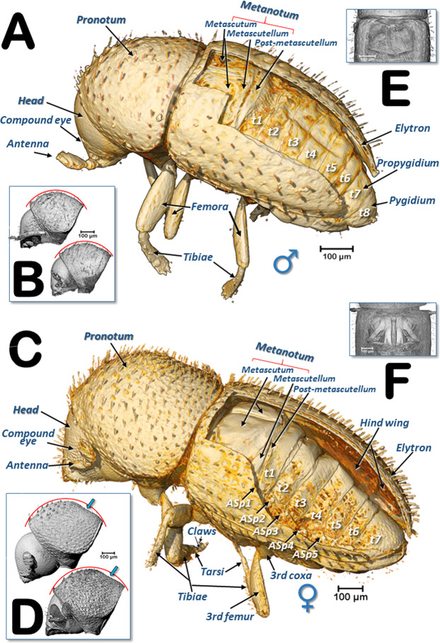
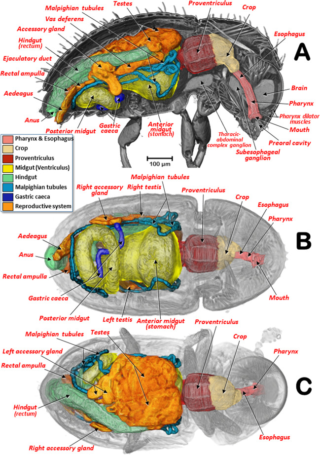
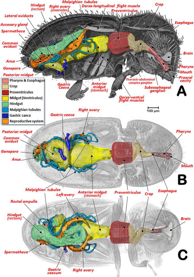
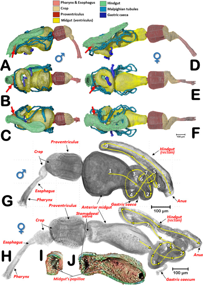
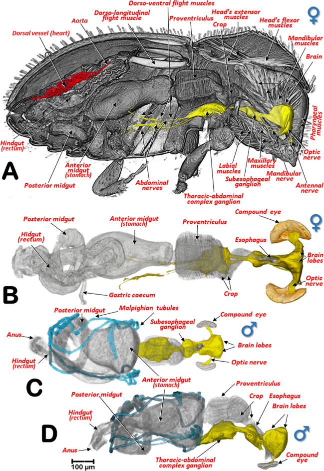
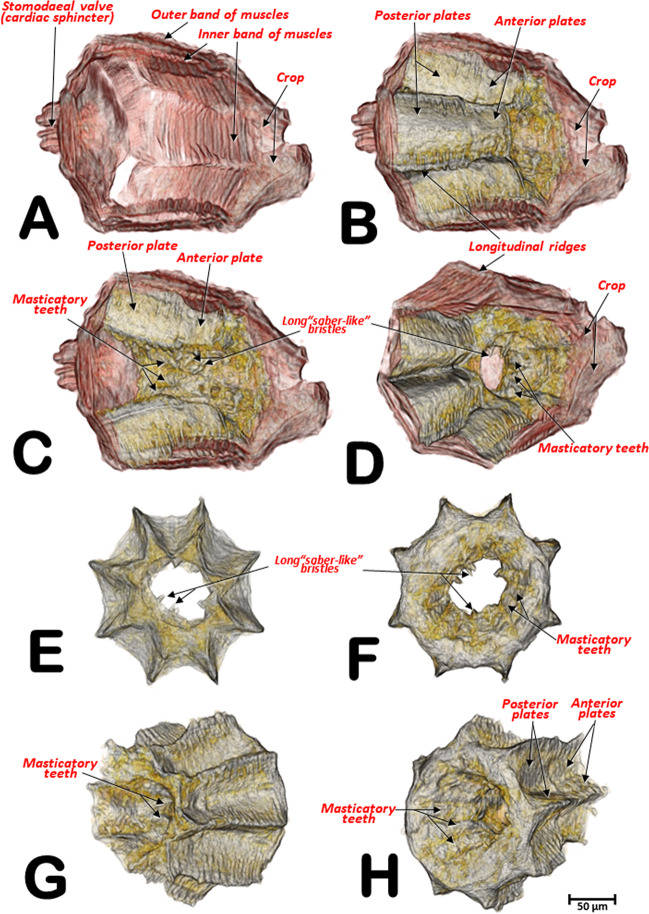
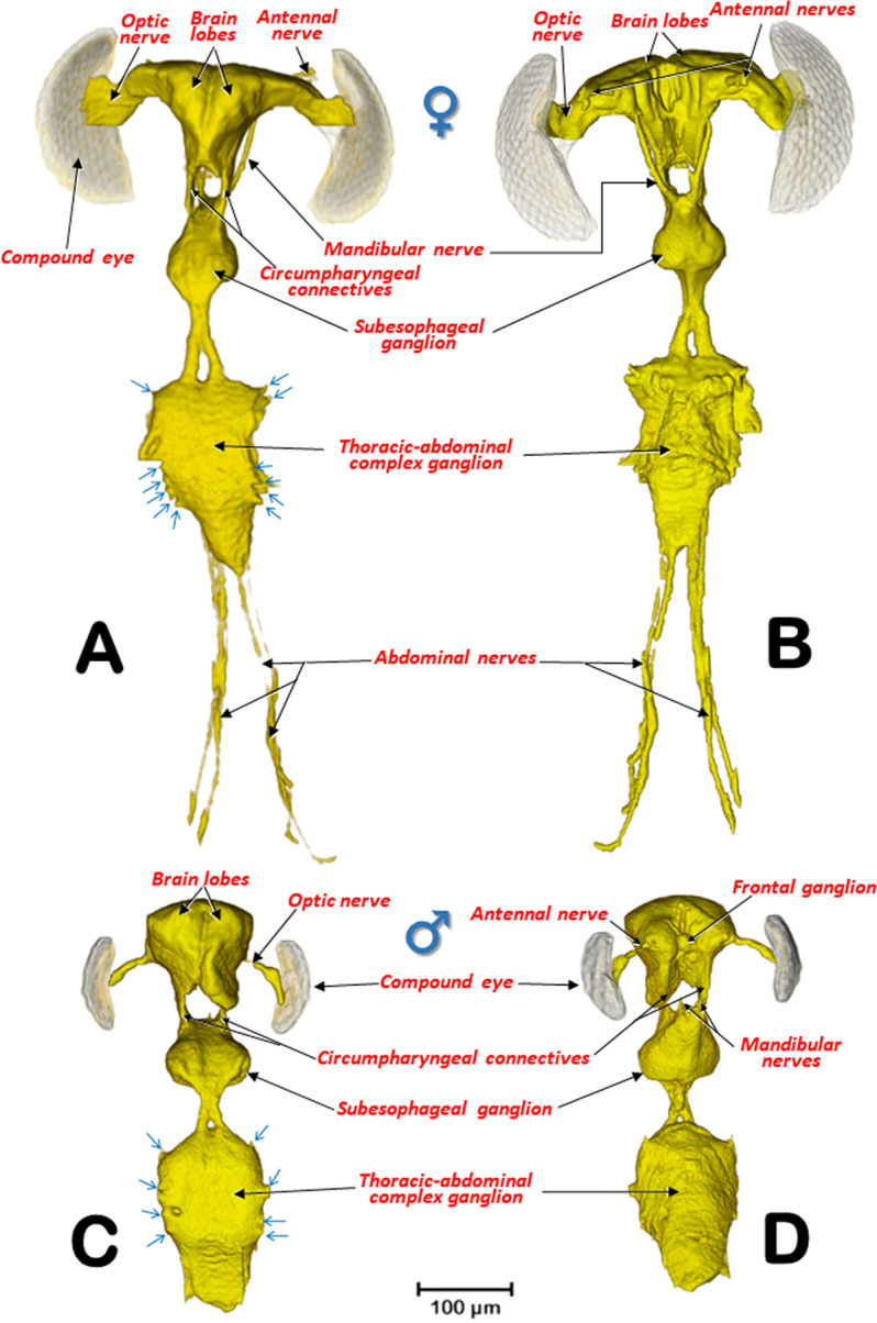
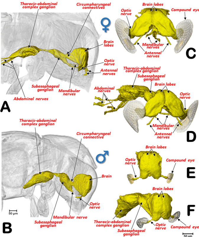
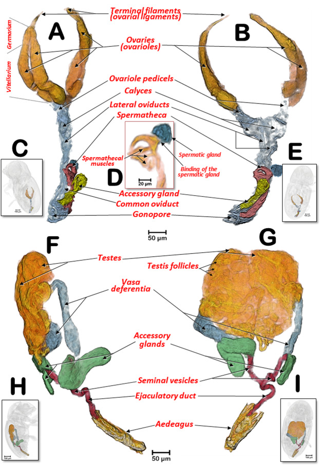
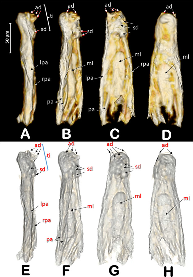
Traditionally, the study of anatomy in insects has been based on dissection techniques. Micro-computed tomography (micro-CT) is an X-ray based technique that allows visualization of the internal anatomy of insects in situ and does not require dissections. We report on the use of micro-CT scans to study, in detail, the internal structures and organs of the coffee berry borer (Hypothenemus hampei), the most damaging insect pest of coffee worldwide. Detailed images and videos allowed us to make the first description of the aedeagus and the first report of differences between the sexes based on internal anatomy (flight musculature, midgut shape, hindgut convolutions, brain shape and size) and external morphology (lateral outline of the pronotum and number of abdominal tergites). This study is the first complete micro-CT reconstruction of the anatomy of an insect and is also the smallest insect to have been evaluated in this way. High quality rendered images, and additional supplementary videos and 3D models are suitable for use with mobile devices and are useful tools for future research and as teaching aids.


Conflict of interest statement

The authors declare no competing interests.

Figures

Figure 1
Figure 1
Left dorso-lateral views of a male (A) and a female (C) coffee berry borer. Elytra have been removed using software thereby revealing the dorsal part of the thoracic and abdominal segments. Abdominal tergites (t) and the position of the abdominal respiratory spiracles (ASp) are labelled sequentially (terminology after Hopkins). Insets (B,D) show a uniformly curved pronotal disc in males (B) in contrast to a concave depression on the posterior pronotal third in females (D; blue arrows). Note different scales were used for each sex. The meso- and metanotum were removed by software to observe the reduced flight muscles in the male (E) in contrast to conspicuous flight muscles in the female (F).
Figure 2
Figure 2
Internal anatomy of a male coffee berry borer: right-lateral (A), ventral (B), and dorsal (C) views. To enhance the actual anatomical position of the different organs, the body (except the internal organs) have been rendered transparent (B,C) by depleting the opacity values using Amira software.
Figure 3
Figure 3
Internal anatomy of a female coffee berry borer: right-lateral (A), ventral (B), and dorsal (C) views. To enhance the actual anatomical position of the different organs, the body (except the internal organs) has been rendered transparent (B,C) by depleting the opacity values using Amira software.
Figure 4
Figure 4
Alimentary canal of a male (A,B,C) and a female coffee berry borer (D,E,F): right-lateral (A,D), ventral (B,E), and dorsal (C,F) views. Red arrows indicate the rectal ampulla. Lateral views of the male (G) and female (H) alimentary canals with the trajectories and convolutions of the midgut and hindgut shown in yellow. Each time the digestive tracts began a new trajectory curve, the new tract is marked with an incremental number. Anterior-transversal (I) and sagittal (J) cuts close to the stomodaeal valve of the midgut reveal the internal papillae.
Figure 5
Figure 5
Right-sagittal view of the internal anatomy of the female, showing the main musculature, dorsal vessel and nervous system (A), and a dorsal view of the digestive and nervous systems (B). Dorsal (C) and right-lateral (D) views of a male focusing on the positioning of the nervous system in relation to the digestive system.
Figure 6
Figure 6
Details of the proventriculus: micro-CT volume-rendered images (A–F) and light microscopy photographs (G,H). Right-lateral view (A,G); posterior view showing the stomodaeal valve (=cardiac sphincter) (B). Progressive transverse slices (thickness ca. 20 µm) from the basal to the apical zone, each marked with arrows numbered 1 to 3 (A,C,D,E); sagittal mid-slice (F); postero-lateral view (H). Most of the nomenclature after Eaton.
Figure 7
Figure 7
Micro-CT volume rendered images of the internal structures of the proventriculus from different perspective views: right-lateral (A,B,C), right-antero-posterior (D), anterior (E), posterior (F), left antero-posterior (G) and right-postero-anterior (H). Different perspectives of the grinding sclerotized complex isolated (E–H). A: a superficial window was opened, and the grinding sclerotized complex removed; B,D: as A but including the grinding complex; C: as B but the grinding complex has been superficially sagittally cut, eliminating one side of the octagonal structure.
Figure 8
Figure 8
Nervous systems of a female (A,B) and a male (C,D) coffee berry borer, showing the main ganglia and nerves (A,C: dorsal; B,D: ventral). The abdominal nerves are not shown in the male. The nerve insertions in the thoracic-abdominal-complex ganglion are indicated with blue arrows. Terminology after Atkins and Chapman and Snodgrass.
Figure 9
Figure 9
Lateral views of the nervous system of a female (A) and a male (B) coffee berry borer allowing comparison of the relative size of the central nervous system in relation to the head. Details in frontal (C,E) and latero-frontal (D,F) views of the central nervous system of a female (C,D) and a male (E,F) focusing on the differences between sexes.
Figure 10
Figure 10
Female (A–E) and male (F–I) coffee berry borer reproductive systems, in latero-dorsal (A,C), lateral (E,G) and dorsal (B,D,F,H) views. Details of the distal part of the spermatheca, with the spermathecal muscles and the spermatic gland (D). Figures (C,E,H and I) show the positions of the insect corresponding to figures (A,B,F and G), respectively.
Figure 11
Figure 11
Rendered images of different views of the aedeagus: left-lateral (A,E); left-latero-dorsal (B,F); dorsal (C,G); and ventral (D,H). Images at the bottom have been rendered transparent by software to enhance the view of internal structures. Abbreviations: ad = apical denticles (titillators); lpa = left parameres; rpa = right parameres; ml = median lobe (‘penis’); pa = paramere apophysys; rpa = right paramere; sd = subapical dorsal denticles; ti = titillators. Terminology after Lindroth and Palmen.

References

    1. Vega FE. The rise of coffee. Am. Sci. 2008;96:138–145.
    1. Wintgens, J. N. (Ed.). Coffee: Growing, Processing, Sustainable Production.Weinheim: Wiley-VCH (2004).
    1. Ziska, L. H., et al. Climate change, carbon dioxide, and pest biology, managing the future: Coffee as a case study. Agronomy8, 152 (2018).
    1. Davis AP, et al. High extinction risk for wild coffee species and implications for coffee sector sustainability. Sci. Adv. 2019;5:eaav3473. - PMC - PubMed
    1. Vega, F. E., Infante, F. & Johnson, A. J. The genus Hypothenemus, with emphasis on H. hampei, the coffee berry borer. In: Bark Beetles: Biology and Ecology of Native and Invasive Species (F. E., Vega and R. W., Hofstetter, Eds), pp. 427–494. San Diego: Academic Press (2015).

Publication types