. 2019 Nov;575(7784):669-673.
doi: 10.1038/s41586-019-1769-z. Epub 2019 Nov 20.

NLRP3 inflammasome activation drives tau pathology

Affiliations

NLRP3 inflammasome activation drives tau pathology

Christina Ising et al. Nature. 2019 Nov.

Abstract

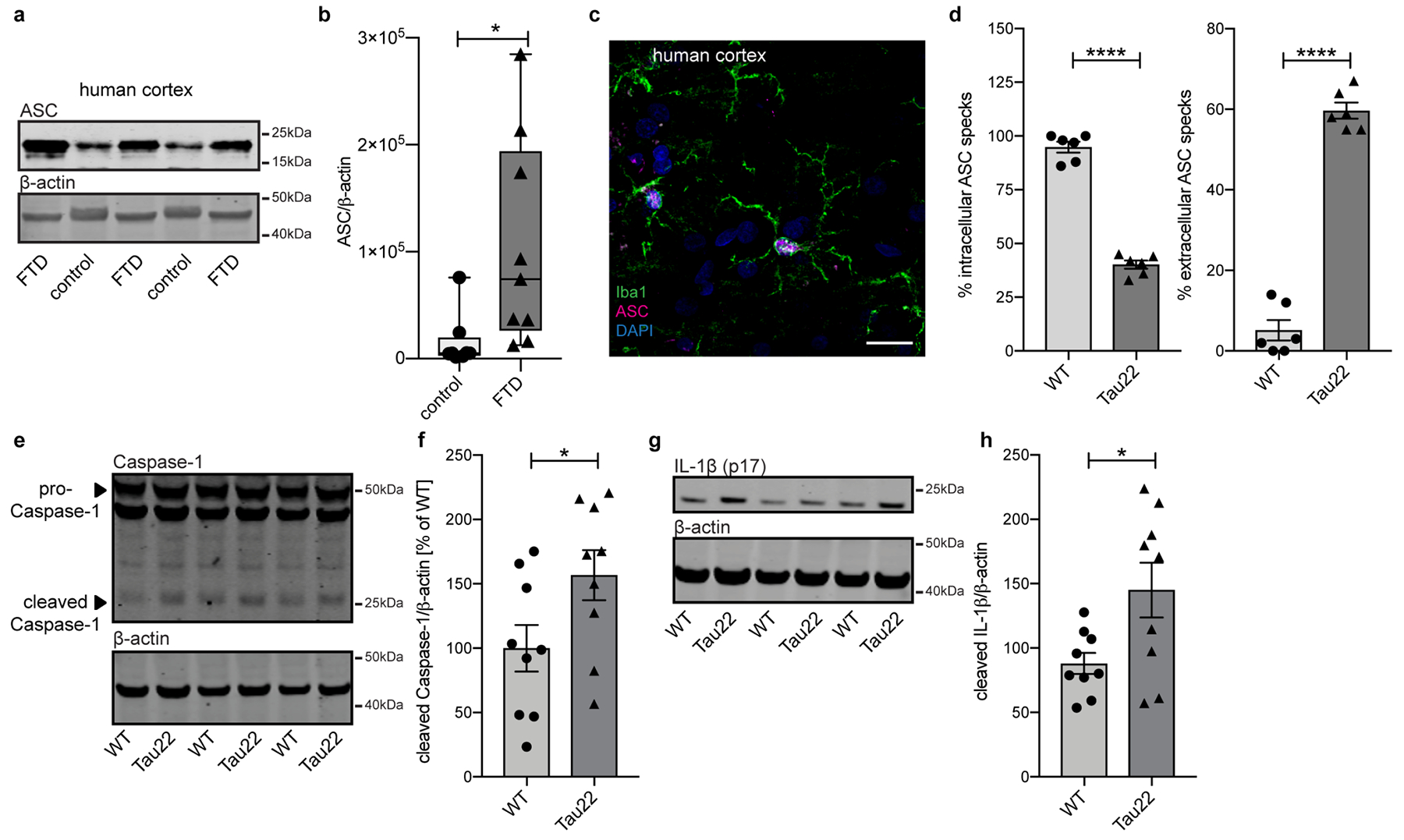
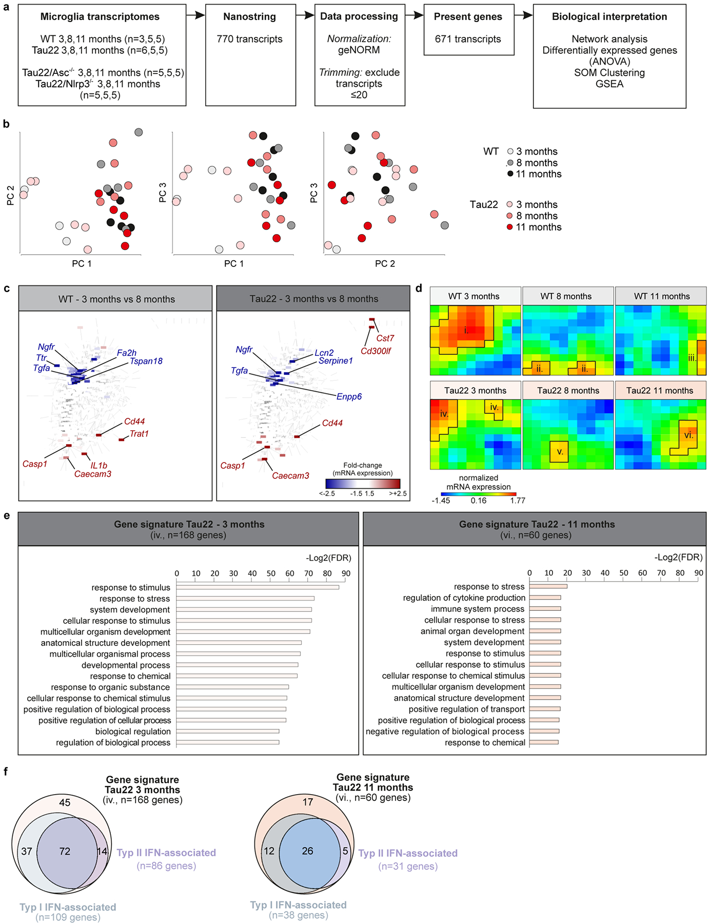
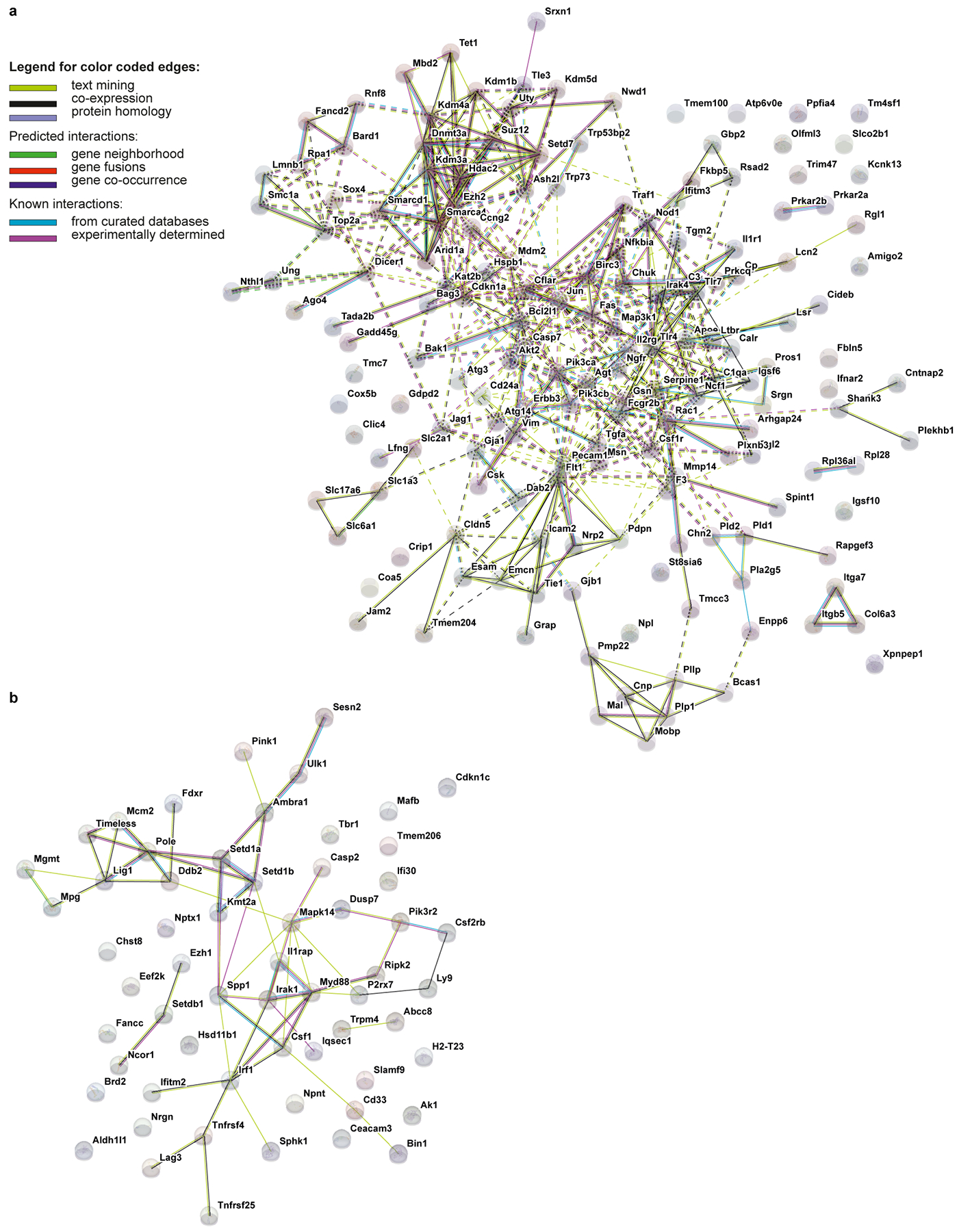
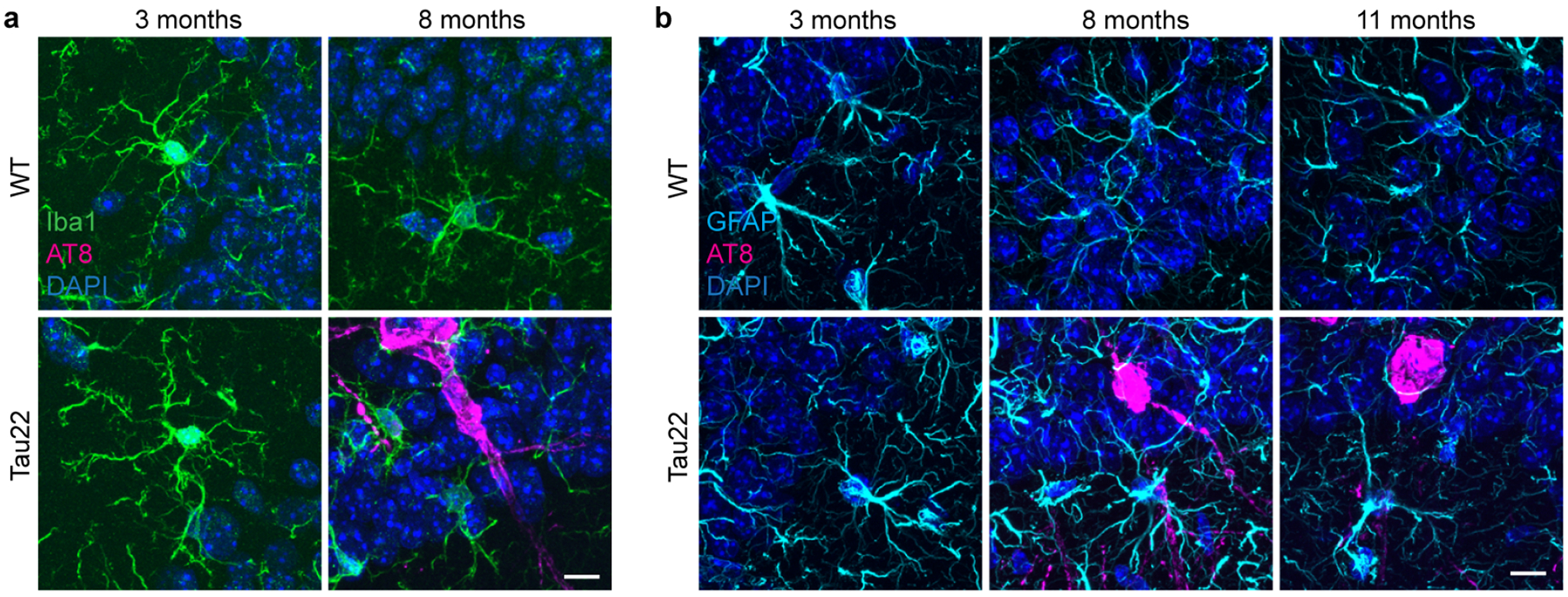
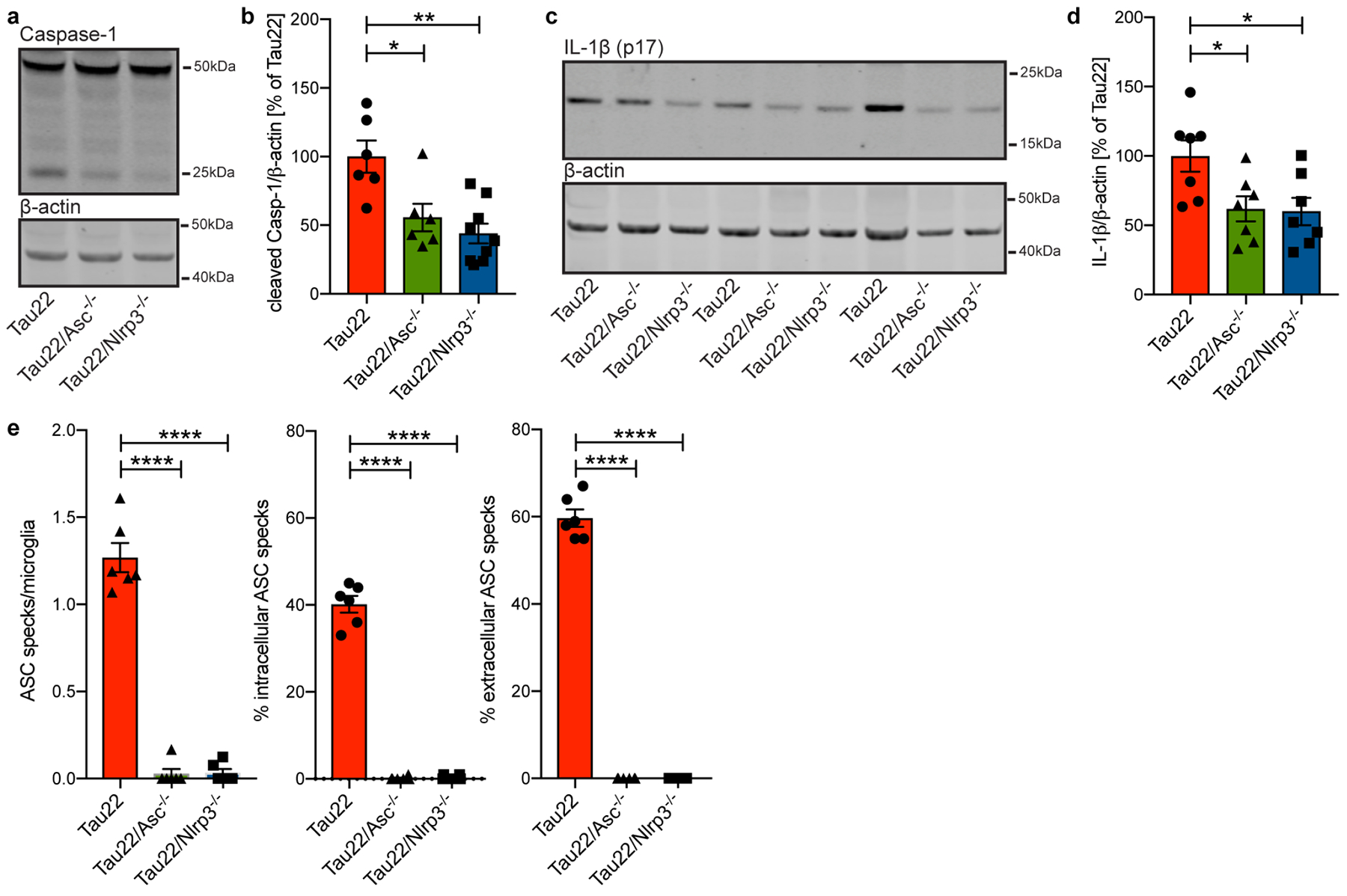
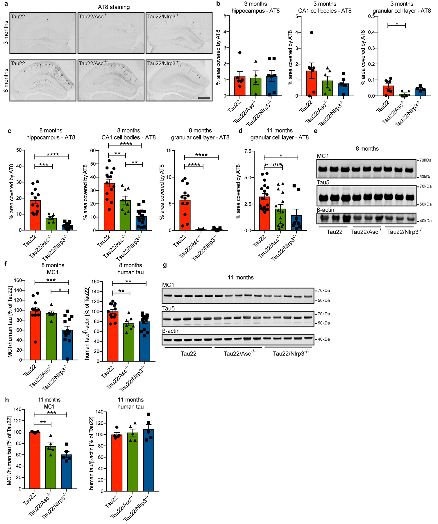
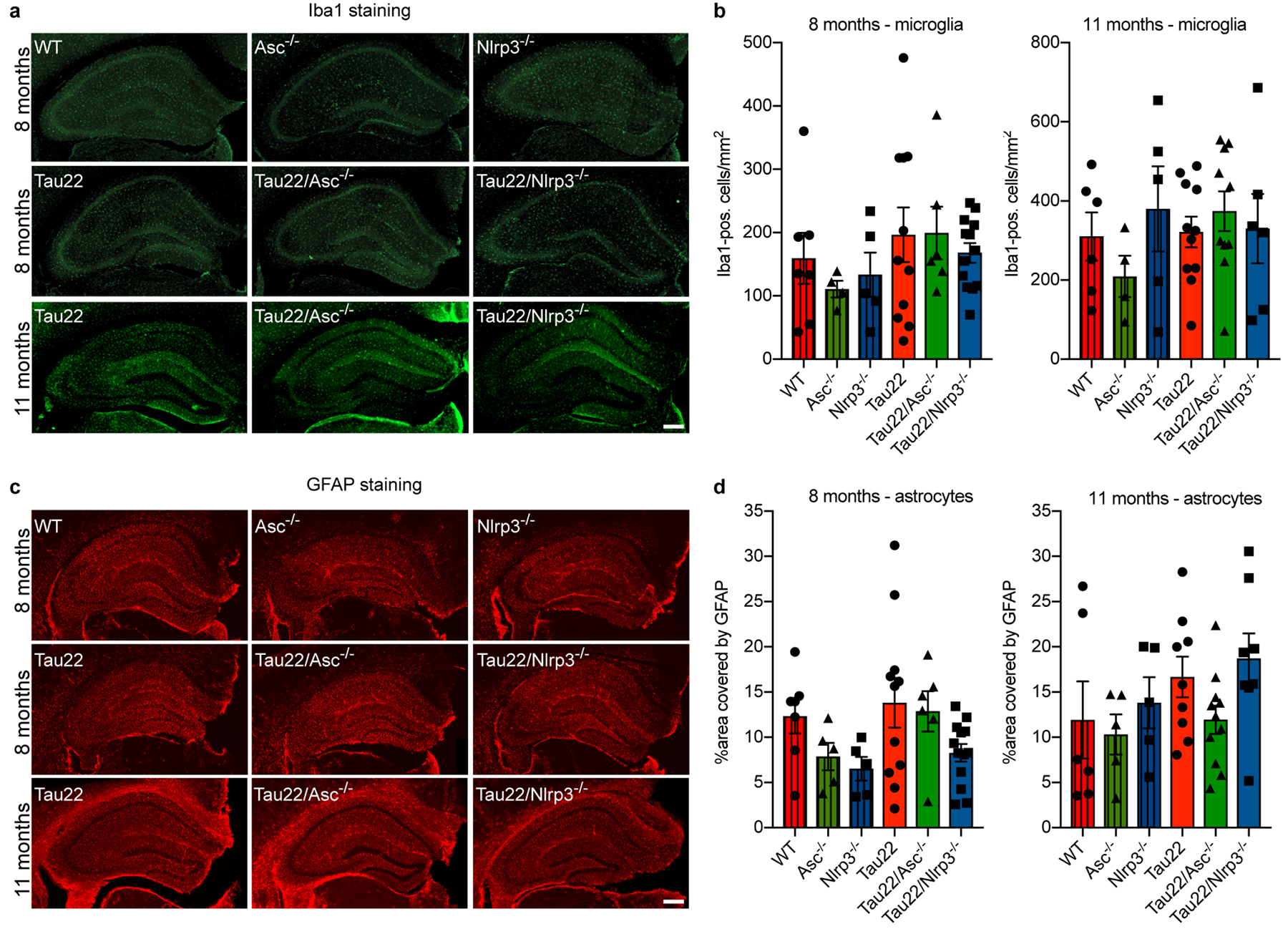
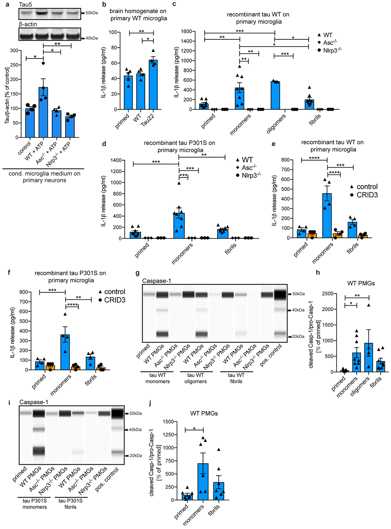
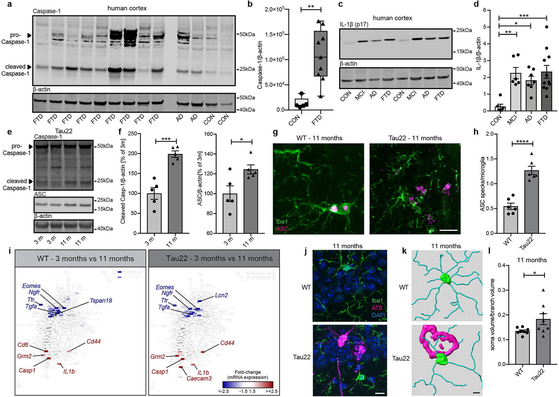
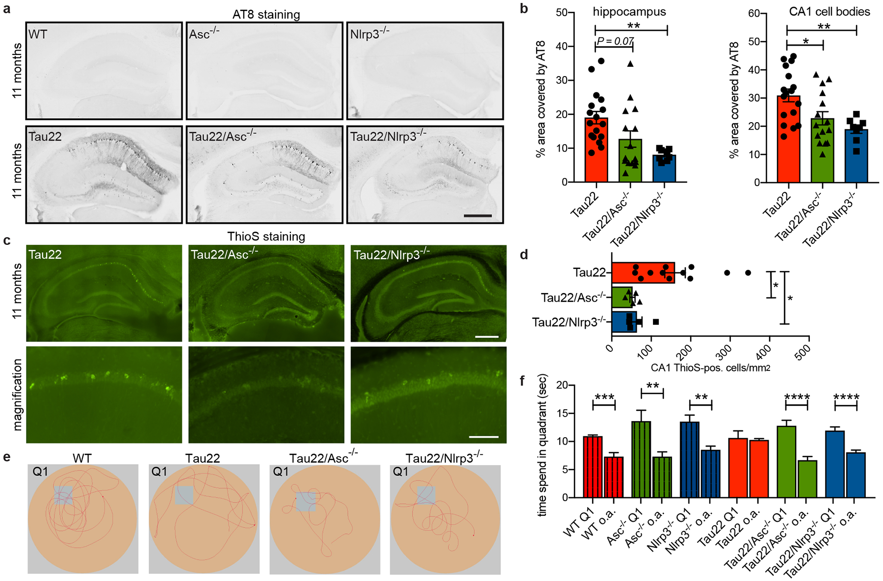
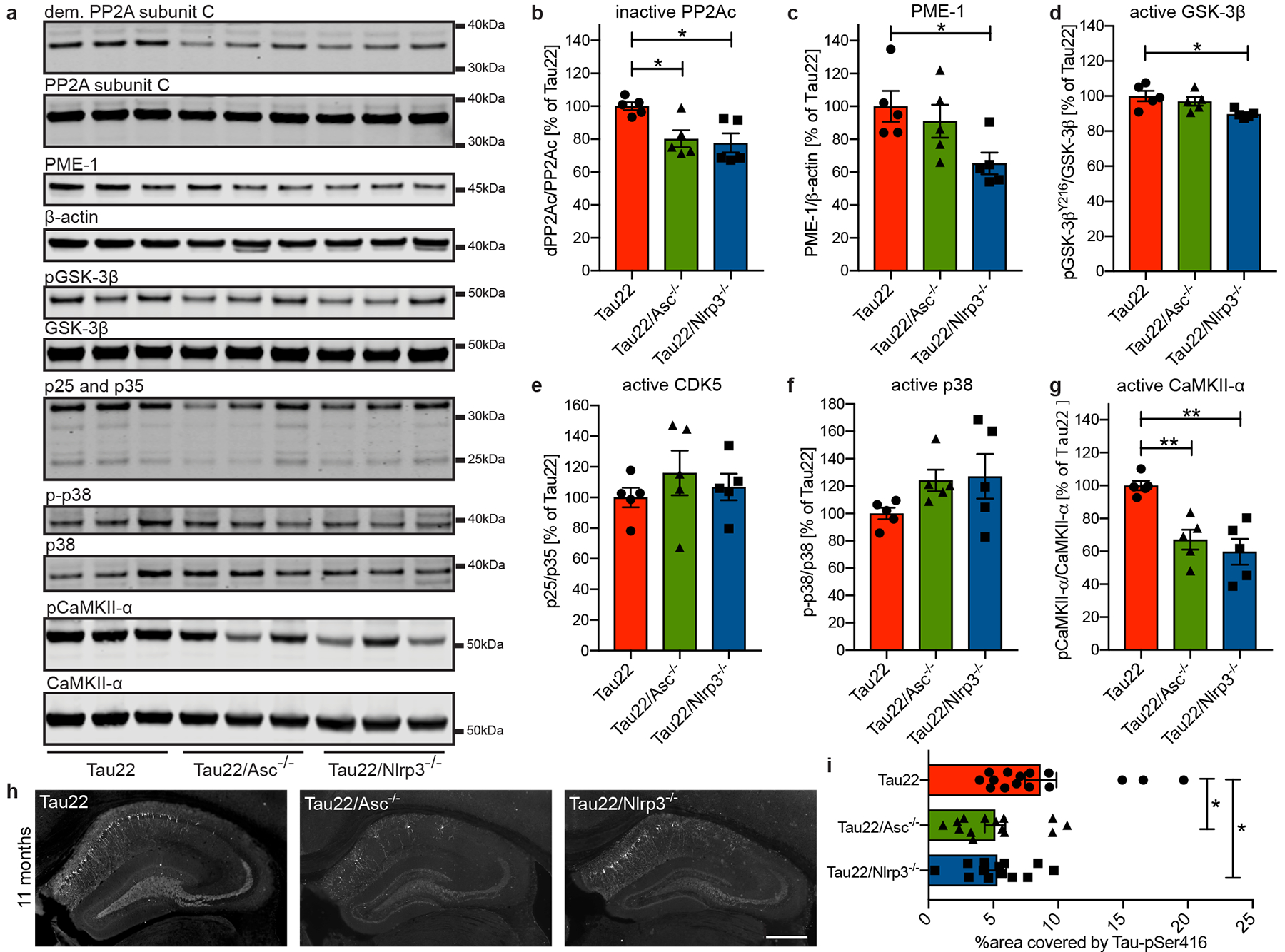
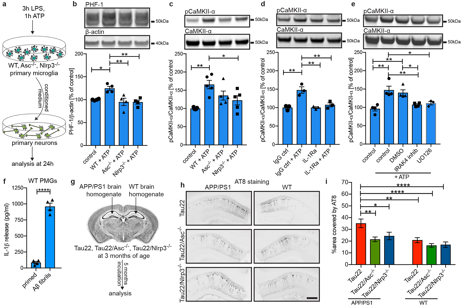
Alzheimer's disease is characterized by the accumulation of amyloid-beta in plaques, aggregation of hyperphosphorylated tau in neurofibrillary tangles and neuroinflammation, together resulting in neurodegeneration and cognitive decline1. The NLRP3 inflammasome assembles inside of microglia on activation, leading to increased cleavage and activity of caspase-1 and downstream interleukin-1β release2. Although the NLRP3 inflammasome has been shown to be essential for the development and progression of amyloid-beta pathology in mice3, the precise effect on tau pathology remains unknown. Here we show that loss of NLRP3 inflammasome function reduced tau hyperphosphorylation and aggregation by regulating tau kinases and phosphatases. Tau activated the NLRP3 inflammasome and intracerebral injection of fibrillar amyloid-beta-containing brain homogenates induced tau pathology in an NLRP3-dependent manner. These data identify an important role of microglia and NLRP3 inflammasome activation in the pathogenesis of tauopathies and support the amyloid-cascade hypothesis in Alzheimer's disease, demonstrating that neurofibrillary tangles develop downstream of amyloid-beta-induced microglial activation.


Conflict of interest statement

Competing interests

M.T.H. serves as advisory board member at IFM Therapeutics and Alector. All other authors declare no competing interests.

Figures

Extended Data Figure 1.
Extended Data Figure 1.. The NLRP3 inflammasome is activated in Tau22 mice.
a, Immunoblot analysis of ASC and β-actin in human cortex of FTD and control patients. b, Quantification of data from a. Box plots show 25/75 percentile. n=8 for controls, n=9 for FTD, *P=0.0239. c, Immunohistochemical staining of human cortex of a FTD patient for microglia and ASC. n=3. Scale bar, 20μm. d, Quantification of % of intracellular ASC and % of extracellular ASC specks from staining shown in Fig 1g. n=6 mice per group, ****P<0.0001. e, Immunoblot analysis of hippocampus samples of 8 month old wildtype (WT) and Tau22 mice stained for Caspase-1 and β-actin. f, Quantification of data from e. n=9 per group, *P=0.0489. g, Immunoblot analysis of hippocampus samples of 8 month old WT and Tau22 mice stained for IL-1β (p17) and β-actin. h, Quantification of data from g. n=9 per group, *P=0.0236. For gel source data, see Supplementary Figure 1. All graphs are presented as mean ± SEM and were analyzed by two-tailed unpaired t-test.
Extended Data Figure 2:
Extended Data Figure 2:. Gene signatures in WT and Tau22 mice identified by NanoString analysis.
a, Workflow for NanoString analysis. b, 2D PC analysis of WT and Tau22 mice at 3, 8 and 11 months of age. c, Gene network analysis of regulated genes at 3 vs. 8 months in WT and Tau22 mice identified by NanoString analysis. d, SOM clustering of WT and Tau22 mice at 3, 8 and 11 months of age with definition of cluster i. - vi. e, Gene signatures in 3 month old Tau22 mice defined by cluster iv. and in 11 month old Tau22 mice defined by cluster vi. Fisher’s exact test followed by a correction for multiple testing. f, Interferome Venn diagrams based on cluster iv. and vi. in Tau22 mice.
Extended Data Figure 3:
Extended Data Figure 3:. STRING network analysis of Tau22 mice.
a-b, Networks visualizing the functional protein association for gene signatures in Tau22 mice at 3 (a) and 11 (b) months of age. Nodes in the network represent proteins. Edges represent protein-protein interactions, which depending on the color indicate known or predicted interactions.
Extended Data Figure 4:
Extended Data Figure 4:. Astrocyte morphology does not change in Tau22 mice.
a, Immunohistochemical staining for microglia (Iba1) and phosphorylated Tau (AT8) in WT and Tau22 mice at 3 and 8 months of age. n=8. Scale bar, 10μm. b, Immunohistochemical staining for astrocytes (GFAP) and phosphorylated Tau (AT8) in WT and Tau22 mice at 3, 8 and 11 months of age. n=8. Scale bar, 10μm.
Extended Data Figure 5:
Extended Data Figure 5:. Knockout of Asc or Nlrp3 efficiently inhibits NLRP3 inflammasome function.
a, Immunoblot analysis of hippocampus samples from 11 month old mice stained for Caspase-1 and β-actin. b, Quantification of data from a. n=6 for Tau22 and Tau22/Asc−/−, n=9 for Tau22/Nlrp3−/−, *P=0.0156, **P=0.0012. c, Immunoblot analysis of hippocampus samples of 11 month old mice stained for IL-1β (p17) and β-actin. d, Quantification of data from c. n=7 for all groups. Tau22 vs. Tau22/Asc−/−: *P=0.0399, Tau22 vs. Tau22/Nlrp3−/−: *P=0.0310. e, Quantification of number of ASC specks/microglia, % of intracellular ASC specks and % of extracellular ASC specks in hippocampus sections of 11 month old mice. n=6 mice per group, ****P<0.0001 for all comparisons. For gel source data, see Supplementary Figure 1. All graphs are presented as mean ± SEM and were analyzed by one-way ANOVA followed by Tukey’s test.
Extended Data Figure 6:
Extended Data Figure 6:. Tau pathology is reduced in inflammasome-knockout mice.
a, Immunohistochemical staining for phosphorylated tau (AT8) in mouse hippocampi. Scale bar, 500μm. b, Quantification of AT8 in hippocampus, CA1 cell body layer and granular cell layer in the dentate gyrus of 3 month old mice shown in a (n=6 for hippocampus Tau22 and CA1 and dentate gyrus Tau22/Asc−/−, n=4 for hippocampus Tau22/Asc−/− and dentate gyrus Tau22/Nlrp3−/−, n=7 for hippocampus Tau22/Nlrp3−/− and CA1 and dentate gyrus Tau22, n=5 for CA1 Tau22/Nlrp3−/−. *P=0.0181. c, Quantification of AT8 in hippocampus, CA1 cell body layer and granular cell layer in the dentate gyrus of 8 month old mice shown in a. Hippocampus: n=12 for Tau22, n=6 for Tau22/Asc−/−, n=13 for Tau22/Nlrp3−/−. ***P=0.0004 and ****P<0.0001. CA1: n=14 for Tau22, n=8 for Tau22/Asc−/−, n=15 for Tau22/Nlrp3−/−.Tau22 vs. Tau22/Asc−/−: **P=0.0052, Tau22 vs. Tau22/Nlrp3−/−: ****P<0.0001, Tau22/Asc−/−: vs. Tau22/Nlrp3−/−: **P=0.0075. Dentate gyrus: n=12 for Tau22, n=6 for Tau22/Asc−/−, n=13 for Tau22/Nlrp3−/−), ****P<0.0001. d, Quantification of AT8 in granular cell layer in the dentate gyrus of 11 month old mice shown in Fig. 2a. n=17 for Tau22, n=14 for Tau22/Asc−/−, n=8 for Tau22/Nlrp3−/−, *P=0.0196. e, Immunoblot analysis of sarkosyl-soluble fraction of hippocampi from 8 month old Tau22, Tau22/Asc−/− and Tau22/Nlrp3−/− mice stained for misfolded tau (MC1), total tau (Tau5) and β-actin. f, Quantification of data from e. MC1: n=12 for Tau22, n=6 for Tau22/Asc−/−, n=13 for Tau22/Nlrp3−/− with Tau22 vs. Tau22/Nlrp3−/−: ***P=0.0009 and Tau22/Asc−/− vs. Tau22/Nlrp3−/−: *P=0.0190. Human tau: n=13 for Tau22, n=7 for Tau22/Asc−/−, n=14 for Tau22/Nlrp3−/−. Tau22 vs. Tau22/Asc−/−: **P=0.0047, Tau22 vs. Tau22/Asc−/−: **P=0.0037. g, Immunoblot detection of misfolded tau (MC1), total tau (Tau5) and β-actin in sarkosyl-soluble fraction of mouse hippocampi at 11 months of age. h, Quantification of data from g. n=5, **P=0.0056, ***P=0.0001. For gel source data, see Supplementary Figure 1. All graphs are presented as mean ± SEM and were analyzed by one-way ANOVA followed by Tukey’s test.
Extended Data Figure 7:
Extended Data Figure 7:. Microglia and astrocyte numbers are unaltered in Tau22/Asc−/− and Tau22/Nlrp3−/− mice.
a, Immunohistochemical staining of hippocampus of mice with the indicated genotypes and at the indicated ages for microglia (Iba1). Scale bar, 250μm. b, Quantification of Iba1-positive cells in the hippocampus as seen in a at 8 (left) and 11 months of age (right). n=7 for 8 months WT, n=6 for 11 months WT, n=4 for 8 and 11 months Asc−/−, n=5 for 8 and 11 months Nlrp3−/−, n=11 for 8 and 11 months Tau22, n=6 for 8 months Tau22/Asc−/−, n=10 for 11 months Tau22/Asc−/−, n=13 for 8 months Tau22/Nlrp3−/−, n=6 for 11 months Tau22/Nlrp3−/−. c, Immunohistochemical staining of hippocampus of mice with the indicated genotypes and at the indicated ages for astrocytes (GFAP). Scale bar, 250μm. d, Quantification of GFAP in the hippocampus as seen in c at 8 (left) and 11 months of age (right). n=7 for 8 months WT, n=6 for 11 months WT, n=5 for 8 and 11 months Asc−/− and Nlrp3−/−, n=11 for 8 months Tau22, n=9 for 11 months Tau22, n=6 for 8 months Tau22/Asc−/−, n=11 for 11 months Tau22/Asc−/−, n=13 for 8 months Tau22/Nlrp3−/−, n=8 for 11 months Tau22/Nlrp3−/−). For gel source data, see Supplementary Figure 1. All graphs are presented as mean ± SEM and were analyzed by one-way ANOVA followed by Tukey’s test.
Extended Data Figure 8:
Extended Data Figure 8:. Gene signatures in Tau22 and Tau22/Nlrp3−/− mice identified by NanoString analysis.
a, 2D PC analysis of Tau22 and Tau22/Nlrp3−/− mice at 3, 8 and 11 months of age. n= 5 independent samples for each group. b, Number of induced or suppressed genes comparing Tau22 vs. Tau22/Nlrp3−/− at 3, 8 and 11 months. c, Gene plots of Tau22 vs Tau22/Nlrp3−/− at 3, 8 and 11 months. d, Heatmap comparing significantly changed genes in Tau22 vs. Tau22/Nlrp3−/− mice at various ages, identified by NanoString analysis.
Extended Data Figure 9:
Extended Data Figure 9:. Nlrp3-knockout does not affect the microbiome of Tau22 mice.
a, Amount of bacteria in stool samples obtained from the colon of 11 month old WT, Nlrp3−/−, Tau22 and Tau22/Nlrp3−/− mice. n=6 for WT, n=4 for Nlrp3−/−, Tau22, Tau22/Nlrp3−/−. b, Cecum weight of 11 month old WT, Nlrp3−/−, Tau22 and Tau22/Nlrp3−/− mice. n=8 for WT, n=5 for Nlrp3−/−, n=4 for Tau22, n=6 for Tau22/Nlrp3−/−. c, Colon length of 11 month old WT, Nlrp3−/−, Tau22 and Tau22/Nlrp3−/− mice. n=8 for WT, n=5 for Nlrp3−/−, n=4 for Tau22, n=6 for Tau22/Nlrp3−/−, *P=0.0350. d-f, IL-1β, IL-6 and TNFα levels in colon samples of 11 month old WT, Nlrp3−/−, Tau22 and Tau22/Nlrp3−/− mice. n=8 for WT, n=4 for Nlrp3−/−, n=5 for IL-1β and IL-6 in Tau22, n=4 for TNFα in Tau22, n=6 for Tau22/Nlrp3−/−. g-i, IL-1β, IL-6 and TNFα levels in medial intestine samples of 11 month old WT, Nlrp3−/−, Tau22 and Tau22/Nlrp3−/− mice. n=8 for WT, n=4 for Nlrp3−/−, n=4 for IL-1β in Tau22, n= 5 for IL-6 and TNFα in Tau22, n=6 for IL-1β and TNFα in Tau22/Nlrp3−/−, n=5 for IL-6 in Tau22/Nlrp3−/−. j-l, IL-1β, IL-6 and TNFα levels in spleen samples of 11 month old WT, Nlrp3−/−, Tau22 and Tau22/Nlrp3−/− mice. n=8 for WT, n=4 for Nlrp3−/−, n=5 for Tau22, n=6 for Tau22/Nlrp3−/−. m-o, IL-1β, IL-6 and TNFα levels in serum samples of 11 month old WT, Nlrp3−/−, Tau22 and Tau22/Nlrp3−/− mice. n=8 for WT, n=4 for Nlrp3−/− and Tau22, n=6 for Tau22/Nlrp3−/−, *P=0.0281. All graphs are presented as mean ± SEM and were analyzed by one-way ANOVA followed by Tukey’s test.
Extended Data Figure 10:
Extended Data Figure 10:. Tau can activate the NLRP3 inflammasome
a, Immunoblot analysis and quantification of total tau in primary neurons after treatment with conditioned medium from primary WT microglia (control), LPS/ATP-activated WT, Asc- or Nlrp3-knockout microglia (WT+ATP, Asc−/−+ATP or Nlrp3−/−+ATP). n=4 for each group. Control vs. WT+ATP: *P=0.0252, WT+ATP vs. Asc−/−+ATP: *P=0.0148, WT+ATP vs. Nlrp3−/−+ATP: **P=0.0029. b, IL-1β levels in conditioned medium of primary WT microglia primed with LPS and treated with hippocampus homogenate from either 11 month old WT or Tau22 mice. n=5 for primed, n=4 for WT and Tau22 homogenate treated microglia. Primed vs. Tau22: **P=0.0092, WT vs. Tau22: *P=0.0276. c, IL-1β levels in conditioned medium of primary WT, Asc−/− and Nlrp3−/− microglia primed with LPS and treated with different forms of 2μM recombinant wildtype tau (tau WT). n=3 for Asc−/− and Nlrp3−/− microglia treatments and WT oligomer treatment, n=8 for all other WT treatments. WT primed vs. WT monomers: **P=0.0011, WT primed vs. WT oligomers: ***P=0.0007, WT monomers vs. Asc−/− and Nlrp3−/− monomers: **P=0.0011, WT monomers vs. WT fibrils: *P=0.0388, WT oligomers vs. Nlrp3−/− oligomers: ***P=0.0004, WT oligomers vs. WT fibrils: *P=0.0112. d, IL-1β levels in conditioned medium of primary WT, Asc−/− and Nlrp3−/− microglia primed with LPS and treated with different forms of 2μM recombinant tau with a P301S (tau P301S) mutation. n=8 for WT microglia treatments, n=3 for Asc−/− and Nlrp3−/− microglia treatments, ***P=0.0002, **P=0.0018. e, IL-1β levels in conditioned medium of primary WT microglia primed with LPS and treated with different forms of 2μM recombinant tau WT with and without the NLRP3 inhibitior CRID3. n=4 for all groups, ***P=0.0002, ****P<0.0001. f, IL-1β levels in conditioned medium of primary WT microglia primed with LPS and treated with different forms of 2μM recombinant tau P301S with and without CRID3 treatment. n=4 for all groups, **P=0.0037, ***P=0.0005, ****P<0.0001. g, Jess-based analysis of conditioned medium of LPS + tau WT-treated WT microglia stained for Caspase-1. LPS/ATP-treated WT microglia served as positive control. h, Quantification of data from g. n=7 for primed, n=8 for tau monomers and fibrils, n=4 for tau oligomers. *P=0.0458, **P=0.0091. i, Jess-based analysis of conditioned medium of primary WT, Asc−/− and Nlrp3−/− microglia primed with LPS and treated with the indicated forms of tau P301S. LPS/ATP-treated WT microglia served as positive control. Samples were stained for Caspase-1. j, Quantification of data from i. n=7 for primed and fibrils, n=6 for monomers, *P=0.0128. For gel source data, see Supplementary Figure 1. All graphs are presented as mean ± SEM and were analyzed by one-way (a, b, h, j) or two-way ANOVA (c-f) followed by Tukey’s test.
Figure 1:
Figure 1:. NLRP3 inflammasome is activated in FTD patients and Tau22 mice
a, Immunoblot detection of Caspase-1 and β-actin in cortex of frontotemporal dementia patients (FTD), Alzheimer’s disease patients (AD) and controls (CON). b, Quantification of CON and FTD data from a. Box plots show 25/75 percentile. n=5 for CON, n=9 for FTD, **P=0.0018. c, Immunoblot analysis of IL-1β (p17) and β-actin in cortex of CON, mild cognitive impairment (MCI), AD and FTD patients. d, Quantification of data from c. n=6 for CON and MCI, n=7 for AD, n=11 for FTD. **P=0.0049, *P=0.0269, ***P=0.0009. e, Immunoblot detection of Caspase-1, ASC and β-actin in hippocampi of Tau22 mice (3 and 11 months). f, Quantification of data from e. n=5, ***P=0.0004, *P=0.0281. g, Immunohistochemical staining for microglia and ASC close to the CA1 region in WT and Tau22 mice. Scale bar, 10μm. h, Quantification of ASC specks/microglia from staining in g. n=6, ****P<0.0001. i, Gene network analysis of regulated genes at 3 vs. 11 months in WT and Tau22 mice identified by NanoString analysis. j, Immunohistochemical staining for microglia and phosphorylated Tau (AT8) in WT and Tau22 mice. n=8. Scale bar, 10μm. k, Reconstruction of microglia (green/blue) in WT and Tau22 mice close to an AT8-positive neuron (magenta). Scale bar, 20 μm. l, Quantification of soma to branch volume of microglia located close to the CA1 region in WT or microglia in contact with AT8-positive cells in Tau22 mice. n=8, *P=0.0491. For gel source data, see Supplementary Figure 1. Data are mean ± SEM and were analyzed by two-tailed unpaired t-test (b, f, h, l) or one-way ANOVA with Tukey’s test (d).
Figure 2:
Figure 2:. Loss of NLRP3 inflammasome function decreases tau pathology and improves cognition
a, Immunohistochemical staining for phosphorylated tau (AT8) in mouse hippocampi. Scale bar, 500μm. b, Quantification of AT8 in hippocampus and CA1 region shown in a. n=17 for Tau22, n=15 for Tau22/Asc−/−, n=8 for Tau22/Nlrp3−/−. Hippocampus: **P=0.0055, CA1: *P=0.0278, **P=0.0059. c, Staining with thioflavine S (aggregated Tau) of mouse hippocampi. Scale bar, 500μm (top) and 100μm (bottom). d, Quantification of thioflavine S-positive cells in CA1 region shown in c. n=12 for Tau22, n=5 for Tau22/Asc−/− and Tau22/Nlrp3−/−. Tau22 vs. Tau22/Asc−/−: *P=0.0240, Tau22 vs. Tau22/Nlrp3−/−: *P=0.0444. e, Example of movement of mice in Morris water maze test (MWM). f, Quantification of time spend in quadrant 1 (Q1) vs. all other quadrants (a.o.) in MWM. n=12 for WT, Nlrp3−/−, Tau22/Asc−/−, Tau22/Nlrp3−/−, n=14 for Asc−/−, n=16 for Tau22. ***P=0.0001, ****P<0.0001, Asc−/−: **P=0.0065, Nlrp3−/−: **P=0.0012. For gel source data, see Supplementary Figure 1. Data are mean ± SEM and were analyzed by one-way ANOVA with Tukey’s test (b, d) or two-tailed unpaired t-test (f).
Figure 3:
Figure 3:. Nlrp3- and Asc-KO inhibits CaMKII-α and promotes phosphatase activity
a, Immunoblot analysis of mouse hippocampi (11 months) stained for demethylated PP2A subunit C (dem. PP2A subunit C), total PP2A subunit C, PP2A methylesterase (PME-1), β-actin, GSK-3β phosphorylated at Tyr216 (pGSK-3β), total GSK-3beta, p25/p35, phosphorylated p38 (p-p38), total p38, Calmodulin dependent protein kinase II alpha phosphorylated at Thr286 (pCaMKII-α) and total CaMKII-α. b - g, Quantification of the enzyme activities/abundance shown in a. n=5. PP2Ac: Tau22 vs. Tau22/Asc−/−: *P= 0.0286, Tau22 vs. Tau22/Nlrp3−/−: *P=0.0144, PME1: *P=0.0398, GSK-3β: *P=0.0205, CaMKII-α: Tau22 vs. Tau22/Asc−/−: **P=0.0055, CaMKII-α Tau22 vs. Tau22/Nlrp3−/−: **P=0.0012. h, Immunohistochemical staining for Tau phosphorylated at Serin 416 (Tau-pSer416) in mouse hippocampi. Scale bar, 500μm. i, Quantification of Tau-pSer416 in CA1 region shown in h. n=15 for Tau22, n=14 for Tau22/Asc−/−, n=13 for Tau22/Nlrp3−/−. Tau22 vs. Tau22/Asc−/−: *P=0.0314, Tau22 vs. Tau22/Nlrp3−/−: *P=0.0476. For gel source data, see Supplementary Figure 1. Data are mean ± SEM and were analyzed by one-way ANOVA with Tukey’s test.
Figure 4:
Figure 4:. Inflammasome function is necessary for Aβ-induced tau pathology
a, Schematic illustrating the experimental set-up used for experiments shown in b - e. b - c, Immunoblot analysis and quantification of tau phosphorylated at Ser396/Ser404 (PHF-1) and pCaMKII-α in treated neurons (primary WT microglia (control), LPS+ATP-activated WT microglia (WT+ATP), LPS+ATP-activated Asc- or Nlrp3-knockout microglia (Asc−/−+ATP or Nlrp3−/−+ATP)). n=4 in b, with ctrl vs. WT+ATP: *P=0.0235, WT+ATP vs. Asc−/−+ATP: **P=0.0072, WT+ATP vs. Nlrp3−/−+ATP: **P=0.0064. n=5 in c with **P=0.0022, *P=0.0454. d, Immunoblot analysis and quantification of pCaMKII-α in neurons treated with an IL-1 receptor antagonist (IL-1Ra) or the corresponding isotype control in addition to conditioned medium from WT microglia. n=3, IgG ctrl vs. IgG ctrl+ATP and vs. IL-1ra: **P=0.0015, IgG ctrl+ATP vs. IL-1ra+ATP: **P=0.0044. e, Immunoblot analysis and quantification of pCaMKII-α in neurons after treatment with DMSO, IRAK4 inhibitor PF06650833 or MEK1/2 inhibitor UO126 in addition to conditioned medium from WT microglia. n=3 for UO126, n=4 for all other groups, ctrl vs. ctrl+ATP: **P=0.0011, ctrl vs. DMSO+ATP: **P=0.0040, ctrl+ATP vs. IRAK4 inhib+ATP: **P=0.0087, ctrl+ATP vs. UO126+ATP: *P=0.0309, DMSO+ATP vs. IRAK4 inhibitor: *P=0.0338. f, IL-1β levels in conditioned medium of primary WT microglia treated with LPS and 10μM Aβ fibrils. n=4, ****P<0.0001. g, Schematic for injection model. h, Immunohistochemical staining for phosphorylated tau (AT8) of CA1 region of mice injected with either APP/PS1 or WT brain homogenates. Scale bar, 250μm. i, Quantification of AT8 in CA1 region of injected mice shown in h. n=17 sections of n=6 mice for Tau22, n=30 sections of n=6 mice for Tau22/Asc−/−, n=25 sections of n=6 mice for Tau22/Nlrp3−/−. *P=0.0490, ****P<0.0001, Tau22+APP/PS1 vs. Tau22/Asc−/−+APP/PS1: **P=0.0034, Tau22+APP/PS1 vs. Tau22+WT: **P=0.0071. For gel source data, see Supplementary Figure 1. Data are mean ± SEM and were analyzed by one-way ANOVA with Tukey’s test (b-e, i) or two-tailed unpaired t-test (f).

Comment in

References

    1. Ising C & Heneka MT Functional and structural damage of neurons by innate immune mechanisms during neurodegeneration. Cell Death Dis. 9, 120 (2018). - PMC - PubMed
    1. Heneka MT, McManus RM & Latz E Inflammasome signalling in brain function and neurodegenerative disease. Nat. Rev. Neurosci 19, 610–621 (2018). - PubMed
    1. Venegas C et al. Microglia-derived ASC specks cross-seed amyloid-β in Alzheimer’s disease. Nature 552, 355–361 (2017). - PubMed
    1. Halle A et al. The NALP3 inflammasome is involved in the innate immune response to amyloid-beta. Nat. Immunol 9, 857–865 (2008). - PMC - PubMed
    1. Heneka MT et al. NLRP3 is activated in Alzheimer’s disease and contributes to pathology in APP/PS1 mice. Nature 493, 674–678 (2013). - PMC - PubMed

Publication types

MeSH terms

Substances