

Adult-onset neuronal intranuclear inclusion disease presenting with typical MRI changes
- PMID: 31749292
- PMCID: PMC6908888
- DOI: 10.1002/brb3.1477
Adult-onset neuronal intranuclear inclusion disease presenting with typical MRI changes
Abstract
Background: This study aims to analyze the clinical, imaging, electrophysiological, and dermatopathological features of a patient with adult-onset neuronal intranuclear inclusion disease (NIID) and to explore the diagnostic methods of adult-onset NIID.
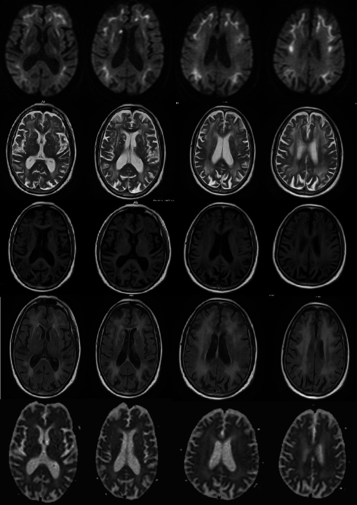
Case presentation: We here report a 63-year-old male with recurrent acute encephalopathy syndrome and autonomic nervous system damage syndrome characterized by sexual dysfunction and urinary and fecal dysfunction. Cranial diffusion-weighted magnetic resonance imaging (DWI) demonstrated symmetrically distributed strip-shaped high-intensity signal in bilateral fronto-occipital-parietal cortical-medullary junction. Electrophysiological test revealed that the main site of injury was myelin sheath in both motor and sensory nerves. Skin biopsy revealed eosinophilic spherical inclusion bodies in the nucleus of sweat gland epithelial cells.
Conclusion: This case suggests that adult NIID is a chronic neurodegenerative disease with high clinical heterogeneity. Subcortical strip-shaped high-intensity signal on DWI has high diagnostic significance. Eosinophilic intranuclear inclusion bodies detected by skin biopsy contribute to diagnosis.
Keywords: acute encephalopathy syndrome; magnetic resonance imaging; neuronal intranuclear inclusion disease.
© 2019 The Authors. Brain and Behavior published by Wiley Periodicals, Inc.
Conflict of interest statement
The authors have declared no conflicts of interest.
Figures


References
-
- Chen, W. A. , Li, X. , Zhu, W. Q. , Zhang, Y. , & Zhang, Z. Q. (2018). Adult‐onset neuronal intranuclear inclusion disease: A case report and review of literature. Zhonghua Shenjingke Zazhi, 51, 905–908.
-
- Hirose, B. , Hisahara, S. , Uesugi, H. , Sone, J. , Sobue, G. , & Shimohama, S. (2018). Sporadic adult‐onset neuronal intranuclear inclusion disease with abnormal electroretinogram, nerve conduction studies and somatosensory evoked potential. Rinsho Shinkeigaku, 58, 407–410. 10.5692/clinicalneurol.cn-001154 - DOI - PubMed
Publication types
MeSH terms
Supplementary concepts
LinkOut - more resources
Full Text Sources
Medical
