. 2019 Oct 22:13:2071-2073.
doi: 10.2147/OPTH.S227318. eCollection 2019.

Utility Of Three-Dimensional Heads-Up Surgery In Cataract And Minimally Invasive Glaucoma Surgeries

Affiliations

Utility Of Three-Dimensional Heads-Up Surgery In Cataract And Minimally Invasive Glaucoma Surgeries

Hisato Ohno. Clin Ophthalmol. .

Abstract

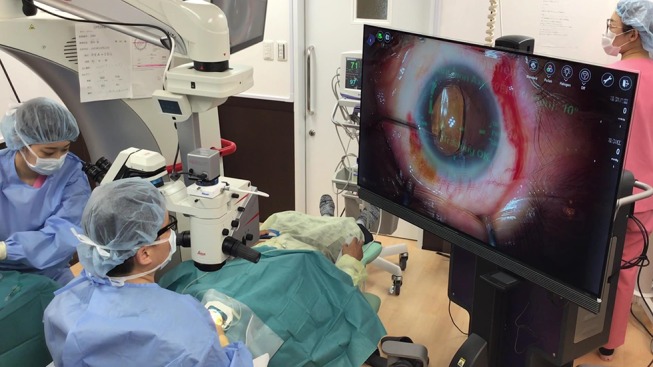
Purpose: This study aimed to assess the utility of the three-dimensional (3D) heads-up visualization system for minimal incision cataract surgery (MICS) and minimally invasive glaucoma surgeries (MIGSs).

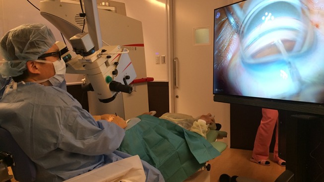
Methods: Toric intraocular lens (IOL) implantation with phacoemulsification and trabecular microbypass stent implantation in patients with cataract and open-angle glaucoma were performed using the heads-up 3D visualization system combined with surgical navigation rather than the conventional microscope.

Results: This procedure was found to have the following advantages: the ability to clearly observe the anterior chamber angle image without requiring frequent focus adjustment owing to the extended depth of field and emphasized stereoscopic effect provided by this system and maintain the surgeon's posture.

Conclusion: The feasibility and comfort of this system are greater than those of the conventional microscopic for performing MICS and MIGS.

Keywords: open-angle glaucoma; phacoemulsification; toric intraocular lens.


Conflict of interest statement

The author reports no conflicts of interest in this work.

Figures

Figure 1
Figure 1
Toric IOL implantation by NGENUITY 3D Visualization System combined with VERION image guided system.
Figure 2
Figure 2
In HUS, the surgeon’s posture is maintained even if the microscope and patient’s face are tilted to acquire a good view of the anterior chamber angle when using a gonioprism.

References

    1. Eckardt C, Paulo EB. Heads-up surgery for vitreoretinal procedures: an experimental and clinical study. Retina. 2016;36(1):137–147. doi:10.1097/IAE.0000000000000689 - DOI - PubMed
    1. Adams MK, Thornton S, Regillo CD, Park C, Ho AC, Hsu J. Minimal endoillumination levels and display luminous emittance during three-dimensional heads-up vitreoretinal surgery. Retina. 2017;37(9):1746–1749. doi:10.1097/IAE.0000000000001420 - DOI - PubMed
    1. Freeman WR, Chen KC, Ho J, et al. Resolution, depth of field, and physician satisfaction during digitally assisted vitreoretinal surgery. Retina. 2019;39:1768–1771. doi:10.1097/IAE.0000000000002236 - DOI - PMC - PubMed
    1. Webers VSC, Bauer NJC, Visser N, Berendschot TT, van Den Biggelaar FJ, Nuijts RM. Image-guided system versus manual marking for toric intraocular lens alignment in cataract surgery. J Cataract Refract Surg. 2017;43(6):781–788. doi:10.1016/j.jcrs.2017.03.041 - DOI - PubMed
    1. Weinstock RJ, Diakonis VF, Schwartz AJ, Weinstock AJ. Heads-up cataract surgery: complication rates, surgical duration, and comparison with traditional microscopes. J Refract Surg. 2019;35(5):318–322. doi:10.3928/1081597X-20190410-02 - DOI - PubMed

LinkOut - more resources