. 2020 Mar 1;61(3):519-535.
doi: 10.1093/pcp/pcz217.

Nickel Toxicity Targets Cell Wall-Related Processes and PIN2-Mediated Auxin Transport to Inhibit Root Elongation and Gravitropic Responses in Arabidopsis

Affiliations

Nickel Toxicity Targets Cell Wall-Related Processes and PIN2-Mediated Auxin Transport to Inhibit Root Elongation and Gravitropic Responses in Arabidopsis

Alexandra Leškovï et al. Plant Cell Physiol. .

Abstract

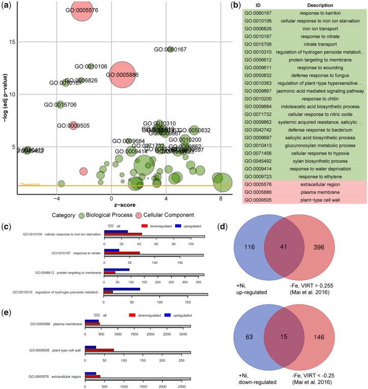
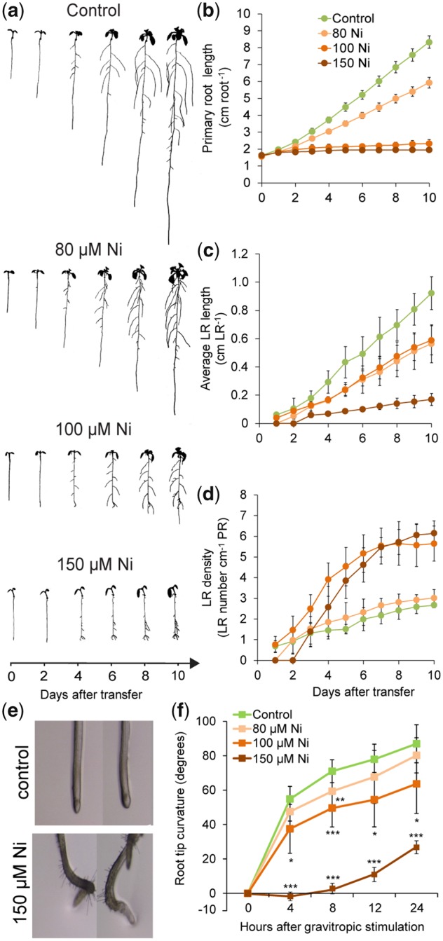
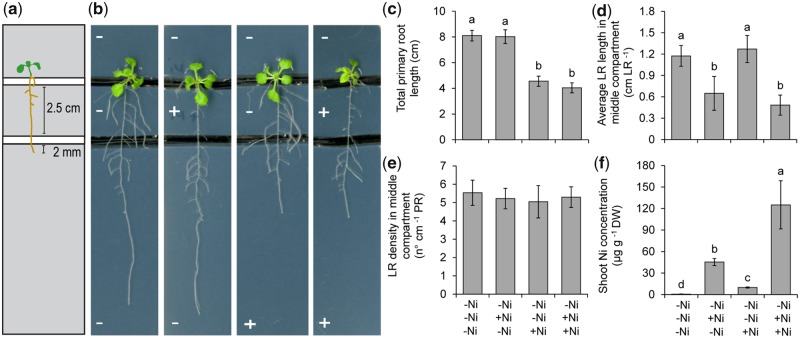
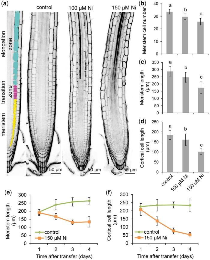
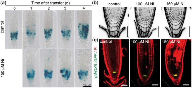
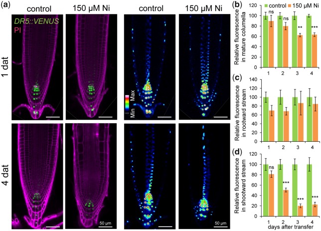
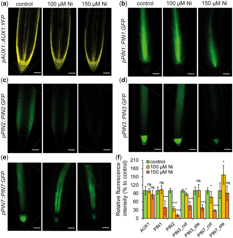
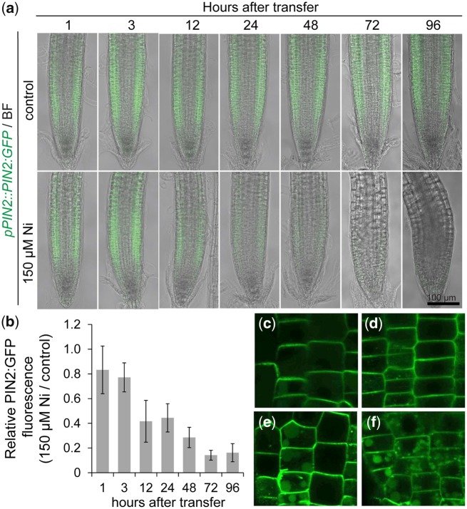
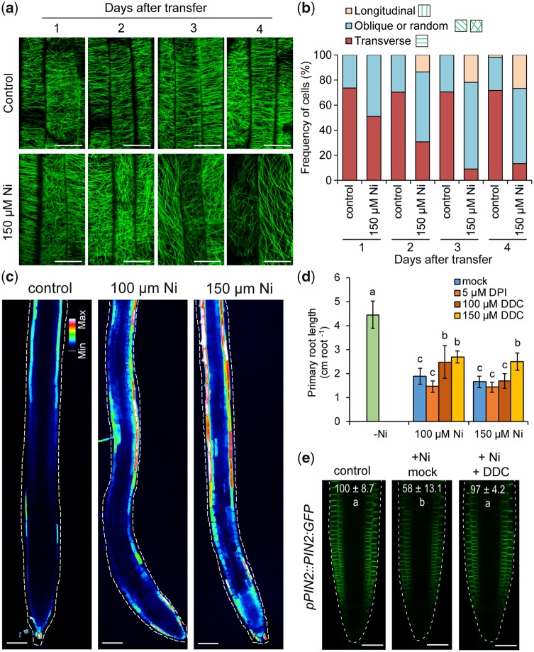
Contamination of soils with heavy metals, such as nickel (Ni), is a major environmental concern due to increasing pollution from industrial activities, burning of fossil fuels, incorrect disposal of sewage sludge, excessive manure application and the use of fertilizers and pesticides in agriculture. Excess Ni induces leaf chlorosis and inhibits plant growth, but the mechanisms underlying growth inhibition remain largely unknown. A detailed analysis of root development in Arabidopsis thaliana in the presence of Ni revealed that this heavy metal induces gravitropic defects and locally inhibits root growth by suppressing cell elongation without significantly disrupting the integrity of the stem cell niche. The analysis of auxin-responsive reporters revealed that excess Ni inhibits shootward auxin distribution. Furthermore, we found that PIN2 is very sensitive to Ni, as the presence of this heavy metal rapidly reduced PIN2 levels in roots. A transcriptome analysis also showed that Ni affects the expression of many genes associated with plant cell walls and that Ni-induced transcriptional changes are largely independent of iron (Fe). In addition, we raised evidence that excess Ni increases the accumulation of reactive oxygen species and disturbs the integrity and orientation of microtubules. Together, our results highlight which processes are primarily targeted by Ni to alter root growth and development.

Keywords: Arabidopsis thaliana; Heavy metals; Polar auxin transport; Reactive oxygen species; Root development; Transcriptome.


Figures

Fig. 1
Fig. 1
Genome-wide transcriptional changes in response to high Ni. (a) GO enrichment analysis of overrepresented categories of genes significantly altered by high Ni. Each circle corresponds to a significantly enriched GO category (adjusted P-value <0.05 indicated by the ‘threshold’ line). Size of circles reflects the number of genes that are associated with each respective GO category, and colors reflect the ontology, where green color indicates biological processes and red color indicates cellular component. z-score indicates whether genes from GO terms were primarily upregulated (positive z-score) or downregulated (negative z-score). (b) List of most significant GO terms. (c) Analysis of differentially expressed genes of selected biological processes in Ni-exposed roots. Shown is the total number of genes belonging to the indicated GO clusters and the number of genes significantly up- or downregulated by Ni. (d) Venn diagrams showing the overlap between genes significantly up- or downregulated in response to Ni [−1.0 > log2FC (+Ni/control) > 1.0; adjusted P-value <0.05] and previously shown to robustly respond to Fe deficiency (−Fe). Control, no Ni added; +Ni, 100 �M Ni added to one-half-strength MS media. (e) Analysis of differentially expressed genes of selected cellular compartments.
Fig. 2
Fig. 2
Ni inhibits root elongation and induces gravitropic defects. Seedlings of A. thaliana accession Col-0 were pre-cultured for 7 d on one-half-strength MS agar medium and then transferred to fresh medium without added Ni (control) or containing the indicated concentrations of Ni. Plants were imaged daily with a scanner. (a) Images of representative plants at the indicated time-points after transfer to treatments. Daily changes in primary root length (b), average lateral root length (c) and lateral root density (d) in response to Ni. Bars represent mean � SD (n = 7 plants). High Ni induces agravitropic root growth (e) and reduces the root response to gravistimulation (f). Seven-day-old seedlings pre-cultured on solid one-half-strength MS medium transferred to control medium or medium containing the indicated concentrations of Ni. Bars represent mean � SD (n = 7 plants). *P <0.05; **P <0.01; ***P <0.001 according to Student’s t-test (pairwise comparison with control at each time-point). LR, lateral root; PR, primary root.
Fig. 3
Fig. 3
Ni exerts a local inhibitory effect on root elongation. (a) Schematic representation of the split-agar setup. Plants were pre-cultured on one-half-strength MS agar medium and transferred to agar plates in which three compartments were spatially separated with a gap. Ten-day-old seedlings were placed in a way that only approximately 2 mm of the apical root zone was in contact with the lowest segment, while shoots were placed on the uppermost, Ni-free compartment. (b) Appearance of A. thaliana plants accession Col-0 on horizontally split agar plates with differential Ni supply to roots. − and + indicate medium without added Ni and medium containing 150 �M Ni, respectively. Plants were photographed after 7 d and representative plants are shown. Total primary root length along three compartments (c), average lateral root length (d) and lateral root density in the middle compartment (e) and Ni concentrations in whole shoots (f). Bars represent mean � SD (n =15 for root traits and n =6 replicates containing five shoots each for elemental 1analysis). Different letters indicate significant differences according to Tukey’s multiple test at P <0.05. DW, dry weight; LR, lateral root; PR, primary root.
Fig. 4
Fig. 4
Effect of Ni on cell elongation. (a) Longitudinal view of representative roots of A. thaliana accession Col-0 exposed to different Ni treatments showing tissue organization and developmental zones. Seven-day-old seedlings pre-cultured on one-half-strength MS agar medium were transferred to fresh medium without added Ni (control) or containing the indicated concentrations of Ni. After 4 d, roots were stained with propidium iodide and imaged under a confocal laser scanning microscope. Cells highlighted in different colors belong to the indicated root zones. Meristem cell number (b), meristem length (c) and average cell size (length) of mature cells of the cortical cell layer (d) of roots exposed for 4 d to control or the indicated Ni concentrations, as detailed in (a). Different letters indicate significant differences according to Tukey’s multiple test at P <0.05. Time-course changes in meristem length (e) and cell length of mature cells of the cortical cell layer (f) under control or 150 �M Ni. Values represent the mean � SD of 15 measurements.
Fig. 5
Fig. 5
Effect of Ni on mitotic activity and the integrity of the root apical meristem. (a) Time-course changes in cell division activity in primary roots as revealed by pCYCB1;1-dependent GUS assay. Seven-day-old seedlings pre-cultured on one-half-strength MS agar medium were transferred to fresh medium without added Ni (control) or containing 150 �M Ni. GUS activity was assessed at the indicated time-points, and representative images of 15 plants are shown. Close-up view of apical meristems (b) and pWOX5::GFP in the quiescent center (c) in response to external Ni. Seedlings pre-cultured for 7 d on one-half-strength MS medium were transferred to control medium or medium supplemented with 100 and 150 �M Ni. Representative images of eight plants are shown. Scale bars: 50 �m.
Fig. 6
Fig. 6
Ni inhibits auxin responses in outer cell layers. (a) Localization of DR5::VENUS expression in root tips of plants grown for 1 or 4 d on one-half-strength MS agar medium without (control) or with 150 �M Ni. Shown are representative images of DR5::VENUS distribution in propidium iodide-stained roots and pseudo-colored confocal images of DR5::VENUS expression intensity. Quantification of DR5::VENUS signal intensities in mature columella cells (b), in inner cell layers where rootward auxin stream takes place (c) and in outer cell layers engaged with shootward auxin flow (d). Values represent the mean � SE of 10 measurements per condition and time-point. **P <0.01; ***P <0.001; ns, not significant (P >0.05) according to Student’s t-test (pairwise comparison with control at each time-point).
Fig. 7
Fig. 7
Ni affects differentially the protein levels of different auxin transporters. Distribution of AUX1 (a), PIN1 (b), PIN2 (c), PIN3 (d) and PIN7 (e) in response to Ni. Translational fusion lines of the indicated auxin transporters were pre-cultured on one-half-strength MS agar medium for 7 d and then transferred for 5 d to fresh medium containing the indicated treatments. Scale bars: 50 �m. (f) Relative quantification of the fluorescence signals. Data represent the mean � SD (n =7–9 roots). *P <0.05; ***P <0.001; ns, not significant (P >0.05) according to Student’s t-test (pairwise comparison with control). Col, fluorescence in columella cells; ste, fluorescence in root stele.
Fig. 8
Fig. 8
Ni rapidly inhibits PIN2 accumulation and alters PIN2 membrane distribution. (A and B) Time-course analysis of pPIN2::PIN2:GFP accumulation in response to elevated Ni. Representative images (a) and quantification of the GFP fluorescence signal (b). Values are mean � SD of 10 measurements. Seven-day-old seedlings pre-cultured on one-half-strength MS agar medium were transferred to fresh medium without added Ni (control) or containing 150 �M Ni. (c–f) Close-up view of PIN2:GFP protein localization in cell membranes and in intracellular agglomerates. Pictures were taken after 12 h incubation in 0 (c), 50 (d), 100 (E) or 200 �M Ni (f). BF, bright-field.
Fig. 9
Fig. 9
Ni induces microtubule re-orientation and ROS accumulation. (a) Ni-induced changes in microtubule integrity and orientation in epidermal cells of the elongation zone assessed with the microtubular marker line 35S::GFP:MAP4. Confocal images were taken at the indicated time-points after transfer to treatments. Note that after 3 d and especially 4 d of exposure to high Ni, microtubule density is decreased. (b) Frequency of different microtubule orientation patterns in epidermal cells of the elongation zone (n >20 cells per condition and time-point). (c) Carboxy-H2DCFDA staining of ROS in roots exposed to the indicated Ni concentrations for 4 d. Representative false color confocal images are shown. To allow comparison, images were taken with the same microscopic settings. Effect of the NADPH oxidase and superoxide dismutase inhibitors DPI and DDC, respectively, on Ni-induced primary root arrest (d) and PIN2 accumulation in root tips (e). Seven-day-old seedlings pre-cultured on one-half-strength MS agar medium were transferred for 4 d to fresh media without added Ni (control) or containing the indicated concentrations of Ni and ROS inhibitors. In (d), data represent mean � SD (n = 12 roots). In (e), values indicate relative PIN2-GFP signal intensities (% compared with control) after 12 h treatment. Data are mean � SD (n = 7–8 roots). +Ni, 150 �M Ni; +DDC, 150 �M. Different letters indicate significant differences according to Tukey’s multiple test at P <0.05. Scale bars: 20 �m (a), 100 �m (c) and 50 �m (e).

Comment in

References

    1. Alexa A., Rahnenfuhrer J. (2019) topGO: Enrichment Analysis for Gene Ontology. R Package Version 2.36.0.
    1. Ambrose C., Ruan Y., Gardiner J., Tamblyn L.M., Catching A., Kirik V. (2013) CLASP interacts with Sorting Nexin 1 to link microtubules and auxin transport via PIN2 recycling in Arabidopsis thaliana. Dev. Cell 24: 649–659. - PubMed
    1. Baskin T.I. (2005) Anisotropic expansion of the plant cell wall. Annu. Rev. Cell Dev. Biol. 21: 203–222. - PubMed
    1. Becher M., Talke I.N., Krall L., Kr�mer U. (2004) Cross-species microarray transcript profiling reveals high constitutive expression of metal homeostasis genes in shoots of the zinc hyperaccumulator Arabidopsis halleri. Plant J. 37: 251–268. - PubMed
    1. Beneš I., Schreiber K., Ripperger H., Kircheiss A. (1983) Metal complex formation by nicotianamine, a possible phytosiderophore. Experientia 39: 261–262.

MeSH terms