Review
. 2020 Apr 1;100(2):695-724.
doi: 10.1152/physrev.00013.2019. Epub 2019 Nov 21.

Stiffness Sensing by Cells

Affiliations
Review

Stiffness Sensing by Cells

Paul A Janmey et al. Physiol Rev. .

Abstract

Physical stimuli are essential for the function of eukaryotic cells, and changes in physical signals are important elements in normal tissue development as well as in disease initiation and progression. The complexity of physical stimuli and the cellular signals they initiate are as complex as those triggered by chemical signals. One of the most important, and the focus of this review, is the effect of substrate mechanical properties on cell structure and function. The past decade has produced a nearly exponentially increasing number of mechanobiological studies to define how substrate stiffness alters cell biology using both purified systems and intact tissues. Here we attempt to identify common features of mechanosensing in different systems while also highlighting the numerous informative exceptions to what in early studies appeared to be simple rules by which cells respond to mechanical stresses.

Keywords: cell mechanics; cytoskeleton; mechanobiology; mechanotransduction; substrate stiffness; tissue mechanics; viscoelasticity.


Conflict of interest statement

No conflicts of interest, financial or otherwise, are declared by the authors.

Figures

None
Graphical abstract
FIGURE 1.
FIGURE 1.
Stress σ0 and strain γ0 amplitudes against time in an oscillatory deformation. The stress and strain signals are phase shifted by an angle δ.
FIGURE 2.
FIGURE 2.
Spread cell area as a function of ligand density on soft, stiff, and rigid substrates 4 h after plating. [From Engler et al. (59), with permission from Elsevier.]
FIGURE 3.
FIGURE 3.
Adherent area of primary murine portal fibroblasts and hepatic stellate cells on collagen I-coated polyacrylamide gels of different stiffness. The panel above shows images of fibroblasts on each of the stiffnesses. [From Li et al. (137), with permission from John Wiley and Sons.]
FIGURE 4.
FIGURE 4.
Integrin ligand dependence of response to substrate stiffness. Area of LBC3 human glioma cells on polyacrylamide gels coated with collagen I or laminin compared with area on glass after 24 h. [From Pogoda et al. (173).]
FIGURE 5.
FIGURE 5.
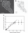
Force measurements on individual 3T3 fibroblast cells using a fibronectin-coated microfabricated pillar array. A: transmission light microscopy image of a cell deforming pillars. The cell border is represented by the white line. Scale bar = 10 μm. B: force field exerted by the cell obtained from the deflection of the underlying pillars. C: mean traction forces exerted by individual fibroblasts as a function of substrate rigidity. [From Ghibaudo et al. (75).]
FIGURE 6.
FIGURE 6.
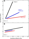
Cell stiffness tracks substrate stiffness for some but not all cell types. Cortical cell stiffness measured by micropipette aspiration (circles), magnetic bead twisting cytometry (MTC; diamonds), or atomic force microscopy (AFM; triangles). Cell cortical stiffness measured by micropipette aspiration or AFM scales with substrate stiffness for adult human bone marrow stromal cells (hMSC; dark blue) but not for murine embryonic stem cells (ESC; red). Inhibition of myosin by blebbistatin abrogates stiffness response of hMSC (light blue). [From Engler et al. (60), with permission from Elsevier; and Poh et al. (176), with permission from Elsevier.]
FIGURE 7.
FIGURE 7.
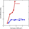
Relation of cell stiffness to spread area for NIH 3T3 fibroblasts and LBC3 glioma cells grown on gel substrates of different stiffness. [Adapted from Solon et al. (200), with permission from Elsevier; and Pogoda et al. (173).]
FIGURE 8.
FIGURE 8.
Melanoma stiffness measured by atomic force microscopy. The effective Young’s moduli of A7 (A) and M2 (B) melanoma cells cultured for 24 h on polyacrylamide gels laminated with collagen I (black), fibronectin (red), or mixture of collagen I and fibronectin (blue). [From Byfield et al. (30), with permission from Elsevier.]
FIGURE 9.
FIGURE 9.
Both viscous and elastic moduli change with substrate stiffness. Shear storage and loss moduli of T24 cells measured in the perinuclear region on gels with effective Young's moduli = 8 and 28 kPa. [From Abidine et al. (1), with permission from Elsevier.]
FIGURE 10.
FIGURE 10.
Stiffness of human bone marrow stromal cells (hMSCs) as functions of projected area and substrate stiffness. hMSCs are plated on microposts of different spring constants with various sizes of fibronectin patterns. For tall posts (3.8 nN/mm), cell stiffness remains constantly soft; for medium (18 nN/mm) and short (1,500 nN/mm) posts, cell stiffness increases with projected area. [From Tee et al. (207), with permission from Elsevier.]
FIGURE 11.
FIGURE 11.
Different classes of soft substrates with variable elastic and viscous properties. The simplest type of soft material is purely elastic, made from polymer chains that are covalently attached to each other to make a continuous network and which are embedded in a solvent of low viscosity. A common example is the polyacrylamide gel, which has a shear storage modulus much larger than its loss modulus and can be modeled as a simple spring (top). As a result, when such materials are subjected to a sudden stress (shown in green), they immediately respond to reach a time-independent level of strain; when the stress is removed, they recover their initial state before stress was applied. If the gel network is only partially connected by covalent bonds or is crosslinked by covalent and noncovalent bonds placed in series, the rheological response can be modeled as a spring in series with a dissipative dashpot (middle). The material continues to deform (creep) in response to a constant stress, like a viscoelastic liquid, and when the stress is removed it does not recover its initial shape and exhibits a plastic deformation. If the dissipative or labile links are placed in parallel with the covalent bonds, the material is a viscoelastic solid (bottom). The material deforms slowly in response to a sudden stress but eventually reaches a constant deformation; when the stress is removed, the sample slowly recovers and eventually reaches its initial unstrained state.
FIGURE 12.
FIGURE 12.
Effect of medium viscosity on cell spreading. Addition of high-molecular-weight polyethylene glycol (PEG) increases medium viscosity and the rate and extent of cell spreading. Blocking integrins with the tripeptide Arg-Gly-Asp (RGD) blocks spreading on fibronectin (Fn). [From Gonzalez-Molina et al. (83), with permission from Elsevier.]
FIGURE 13.
FIGURE 13.
Rigidity-dependent changes in proliferation of diverse cancer cell lines. [From Tilghman et al. (208).]
FIGURE 14.
FIGURE 14.
Mechanical priming of cells. Rat lung fibroblasts were seeded at 2,500 cells/cm2 on collagen-coated silicone substrates and cultured for four passages to model healthy lung tissue (5 kPa), mature fibrotic tissue (100 kPa), and unphysiologically stiff tissue culture plastic (TCP). The cells were then migrated from soft to fibrotic-stiff zones (5T100), or out of stiff fibrotic zones to soft substrates (100T5). Fourteen days (2 passages) later, the myofibroblast phenotype was assaying immunostaining for α-smooth muscle actin (green). Scale bar = 100 mm. [From Balestrini et al. (6), with permission from Oxford University Press.]
FIGURE 15.
FIGURE 15.
Tumor necrosis factor (TNF)-α release by macrophages is regulated by substrate stiffness. Bone marrow-derived macrophages (BMMs) (A) and RAW264.7 cells (B) were plated on fibronectin-coated 1, 20, and 150 kPa polyacrylamide gels for 18 h and stimulated for 24 h with media (−) or 3 μM CpG DNA (+). *Statistically significant; ns, not significant. [From Gruber et al. (85), with permission from Oxford University Press.]
FIGURE 16.
FIGURE 16.
Reciprocal relation between substrate and cell. As a cell encounters a substrate with specific physical properties, it responds by reorganizing its morphology but also by changing processes such as matrix deposition or activation of matrix remodeling enzymes that alter the chemical and physical properties of the substrate.
FIGURE 17.
FIGURE 17.
Cartoon of cell sensing and response to matrix stiffness. As a cell encounters a matrix, it uses actin-rich filopodia to probe the mechanical environment, establishing nascent adhesions at the leading edge. Resistance from a stiff matrix promotes the maturation of nascent adhesions into focal adhesions and spreading of the lamellipodium. If a cell, instead, encounters a compliant matrix, the adhesions disassemble and the filipodia retract. Importantly, these responses are cell-type and ligand-type specific.

References

    1. Abidine Y, Constantinescu A, Laurent VM, Sundar Rajan V, Michel R, Laplaud V, Duperray A, Verdier C. Mechanosensitivity of Cancer Cells in Contact with Soft Substrates Using AFM. Biophys J 114: 1165–1175, 2018. doi:10.1016/j.bpj.2018.01.005. - DOI - PMC - PubMed
    1. Alexander NR, Branch KM, Parekh A, Clark ES, Iwueke IC, Guelcher SA, Weaver AM. Extracellular matrix rigidity promotes invadopodia activity. Curr Biol 18: 1295–1299, 2008. doi:10.1016/j.cub.2008.07.090. - DOI - PMC - PubMed
    1. Ananthanarayanan B, Kim Y, Kumar S. Elucidating the mechanobiology of malignant brain tumors using a brain matrix-mimetic hyaluronic acid hydrogel platform. Biomaterials 32: 7913–7923, 2011. doi:10.1016/j.biomaterials.2011.07.005. - DOI - PMC - PubMed
    1. Andresen Eguiluz RC, Kaylan KB, Underhill GH, Leckband DE. Substrate stiffness and VE-cadherin mechano-transduction coordinate to regulate endothelial monolayer integrity. Biomaterials 140: 45–57, 2017. doi:10.1016/j.biomaterials.2017.06.010. - DOI - PMC - PubMed
    1. Andriani Y, Chua JM, Chua BY, Phang IY, Shyh-Chang N, Tan WS. Polyurethane acrylates as effective substrates for sustained in vitro culture of human myotubes. Acta Biomater 57: 115–126, 2017. doi:10.1016/j.actbio.2017.04.022. - DOI - PubMed

Publication types

LinkOut - more resources