. 2020 Jan;22(1):1-9.
doi: 10.1016/j.neo.2019.08.002. Epub 2019 Nov 18.

Liver X Receptor Agonism Sensitizes a Subset of Hepatocellular Carcinoma to Sorafenib by Dual-Inhibiting MET and EGFR

Affiliations

Liver X Receptor Agonism Sensitizes a Subset of Hepatocellular Carcinoma to Sorafenib by Dual-Inhibiting MET and EGFR

Weiqing Shao et al. Neoplasia. 2020 Jan.

Abstract

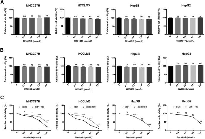
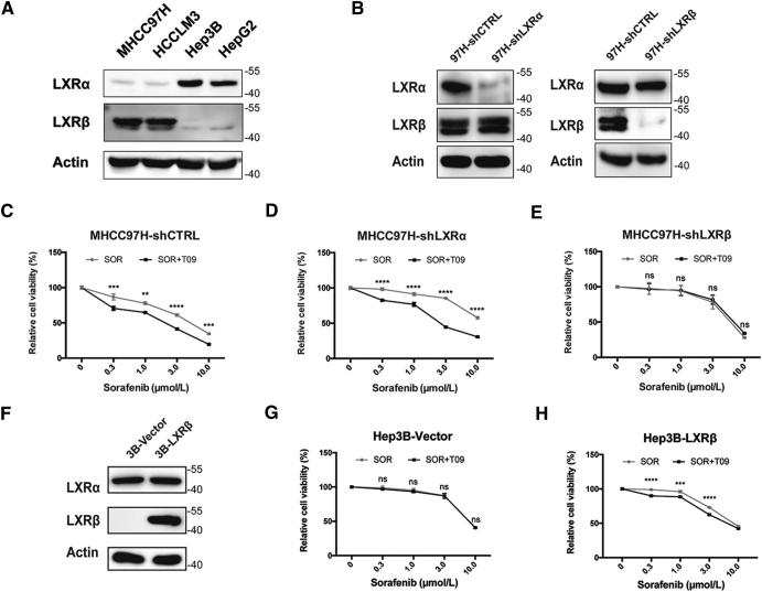
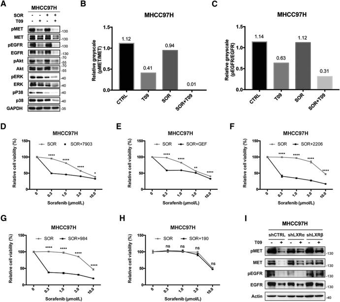
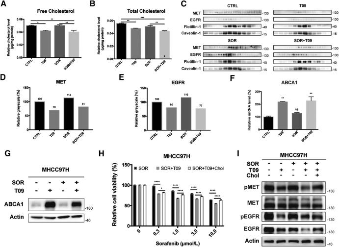
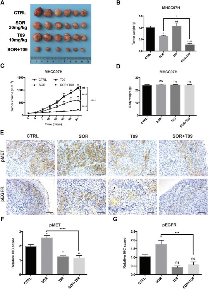
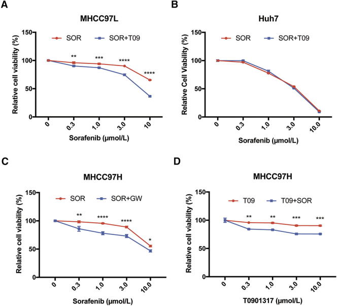
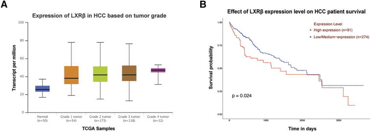
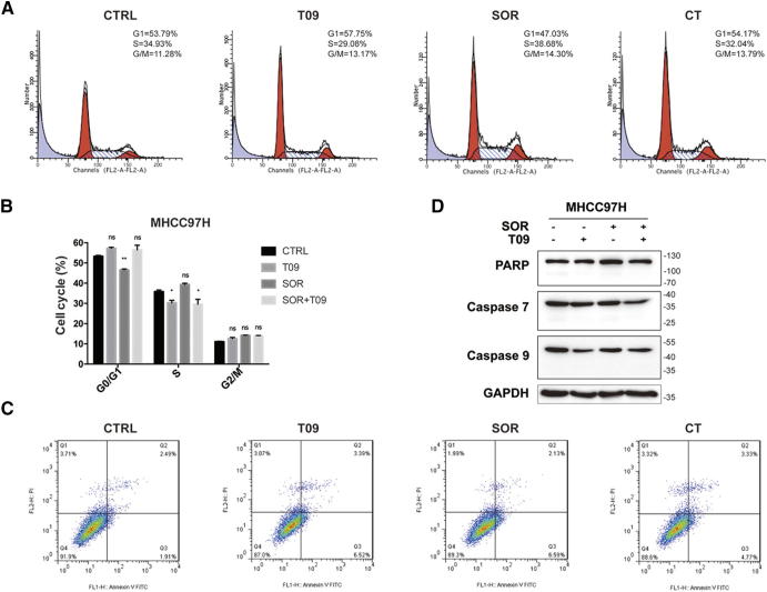
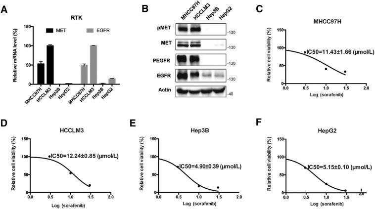
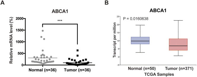
Sorafenib is the first approved systemic therapy for advanced hepatocellular carcinoma (HCC) and is the first-line choice in clinic. Sustained activation of receptor tyrosine kinases (RTKs) is associated with low efficacy of sorafenib in HCC. Activation of liver X receptor (LXR) has been reported to inhibit some RTKs. In this study, we found that the LXR agonist enhanced the anti-tumor activity of sorafenib in a subset of HCC cells with high LXR-β/α gene expression ratio. Mechanically, the activation of LXR suppressed sorafenib dependent recruitment of MET and epidermal growth factor receptor (EGFR) in lipid rafts through cholesterol efflux. Our findings imply that LXR agonist can serve as a potential sensitizer to enhance the anti-tumor effect of sorafenib.


Figures

Fig. 1
Fig. 1
Anti-proliferative effect of LXR agonists with or without sorafenib in human HCC cells. (A) Effects of T0901317 (T09) on in vitro cytotoxicity in MHCC97H, HCCLM3, Hep3B, and HepG2 cells. Cells were incubated in T09 at indicated concentrations for 72 h, then relative viabilities were determined by CCK8 assays. (B) Cells were incubated in GW3965 (GW) at indicated concentrations for 72 h before CCK8 assays. (C) Cells were incubated in sorafenib (SOR) with/without T09 (1 μM) for 72 h before detection. N ≥ 3, *P < .05, ****P < .0001.
Fig. 2
Fig. 2
LXRβ rather than LXRα exerts the synergistic effect. (A) Expression of LXRs in HCC cell lines detected by western blot analysis. (B) Verification of knockdown efficiency of LXRs in MHCC97H cells by western blot. (C-E) Effects of SOR with or without T09 (1 μM) on cell viabilities in MHCC97H-shCTRL (C), MHCC97H-shLXRα (D), and MHCC97H-shLXRβ cells (E). (F) Verification of LXRβ overexpression in Hep3B cells by western blot. (G-H) Effects of SOR with or without T09 (1 μM) on cell viabilities in Hep3B-Vector (G), and Hep3B-LXRβ cells (H). N ≥ 3, **P < .01, ***P < .001, ****P < .0001.
Fig. 3
Fig. 3
T0901317 dual-inhibits MET and EGFR. (A) Effects of T09 with or without SOR on MET/EGFR pathways in MHCC97H cells. Cells were incubated in SOR (3 μM) with/without T09 (1 μM) for 72 h before western blot. (B-C) Quantification of phospho−/non-phospho- MET (B) and EGFR (C) in (A). (D-H) Effects of inhibitors targeting MET (D), EGFR (E), Akt (F), ERK (G), and P38 (H) combined with SOR on proliferation of MHCC97H cells. Cells were incubated in SOR with/without PF-04217903 (7903, 10 nM), Gefitinib (GEF, 10 μM), MK-2206 (2206, 3 μM), SCH772984 (984, 3 μM), or SB202190 (190, 3 μM) for 72 h before CCK8 assays. (I) Effects of T09 on MET, pMET, EGFR, and pEGFR in MHCC97H-shCTRL, MHCC97H-shLXRα, and MHCC97H-shLXRβ cells. N ≥ 3, *P < .05, **P < .01, ****P < .0001.
Fig. 4
Fig. 4
T0901317 promotes cholesterol efflux by up-regulating ABCA1. (A-B) Quantification of free (A) and total (B) cytoplasmic cholesterol in MHCC97H cells after combination therapy. (C) Western blot analysis of MET and EGFR in lipid rafts in MHCC97H cells incubated in SOR with/without T09. (D-E) Quantification of MET (D) and EGFR (E) in lipid rafts in MHCC97H cells in (C). Expression level in CTRL group was standardized as 100%. (F-G) Quantitative real-time PCR (F) or western blot (G) of ABCA1 expression in MHCC97H cells after combination therapy. (H) Relative cell viabilities of MHCC97H cells treated by SOR, SOR with T09, and combination therapy with Cholesterol (Chol). (H) Effects of T09, SOR, and Chol on MET/EGFR pathways in MHCC97H cells. N ≥ 3, *P < .05, **P < .01, ***P < .001, ****P < .0001.
Fig. 5
Fig. 5
T0901317 improves the efficacy of sorafenib in vivo. (A-B) Tumor volume (A) and tumor weight (B) in MHCC97H xenograft mouse model at day 21 (n = 6). (C) Tumor growth curves in mouse model after SOR and/or T09 treatment. (D) Body weight of the implantation mice at day 21. (E) Representative images of immunohistochemistry (IHC) staining for pMET and pEGFR from MHCC97H xenografts (200 × magnification; scale bars, 100 μm). (F-G) Quantitative analysis of IHC scores of pMET (F) and pEGFR (G) in each group in (E). N ≥ 3, *P < .05, **P < .01, ***P < .001, ****P < .0001.
Figure S1
Figure S1
Effects of combination therapy of sorafenib and LXR agonists in HCC cell lines. (A-B) Effects of SOR alone or with T09 on proliferation in MHCC97L (A) or Huh7 cells (B). (C) Effect of combination therapy of SOR and GW in MHCC97H cells. (D) Effect of T09 with/without SOR on proliferation in MHCC97H cells. N ≥ 3, *P < .05, **P < .01, ***P < .001, ****P < .0001.
Figure S2
Figure S2
T0901317 upregulates mRNA level of CYP7A1 in HCC cells. (A-C) mRNA level of CYP7A1 in MHCC97H-shCTRL (A), MHCC97H-shLXRα (B), and MHCC97H-shLXRβ (C) cells. N ≥ 3, **P < .01, ***P < .001, ****P < .0001.
Figure S3
Figure S3
High LXRβ expression correlates with high tumor grades and poor survival of HCC patients. (A) Expression of LXRβ in HCC based on tumor grades. (B) Effect of LXRβ expression level on HCC patient survival.
Figure S4
Figure S4
LXR agonist augments the cytotoxicity of sorafenib without inducing apoptosis or cell cycle arrest. (A) Effects of SOR alone or with T09 on cell cycle in MHCC97H cells as assessed by flow cytometric analysis. (B) Quantification of each cell cycle in (A). (C) Effects of combination therapy on apoptosis in MHCC97H cells as assessed by flow cytometric analysis. (D) Effects of combination therapy on protein levels of apoptotic-related molecules in MHCC97H cells. N ≥ 3, *P < .05, **P < .01.
Figure S5
Figure S5
Over-activation of RTKs correlates with low efficacy of sorafenib (A) Quantitative real-time PCR of MET and EGFR expressions in HCC cells. (B) Western blot analysis of MET and EGFR expressions in HCC cells. (C-F) IC50 value of sorafenib in HCC cells. N ≥ 3.
Figure S6
Figure S6
ABCA1 is down-regulated in HCC tumor tissues. (A) Quantitative real-time PCR of ABCA1 expression in paired tumor and non-tumor tissues from HCC patients (n=36). ABCA1 expression in tumor was standardized as 100%. (B) Expression of ABCA1 in normal and primary tumor tissues of HCC patients from TCGA. N ≥ 3, ***P < .001.

References

    1. Bray F., Ferlay J., Soerjomataram I., Siegel R.L., Torre L.A., Jemal A. Global cancer statistics 2018: GLOBOCAN estimates of incidence and mortality worldwide for 36 cancers in 185 countries. CA Cancer J Clin. 2018;68:394–424. - PubMed
    1. Llovet J.M., Ricci S., Mazzaferro V., Hilgard P., Gane E., Blanc J.F. Sorafenib in advanced hepatocellular carcinoma. N Engl J Med. 2008;359:378–390. - PubMed
    1. Cheng A.L., Kang Y.K., Chen Z., Tsao C.J., Qin S., Kim J.S. Efficacy and safety of sorafenib in patients in the Asia-Pacific region with advanced hepatocellular carcinoma: a phase III randomised, double-blind, placebo-controlled trial. Lancet Oncol. 2009;10:25–34. - PubMed
    1. Chen K.F., Chen H.L., Tai W.T., Feng W.C., Hsu C.H., Chen P.J. Activation of phosphatidylinositol 3-kinase/Akt signaling pathway mediates acquired resistance to sorafenib in hepatocellular carcinoma cells. J Pharmacol Exp Ther. 2011;337:155–161. - PubMed
    1. Rudalska R., Dauch D., Longerich T., McJunkin K., Wuestefeld T., Kang T.W. In vivo RNAi screening identifies a mechanism of sorafenib resistance in liver cancer. Nat Med. 2014;20:1138–1146. - PMC - PubMed

Publication types

MeSH terms