. 2019 Nov 18;11(11):1813.
doi: 10.3390/cancers11111813.

Evaluation of the Accuracy of Liquid-Based Oral Brush Cytology in Screening for Oral Squamous Cell Carcinoma

Affiliations

Evaluation of the Accuracy of Liquid-Based Oral Brush Cytology in Screening for Oral Squamous Cell Carcinoma

Lena Deuerling et al. Cancers (Basel). .

Abstract

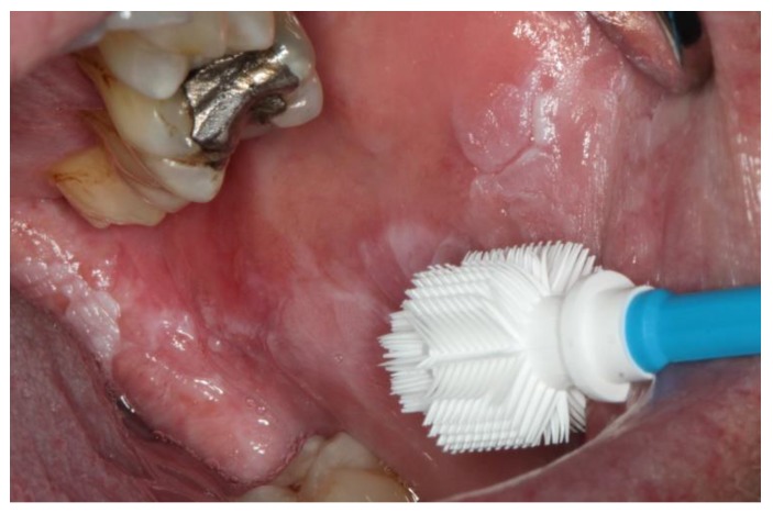
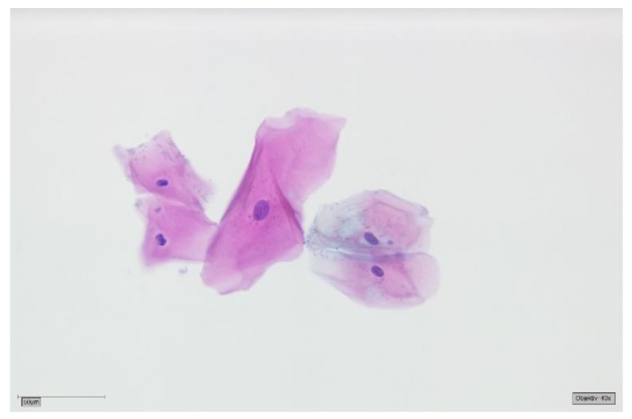
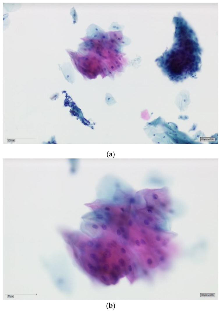
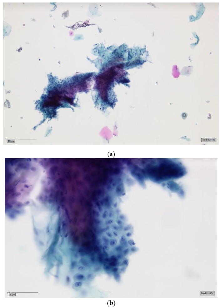
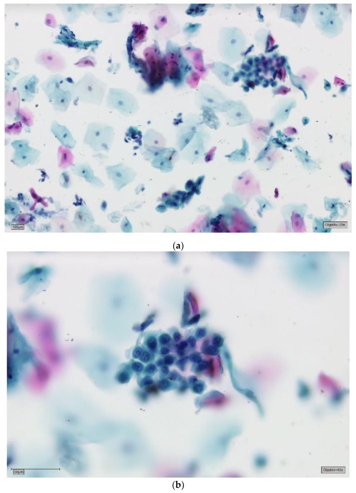
This study evaluates the accuracy of the results of liquid-based oral brush cytology and compares it to the histology and/or the clinical follow-ups of the respective patients. A total of 1352 exfoliated specimens were collected with an Orcellex brush from an identical number of oral lesions, then cytological diagnoses were made using liquid-based cytology. The final diagnoses in the study were 105 histologically proven squamous cell carcinomas (SCCs), 744 potentially malignant lesions and 503 cases of traumatic, inflammatory or benign hyperplastic oral lesions. The sensitivity and specificity of the liquid-based brush biopsy were 95.6% (95% CI 94.5-96.7%) and 84.9% (95% CI 83.0-86.8%), respectively. This led to the conclusion that brush biopsy is potentially a highly sensitive and reliable method to make cytological diagnoses of oral neoplasia. The main advantage of a brush biopsy over a scalpel biopsy is that it is less invasive and is more tolerated by the patients. Therefore, more lesions can be screened and more cancers can be detected at an early stage.

Keywords: brush biopsy; liquid-based cytology; oral cancer; squamous cell carcinoma.


Conflict of interest statement

Torsten W. Remmerbach is CEO of the German Association of Oral Diagnostics (DGOD mbH, Leipzig, Germany).

Figures

Figure 1
Figure 1
Cell collector Orcellex brush in front of a leukoplakia on the buccal mucosa.
Figure 2
Figure 2
Negative for tumour cells—SurePath, staining Papanicolaou, lens 40×. Clinically most probably oral lichen planus (OLP). The background is completely clear. Bacterial flora can still be appreciated clinging to the cell surfaces. A group of five mature squamous cells with reactive changes: cytoplasmic hypereosinophilia and amphophilia, and megalocytosis of the central cell with corresponding mild nuclear enlargement. Small perinuclear halos of the two cells in the left field. All nuclei are round to ovoid with smooth contours and finely granular, evenly dispersed chromatin.
Figure 3
Figure 3
(a) Doubtful for tumour cells—SurePath, staining Papanicolaou, lens 20×. Clinically erosive OLP. The background is mostly clear. In the lower left field is a stromal tissue fragment with mechanically altered nuclei; this correlates with an erosive process. There are several mature squamous cells lying singly and three larger groups of cells. In the upper right field, the basophilic cells are more immature, a sign of regeneration. The group of mature cells in the upper central field shows cytoplasmic hypereosinophilia and amphophilia. There seems to be some degree of anisonucleosis. (b) Doubtful for tumour cells—detail, lens 40×. On high power and in this plane of focus marked variation in nuclear size can be appreciated (factor 2–3). The larger nuclei are rather darker, i.e., more hyperchromatic, and the chromatin is slightly coarse. Nuclear contours are still fairly smooth. These findings are commonly reactive. A mild squamous intraepithelial neoplasia (SIN1) cannot be excluded. We perform DNA-karyometry on these cases. DNA-aneuploidy should prompt invasive biopsy for histology. With DNA-euploidy we would recommend clinical follow-up and repeat brush cytology after 12 months.
Figure 4
Figure 4
(a) Suspicious for tumour cells—SurePath, staining Papanicolaou, lens 10×. Clinically, an ulcerative lesion of the lateral border of the tongue. The background shows some amorphous deposits of partly eosinophilic, partly basophilic material, most obvious in the top central field. The big cell groups form partly sheets, partly three-dimensional crowds. Immaturity, a high nuclear/cytoplasmic (N/C) ratio and anisonucleosis can be suspected in this magnification. (b) Suspicious for tumour cells—detail, lens 40×. On high power there is marked anisonucleosis. The nuclei are haphazardly orientated, the axes of different nuclei are not parallel. Many nuclei show prominent nucleoli and/or irregularities of their borders. Chromatin is frequently irregularly deposited with early condensation along the nuclear membrane. These changes may represent so-called atypical tissue repair. The differential is high grade SIL or invasive SCC. The background may represent the ulcer or tumour diathesis. We would try to confirm the suspicion with DNA-karyometry. A scalpel biopsy must be performed for confirmation.
Figure 5
Figure 5
(a) Positive for tumour cells—SurePath, staining Papanicolaou, lens 20×. Clinically, there is suspicion for a carcinoma. On low power, there is a highly irregular aspect of the slide. In the background there are deposits of amorphous material, sometimes with nuclear fragments. In addition to many mature cells with normal nuclei, there are immature, small epithelial cells, both singly and in small and large groups. Nuclear enlargement and prominent nucleoli can be seen in this magnification. Some bizarre orangeophilic cells represent atypical keratinization. Those cells may have opaque, nearly black nuclei with smudged chromatin. (b) Positive for tumour cells—detail, lens 40×. A loosely cohesive sheet of highly atypical, immature squamous cells. The N/C ratio is markedly increased. Nuclei are highly hyperchromatic and chromatin is coarse. Some cells show an irregular nuclear contour and/or small nucleoli. These findings constitute the cytological diagnosis of a moderately well differentiated, keratinizing squamous cell carcinoma (SCC). Almost all these cases show aneuploidy on DNA-karyometry.

References

    1. Robert Koch-Institut . Krebs in Deutschland 2013/2014. Robert Koch-Institut; Berlin, Germany: 2014.
    1. Gleich L.L., Salamone F.N. Molecular genetics of head and neck cancer. Cancer Control. 2002;9:369–378. doi: 10.1177/107327480200900502. - DOI - PubMed
    1. Ali J., Sabiha B., Jan H.U., Haider S.A., Khan A.A., Ali S.S. Genetic etiology of oral cancer. Oral Oncol. 2017;70:23–28. doi: 10.1016/j.oraloncology.2017.05.004. - DOI - PubMed
    1. Scully C., Field J.K., Tanzawa H. Genetic aberrations in oral or head and neck squamous cell carcinoma 2: Chromosomal aberrations. Oral Oncol. 2000;36:311–327. doi: 10.1016/S1368-8375(00)00021-X. - DOI - PubMed
    1. Scully C., Field J.K., Tanzawa H. Genetic aberrations in oral or head and neck squamous cell carcinoma (SCCHN): 1. Carcinogen metabolism, DNA repair and cell cycle control. Oral Oncol. 2000;36:256–263. doi: 10.1016/S1368-8375(00)00007-5. - DOI - PubMed

LinkOut - more resources