Review
. 2019 Nov 19;8(11):1462.
doi: 10.3390/cells8111462.

Recent Advances in EPAC-Targeted Therapies: A Biophysical Perspective

Affiliations
Review

Recent Advances in EPAC-Targeted Therapies: A Biophysical Perspective

Alveena Ahmed et al. Cells. .

Abstract

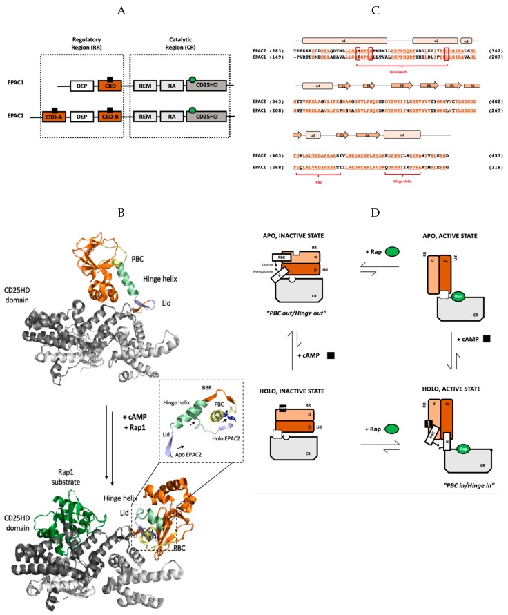
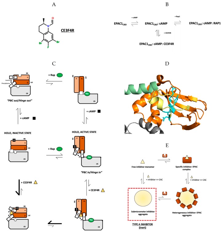
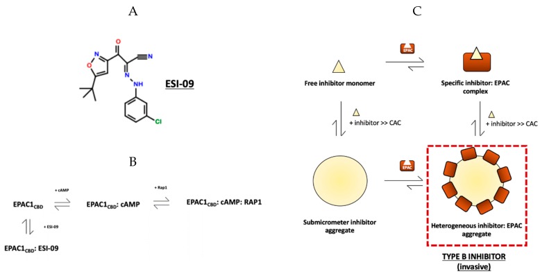
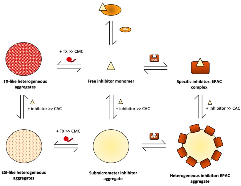
The universal second messenger cAMP regulates diverse intracellular processes by interacting with ubiquitously expressed proteins, such as Protein Kinase A (PKA) and the Exchange Protein directly Activated by cAMP (EPAC). EPAC is implicated in multiple pathologies, thus several EPAC-specific inhibitors have been identified in recent years. However, the mechanisms and molecular interactions underlying the EPAC inhibition elicited by such compounds are still poorly understood. Additionally, being hydrophobic low molecular weight species, EPAC-specific inhibitors are prone to forming colloidal aggregates, which result in non-specific aggregation-based inhibition (ABI) in aqueous systems. Here, we review from a biophysical perspective the molecular basis of the specific and non-specific interactions of two EPAC antagonists-CE3F4R, a non-competitive inhibitor, and ESI-09, a competitive inhibitor of EPAC. Additionally, we discuss the value of common ABI attenuators (e.g., TX and HSA) to reduce false positives at the expense of introducing false negatives when screening aggregation-prone compounds. We hope this review provides the EPAC community effective criteria to evaluate similar compounds, aiding in the optimization of existing drug leads, and informing the development of the next generation of EPAC-specific inhibitors.

Keywords: EPAC; NMR; aggregation-based inhibition; allostery; cAMP; drug design; dynamics; protein-ligand binding; screening; signaling.


Conflict of interest statement

The authors declare no conflict of interest.

Figures

Figure 1
Figure 1
Structure and thermodynamics of the cAMP binding domain (CBD)-binding domain of EPAC. (A) Domain organization of Exchange Protein directly Activated by cAMP (EPAC) isoforms 1 and 2 with catalytic and regulatory regions indicated. cAMP is shown as a black square bound to the CBDs (highlighted in orange) in the regulatory region and the Rap protein substrate is represented by a green circle bound to the CD25 homology domain (colored dark grey), which is responsible for EPAC’s GEF activity. EPAC2 has an additional N-terminal CBD that is dispensable in the regulation of EPAC’s catalytic activity in response to cellular cAMP levels. (B) Global changes in EPAC2 structure upon cAMP binding. In both the apo, inactive state (top) and holo, active state (bottom), unless otherwise specified CBD-B is shown in orange and the catalytic region is shown in grey. Additionally, the PBC, hinge helix, lid region, and CD25HD domain are indicated in yellow, pale green, light blue, and steel grey, respectively; the Rap1 substrate protein is shown in dark green bound to the holo active state and the ligand Sp-cAMPS is indicated in dark blue. Only residues common to both crystal structures are shown in this panel. The zoomed-in view shows the aligned PBC, BBR, hinge helix, and lid of EPAC2 in the unbound apo, inactive state and the Sp-cAMPS bound holo, active state. The direction of movement of the PBC, hinge helix and lid upon ligand binding is represented by arrows. (C) Sequence alignment of the regulatory cAMP-binding domain from EPAC1 and EPAC2 with conserved residues colored in vermillion and underlined. The secondary structure elements (α-helices and β-sheets) are indicated by peach rectangles and orange arrows, respectively. Important structural motifs, namely the PBC, hinge helix and ionic latch, are labelled in red. (D) Schematic representing the four-state thermodynamic cycle of EPAC auto-inhibition and activation in response to cAMP-binding. cAMP shown as a black square and Rap GTPase as green circles; the phosphate-binding cassette and hinge helix are labelled PBC and H, respectively. Being transient species, the EPAC apo, active and holo, inactive states have not been isolated, thus the relative conformation of the PBC and hinge helix remain unknown. As such, no indication of the relative conformations of the PBC and hinge helix is reported in the diagram. Instead, this conformational uncertainty is represented by a dashed white box in place of the PBC and hinge helix in the respective states.
Figure 2
Figure 2
Specific and non-specific interactions of EPAC1CBD and CE3F4R, a novel uncompetitive inhibitor. (A) The molecular structure of CE3F4R. (B) Schematic representing the uncompetitive mechanism of EPAC1 inhibition by CE3F4R. (C) Schematic summarizing the perturbation of the classic four-state thermodynamic cycle of EPAC activation by cAMP by CE3F4R binding, particularly highlighting the stabilization of the mixed holo inactive intermediate with the phosphate-binding cassette (PBC) in the active and hinge helix in the inactive conformation. Relative conformations of the PBC and hinge helix have not yet been elucidated in the holo, inactive and apo, active states and are thus not shown (D) Specific binding site of CE3F4R at the α/β subdomain interface of EPAC1 including residues Y242, I243, D267, and R294, as indicated in cyan, at the β-sheet facing the α-subdomain; the image shows homologous residues in EPAC2. Color scheme followed is consistent with Figure 1B. (E) Proposed thermodynamic cycle encompassing both specific enzyme:inhibitor binding as well as non-specific interactions between the two species as a result of colloidal aggregate formation; CE3F4R, as indicated on the figure, is a type-A inhibitor, forming inert aggregates that do not interact directly with the protein. Instead, they reduce overall inhibitory effect by acting as sinks for monomeric inhibitors (Figure adapted from Boulton, S.; Selvaratnam, R.; Ahmed, R.; Van, K.; Cheng, X.; Melacini, G. Mechanisms of specific versus nonspecific interactions of aggregation-prone inhibitors and attenuators. J. Med. Chem. 2019, 62, 5063–5079. Copyright (2019) American Chemical Society).
Figure 3
Figure 3
Specific and non-specific interactions of EPAC1CBD and ESI-09, a competitive inhibitor. (A) The molecular structure of ESI-09. (B) Schematic representing the competitive mechanism of EPAC1 inhibition by ESI-09. (C) Proposed thermodynamic cycle summarizing specific enzyme:ESI binding in addition to non-specific interactions between the two species as a result of aggregation; ESI-09, as indicated on the figure, is a type-B inhibitor, forming invasive aggregates that non-specifically adsorb protein molecules and may subsequently decrease specific enzyme:ESI-binding by causing protein misfolding, sequestering protein from ligand, as well as other diverse mechanisms (Figure adapted from Boulton, S.; Selvaratnam, R.; Ahmed, R.; Van, K.; Cheng, X.; Melacini, G. Mechanisms of Specific versus Nonspecific Interactions of Aggregation-Prone Inhibitors and Attenuators. J. Med. Chem. 2019, 62, 5063–5079. Copyright (2019) American Chemical Society.).
Figure 4
Figure 4
Specific and non-specific interactions of aggregation-prone inhibitors and aggregation attenuators in the context of EPAC. Aggregation-prone inhibition tends to decrease specific EPAC:ESI interactions. Inert type-A inhibitor aggregates do not interact with the protein receptor, however, they act as sinks for inhibitors, decreasing the free inhibitor concentration which ultimately reduces a compound’s specific inhibitory effect at a particular concentration. In contrast, invasive type-B inhibitor aggregates non-specifically adsorb protein molecules, directly interfering with enzyme:inhibitor interactions. To attenuate the effects of ABI, TX, and HSA are commonly used. HSA directly binds monomeric inhibitors, decreasing free inhibitor concentration, which protects the system from aggregate formation. On the other hand, TX binds ESI aggregates, forming heterogeneous co-aggregates that exhibit inert properties when [TX] >> [ESI] or possibly invasive properties when [ESI] >> TX. Overall, the key thermodynamic parameters needed to evaluate the relative role of specific binding, aggregation and attenuation are the dissociation constants for each of the aforementioned specific interactions, as well as the CAC of the inhibitor and the CMC of the attenuator. Figure adapted from Boulton, S.; Selvaratnam, R.; Ahmed, R.; Van, K.; Cheng, X.; Melacini, G. Mechanisms of Specific versus Nonspecific Interactions of Aggregation-Prone Inhibitors and Attenuators. J. Med. Chem. 2019, 62, 5063–5079. Copyright (2019) American Chemical Society.).

References

    1. Berman H.M., Ten Eyck L.F., Goodsell D.S., Haste N.M., Kornev A., Taylor S.S. The cAMP binding domain: An ancient signaling module. Proc. Natl. Acad. Sci. 2005;102:45–50. doi: 10.1073/pnas.0408579102. - DOI - PMC - PubMed
    1. Berdeaux R., Stewart R. cAMP signaling in skeletal muscle adaptation: Hypertrophy, metabolism, and regeneration. Am. J. Physiol. Endocrinol. Metab. 2012;303:E1–E17. doi: 10.1152/ajpendo.00555.2011. - DOI - PMC - PubMed
    1. Berthet J., Rall T.W., Sutherland E.W. The relationship of epinephrine and glucagon to liver phosphorylase. IV. Effect of epinephrine and glucagon on the reactivation of phosphorylase in liver homogenates. J. Biol. Chem. 1957;224:463–475. - PubMed
    1. Ravnskjaer K., Madiraju A., Montminy M. Role of the cAMP pathway in glucose and lipid metabolism. Handb. Exp. Pharmacol. 2015;233:29–49. - PubMed
    1. Spirli C., Mariotti V., Villani A., Fabris L., Fiorotto R., Strazzabosco M. Adenylyl cyclase 5 links changes in calcium homeostasis to cAMP-dependent cyst growth in polycystic liver disease. J. Hepatol. 2017;66:571–580. doi: 10.1016/j.jhep.2016.10.032. - DOI - PMC - PubMed

Publication types

MeSH terms

Substances

LinkOut - more resources