Comparative Study
. 2019 Nov 19;11(11):2829.
doi: 10.3390/nu11112829.

Investigation of Antibacterial and Antiinflammatory Activities of Proanthocyanidins from Pelargonium sidoides DC Root Extract

Affiliations
Comparative Study

Investigation of Antibacterial and Antiinflammatory Activities of Proanthocyanidins from Pelargonium sidoides DC Root Extract

Aiste Jekabsone et al. Nutrients. .

Abstract

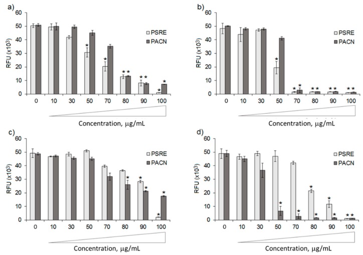
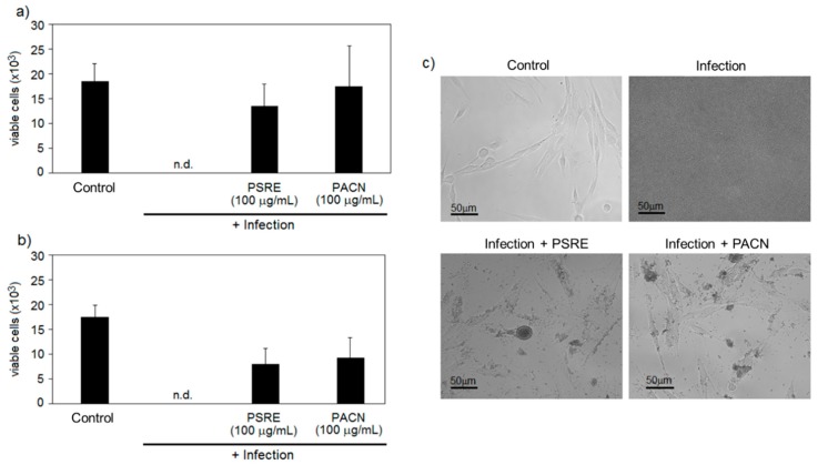
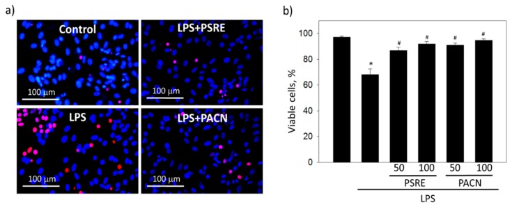
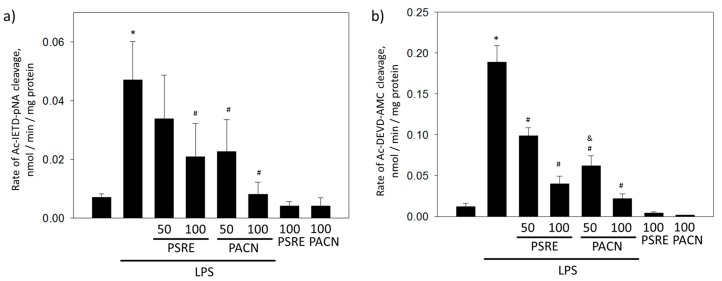
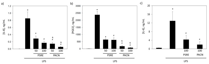
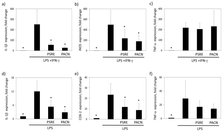
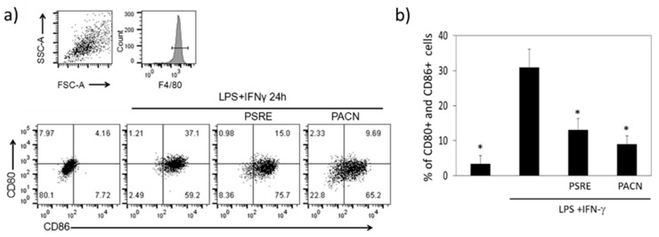
The study explores antibacterial, antiinflammatory and cytoprotective capacity of Pelargonium sidoides DC root extract (PSRE) and proanthocyanidin fraction from PSRE (PACN) under conditions characteristic for periodontal disease. Following previous finding that PACN exerts stronger suppression of Porphyromonas gingivalis compared to the effect on commensal Streptococcus salivarius, the current work continues antibacterial investigation on Staphylococcus aureus, Staphylococcus epidermidis, Aggregatibacter actinomycetemcomitans and Escherichia coli. PSRE and PACN are also studied for their ability to prevent gingival fibroblast cell death in the presence of bacteria or bacterial lipopolysaccharide (LPS), to block LPS- or LPS + IFNγ-induced release of inflammatory mediators, gene expression and surface antigen presentation. Both PSRE and PACN were more efficient in suppressing Staphylococcus and Aggregatibacter compared to Escherichia, prevented A. actinomycetemcomitans- and LPS-induced death of fibroblasts, decreased LPS-induced release of interleukin-8 and prostaglandin E2 from fibroblasts and IL-6 from leukocytes, blocked expression of IL-1β, iNOS, and surface presentation of CD80 and CD86 in LPS + IFNγ-treated macrophages, and IL-1β and COX-2 expression in LPS-treated leukocytes. None of the investigated substances affected either the level of secretion or expression of TNFα. In conclusion, PSRE, and especially PACN, possess strong antibacterial, antiinflammatory and gingival tissue protecting properties under periodontitis-mimicking conditions and are suggestable candidates for treatment of the disease.

Keywords: Pelargonium sidoides DC root extract; bacteriotoxicity; fibroblasts; gene expression; inflammatory cytokines; leukocytes; macrophages; periodontitis; proanthocyanidins.


Conflict of interest statement

The authors declare no conflict of interest.

Figures

Figure 1
Figure 1
Antibacterial activity of Pelargonium sidoides root extract (PSRE) and proanthocyanidins from PSRE (PACN) towards Staphylococcus aureus (a), S. epidermidis (b), Escherichia coli (c) and Aggregatibacter actinomycetemcomitans (d). RFU—relative fluorescence units. Bars represent means and standard deviations of six experimental replicates. * = p < 0.05 vs. untreated control.
Figure 2
Figure 2
The effect of Pelargonium sidoides root extract (PSRE) and proanthocyanidins from PSRE (PACN) on human gingival fibroblast viability in co-culture “race for the surface” assay. Human gingival fibroblasts were infected with A. actinomycetemcomitans (a) or S. aureus (b); bars represent means and standard deviations of six experimental repeats. “Control” represents not infected cells, and “n.d.” means there were no detectable cells in the samples. In (c), there are representative images of human gingival fibroblasts with A. actinomycetemcomitans after 24 h of infection.
Figure 3
Figure 3
The effect of Pelargonium sidoides root extract (PSRE) and proanthocyanidins from PSRE (PACN) on the viability of gingival fibroblasts affected by bacterial lipopolysaccharide (LPS). (a) Representative fluorescent images after cell treatment with LPS and 100 μg/mL PSRE or PACN and nuclear viability staining; propidium iodide-positive necrotic nuclei are red, all nuclei are stained blue with Hoechst33342. (b) Quantitative viability results. Fifty and 100 represent the concentrations (μg/mL). The data are presented as means plus standard deviation of five experiments. *—significant difference compared to the untreated control, and #—significant difference compared to the LPS only treatment, p < 0.05.
Figure 4
Figure 4
The effect of Pelargonium sidoides root extract (PSRE) and proanthocyanidins from PSRE (PACN) on LPS-induced caspase-8 (a) and caspase-3 (b) activation in gingival fibroblasts. Fifty and 100 represent the concentrations expressed in μg/mL. The data are presented as means with standard deviation of seven experimental repeats. *—significant difference compared to untreated control, #—significant difference compared to LPS only treatment and &—significant difference compared to LPS plus 50 μg/mL PSRE treatment, p < 0.05.
Figure 5
Figure 5
The effect of Pelargonium sidoides root extract (PSRE) and proanthocyanidins from PSRE (PACN) on (a) interleukin-8 (IL-8) and (b) prostaglandin E2 (PGE2) secretion from gingival fibroblasts, and (c) interleukin-6 (IL-6) secretion from peripheral blood mononuclear cells after LPS treatment. Fifty and 100 represent the concentrations (μg/mL). The data are presented as means and standard deviations of seven experiments. *—significant difference compared to untreated control, #—significant difference compared to LPS only treatment and &—significant difference compared to LPS plus a 50 μg/mL PSRE treatment, p < 0.05.
Figure 6
Figure 6
The effect of Pelargonium sidoides root extract (PSRE) and proanthocyanidins from PSRE (PACN) on proinflammatory gene expression in bone marrow-derived macrophages (ac) and peripheral blood mononuclear cells (df) after LPS or LPS and IFN- stimulation. (a,d) IL-1β (a,b) iNOS, (c,f) TNF- and (e) COX-2. The data are expressed as a fold change of glucose-6-phosphate isomerase gene transcription and presented as mean ± SD of three independent measurements. *—significantly different from the LPS-treated samples (ANOVA followed by a Tukey’s multiple comparison test, p < 0.05).
Figure 7
Figure 7
Expression of proinflammatory cell surface markers CD80 and CD86 analyzed by flow cytometry 24 h after treating LPS + IFNγ-activated BMDMs with PSRE and PACN. (a) Characteristic mouse macrophage marker F4/80-positive cells were gated for double CD80 and CD86 analysis as a measure of M1 macrophage phenotype (top right small quadrant). Representative plots of a total of three independent experiments in three replicates are presented in the bottom. (b) Mean ± SD of three independent measurements in three parallels. Differences between the measurements were tested using a one-way ANOVA followed by a Tukey’s multiple comparison test. *—significantly different from the LPS and IFN-γ treatment (p < 0.05).

References

    1. Loesche W.J., Grossman N.S. Periodontal Disease as a Specific, albeit Chronic, Infection: Diagnosis and Treatment. Clin. Microbiol. Rev. 2001;14:727. doi: 10.1128/CMR.14.4.727-752.2001. - DOI - PMC - PubMed
    1. Khan S.A., Kong E.F., Meiller T.F., Jabra-Rizk M.A. Periodontal Diseases: Bug Induced, Host Promoted. PLoS Pathog. 2015;11:e1004952. doi: 10.1371/journal.ppat.1004952. - DOI - PMC - PubMed
    1. Soares G.M.S., Figueiredo L.C., Faveri M., Cortelli S.C., Duarte P.M., Feres M. Mechanisms of action of systemic antibiotics used in periodontal treatment and mechanisms of bacterial resistance to these drugs. J. Appl. Oral Sci. 2012;20:295–309. doi: 10.1590/S1678-77572012000300002. - DOI - PMC - PubMed
    1. Pareek V., Gupta R., Panwar J. Do physico-chemical properties of silver nanoparticles decide their interaction with biological media and bactericidal action? A review. Mater. Sci. Eng. C. 2018;90:739–749. doi: 10.1016/j.msec.2018.04.093. - DOI - PubMed
    1. Nakatsuji T., Chen T.H., Narala S., Chun K.A., Two A.M., Yun T., Shafiq F., Kotol P.F., Bouslimani A., Melnik A.V., et al. Antimicrobials from human skin commensal bacteria protect against Staphylococcus aureus and are deficient in atopic dermatitis. Sci. Transl. Med. 2017;9 doi: 10.1126/scitranslmed.aah4680. - DOI - PMC - PubMed

Publication types

MeSH terms

LinkOut - more resources