Review
. 2021 Feb;18(2):178-193.
doi: 10.1080/15476286.2019.1695712. Epub 2019 Dec 3.

The La-related proteins: structures and interactions of a versatile superfamily of RNA-binding proteins

Affiliations
Review

The La-related proteins: structures and interactions of a versatile superfamily of RNA-binding proteins

Anne-Catherine Dock-Bregeon et al. RNA Biol. 2021 Feb.

Abstract

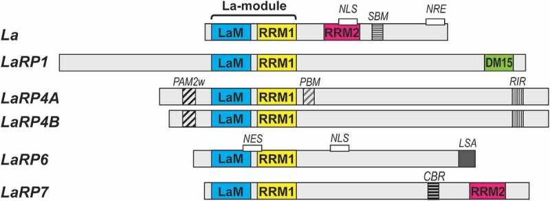
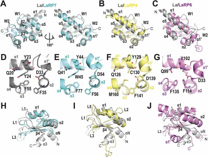
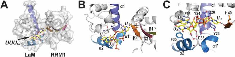
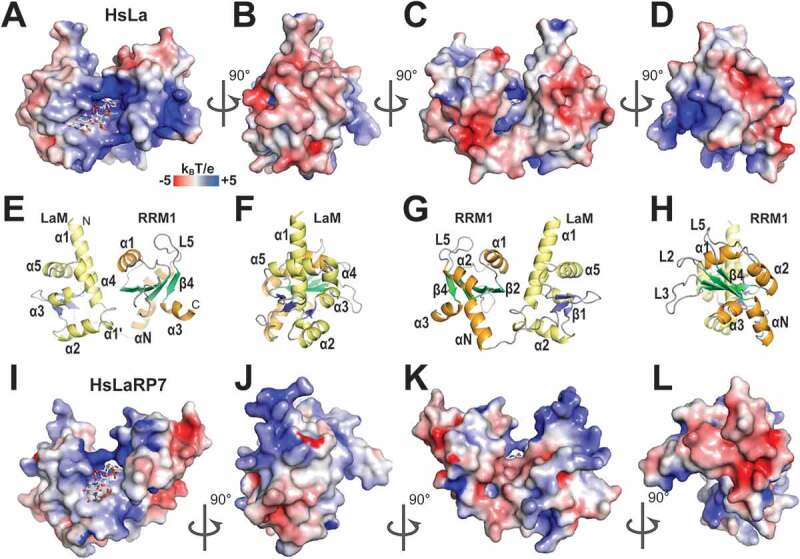
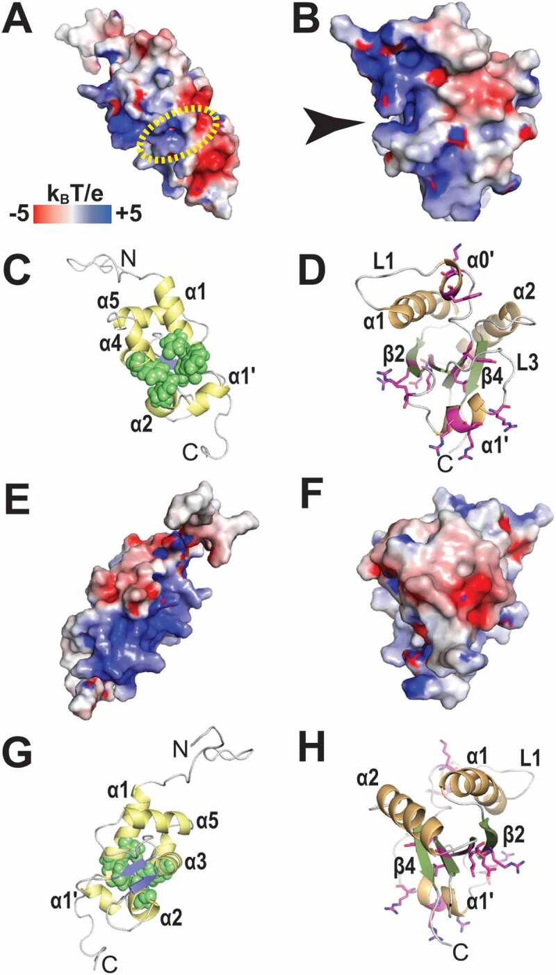
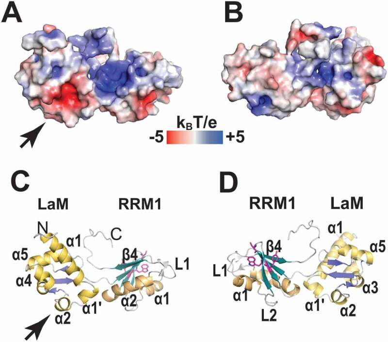
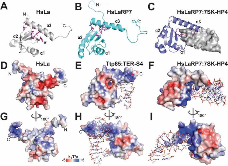
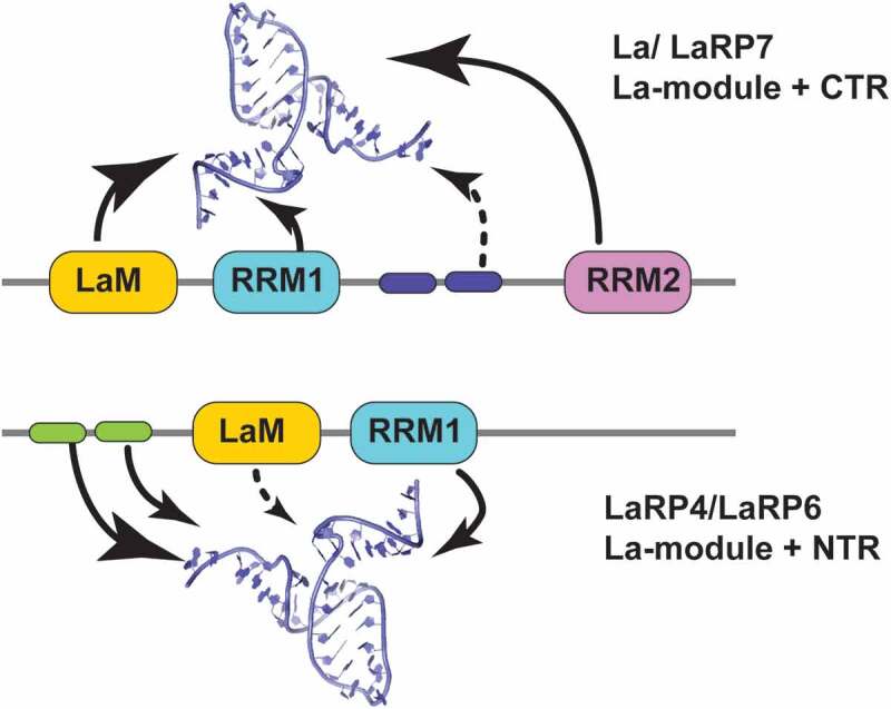
The La-related proteins (LaRPs) are an ancient superfamily of RNA-binding proteins orchestrating the major fates of RNA, from processing and maturation to regulation of mRNA translation. LaRPs are instrumental in modulating complex assemblies where the RNA is bound, folded, processed, escorted and presented to the functional effectors often through recruitment of protein partners. This intricate web of protein-RNA and protein-protein interactions is enabled by the modular nature of the LaRPs, comprising several structured domains connected by flexible linkers, and other sequences lacking recognizable folded motifs. Recent structures, together with biochemical and biophysical studies, have provided insights into how each LaRP family has evolved unique mechanisms of RNA recognition, not only through the conserved RNA-binding unit, the La-module, but also mediated by other family-specific motifs. Furthermore, in a series of unexpected twists and turns, they have revealed that the dynamic and conformational interplay of multi-structured domains and disordered regions operate in unison to achieve RNA substrate discrimination. This review proposes a perspective of our current knowledge of the structure-function relationship of the LaRP superfamily.

Keywords: La-module; La-related proteins; LaRP; RNA biology; RNA-binding proteins; RRM; structure-function relationship.


Conflict of interest statement

No potential conflict of interest was reported by the authors.

Figures

Figure 1.
Figure 1.
Domain organization in LaRP proteins in vertebrates. The domains and motifs are labelled and coloured as follows: La-Motif (LaM, blue), RNA-recognition motif 1 (RRM1, yellow), RNA-recognition motif 2 (RRM2, pink), DM15 domain (green), PAM2w motif (black diagonal stripes), PABP-binding motif (PBM, grey diagonal stripes), RACK-interacting motif (RIR, thin black vertical stripes), short basic motif (SBM, thin black horizontal stripes), La- and S1-associated motif (LSA, solid grey), conserved basic region (CBR, thick black horizontal stripes). Motifs linked to cellular localization are also indicated (NLS, nuclear localization signal; NRE, nuclear retention element; NES, nuclear export signal)
Figure 2.
Figure 2.
Comparison of the building blocks in the structures of LaRPs. (A-C) Superposition of the LaM domains of other LaRPs on HsLa (PDB 2VOD, grey). (A) HsLaRP7 (PDB 4WKR, cyan); (B) HsLaRP4 (PDB 6I9B, yellow); (C) HsLaRP6 (PDB 6MTF, magenta). (D-G) The six conserved residues in the cleft of the LaM domains formed by helices α1, α1ʹ, α2 and α3 in (D) HsLa; (E) HsLaRP7; (F) HsLaRP4; (G) HsLaRP6. (H-J) Superposition of the RRM1 domains of other LaRPs on HsLa (PDB 2VOD, grey). (A) HsLaRP7 (PDB 4WKR, cyan); (B) HsLaRP4 (PDB 6I9B, yellow); (C) HsLaRP6 (PDB 2MTG, magenta). The figure was prepared using PyMOL (https://pymol.org/2/)
Figure 3.
Figure 3.
The 3ʹUUUOH RNA-binding cleft in the La-module of HsLa. (A) Global view of the 3ʹUUUOH binding site in HsLa (2VOD). The La-module is in grey in both surface and cartoon representations with LaM helices α1, α1ʹ and α2 highlighted in different shades of blue. The strands of the RRM1 β-sheet are in shades of olive (β1), brick (β2), purple (β3) and pink (β4). (B) Zoomed-in view in the same orientation showing U−2 residing deep in the binding crevice formed by the LaM and RRM1 interface, while U−1 stacks on α2 of LaM, and U−3 stacks on U−1. (C) Details of the interaction, highlighting the stacking ladder of U−3 and U−1 on F35, and the stacking of U−2 on Y23, as well as the H-bonds of D33 with the two terminal hydroxyls (dotted lines). Dotted lines also identify the specific H-bonds between U−2 to the main chain of I140 in the β2-strand of RRM1 and to the side chain of Q20 from α1 of the LaM domain. The figure was prepared using PyMOL (https://pymol.org/2/)
Figure 4.
Figure 4.
Electrostatic surface potential of the La-modules of HsLa and HsLaRP7. (A) Electrostatic surface potential of HsLa (PDB 2VOD). The RNA 3ʹUUUOH is represented in sticks. (B-D) Same as A but progressively rotated by 90°C. (E-H) Cartoon representations of the views shown in (A-D), with the secondary structures highlighted (LaM helices in yellow; RRM1 helices in orange; LaM β-strands in blue; RRM1 β-strands in green). The bound triplet of uridines is not shown in the cartoon representation. (I-L) Similar views for HsLaRP7 (PDB 4WKR). Electrostatic surface potentials were calculated with APBS [75], ranging from −5 (red) to +5 (blue) kBT/e. The figures were drawn in PyMOL (https://pymol.org/2/)
Figure 5.
Figure 5.
Electrostatic surface potential of the LaM and RRM1 domains of HsLaRP6. (A) Electrostatic surface potential calculated for the LaM domain of HsLaRP6 (PDB 2MTF). The dotted yellow circle indicates where the 3ʹUUUOH triplet would be placed upon superposition with the structure of HsLa in complex with RNA (2VOD). (B) Electrostatic surface potential for the RRM1 domain of HsLaRP6 (PDB 2MTG). The arrow indicates the pocket where the base moiety of U−2 would be found upon superposition with the RRM1 of UUU-bound HsLa. (C) Cartoon representation of the LaM in the same orientation as A, with helices in yellow and strands in blue. Residues that were shown by mutation to be involved in RNA binding [43] are highlighted by green spheres (Q99, F102, Y103, F114, F135). (D) Cartoon representation of the RRM1 in the same orientation as B, with helices in orange and strands in green. Residues that upon mutation did not perturb RNA binding are shown in magenta sticks (L187, Y189, K196, W198, R231, R237, R244, R245, R249, I260, E262). (E-H) Opposite views (+180°) of A-D. Electrostatic surface potentials were calculated with APBS [75] ranging from −5 (red) to +5 (blue) kBT/e. The figures were generated using PyMOL (https://pymol.org/2/)
Figure 6.
Figure 6.
Electrostatic surface potential of the La-module of HsLaRP4A. (A) Electrostatic surface potential for HsLaRP4 (model 1 from PDB 6I9B). The orientation is the same as shown for HsLa in Fig. 3A and the arrowhead indicates where the 3ʹUUUOH triplet would be placed upon superposition with the structure of HsLa in complex with RNA. (B) View turned by 180°. (C-D) Cartoon representations in the same orientation as A and B, respectively, with helices in yellow (LaM) or orange (RRM1) and strands in blue (LaM) or green (RRM1). Three of residues in the RRM1 (W238, Y239 and S236) that showed chemical shift perturbation upon binding of the 15nt oligoA RNA ligand are highlighted in magenta. The electrostatic surface potential was calculated with APBS [75], ranging from −5 (red) to +5 (blue) kBT/e. The figures were generated using PyMOL (https://pymol.org/2/)
Figure 7.
Figure 7.
RRM2 domains in HsLa, HsLaRP7 and Ttp65. (A) Cartoon view of the RRM2 domain of HsLa (PDB 1OWX) showing residues V310, A314 and I318 from the α3 helix and L233, W261, I272, L274 and L306 from the β-sheet in stick representation, engaging at the interface. W261 is labelled by an asterisk. (B) Similar view of the RRM2 in HsLaRP7 apo structure (PDB 5NKW), showing the α3 helix tilted away from the β-sheet surface. Residues at the interface are shown in stick representation (H494 and L524 from the β-sheet, H528, E529, Y532, W533 and I536 from the α3 helix). Compared to HsLa, the interface is more open, partly due to the absence of an equivalent residue to W261 in HsLa. (C) Similar view of the RRM2 in HsLaRP7 bound to the HP4 hairpin (also termed SL4) from 7SK RNA (PDB 6D12), showing the insertion of the apical loop of the RNA hairpin (in grey, surface representation) into the protein. Specific recognition of the RNA is established by to K543 from the α3 helix, R496 from β2 strand, and Y483 from the edge the β-sheet, all shown as magenta sticks. These interactions coincide with a lengthening of the α3 helix (compare to B). (D) Electrostatic potential surface of HsLa RRM2, in the same orientation as in A. (E) Electrostatic potential surface of Ttp65 RRM2 (PDB 4ERD), in a similar orientation as in A, in complex with a fragment of the telomerase TER RNA (TER S4) shown in sticks. The electrostatic potential was calculated from a coordinate file where the selenomethionines (MSE 463 and MSE 527) were changed to methionines, to allow PDB2PQR to assign charges with the PARSE forcefield. (F) The electrostatic potential surface of the HsLaRP7 RRM2, in the same orientation as C, bound to the RNA HP4 from 7SK (residues 303–321), highlighting the residues G312 and G314 nesting in their binding pockets. As in E, the selenomethionines (MSE 475 and MSE 521) were changed for methionines for the calculations. (G) The electrostatic potential surface of HsLa RRM2, same as D but rotated by 180°. (H) Back view of Ttp65 RRM2 (PDB 4ERD) electrostatics potential surface (rotated by 180° from E) showing the α3 helix insertion in the major groove of the RNA double helix. Specific binding is achieved with two residues, G121 and A122, bulged from the helical stack to nest in two pockets. (I) Back view of HsLaRP7 RRM2 electrostatics potential surface (turned by 180° from F), with its bound RNA HP4 (nucleotides 303–321 from human 7SK) showing the α3 helix insertion from the apical loop into the major groove of the RNA hairpin, splaying apart the loop residues G321 and G314. Electrostatic surface potentials were calculated with APBS [75] ranging from −5 (red) to +5 (blue) kBT/e. The figures generated using PyMOL (https://pymol.org/2/)
Figure 8.
Figure 8.
Schematic representation of domain cooperativity within LaRPs. Two types of cooperative mechanisms of the LaRP proteins are proposed to sustain differences in RNA-recognition. In the La/LaRP7 group (top), where the La-module is in the N-terminal part of the protein, binding of selected RNA targets is achieved by the cooperative action of LaM, RRM1 and elements in the C-terminal region (CTR), particularly the RRM2 (large arrows) with possible additional contributions (dotted arrow) of basic sequences in the RRM1-RRM2 linker (blue cartouches). In the LaRP4/LaRP6 group (bottom), the La-module is more centrally located to the molecule, following N-terminal regions (NTRs) of various lengths. In these families, the La-module is working in synergy with sequences in the NTR (green cartouches). This has been demonstrated for HsLaRP4A where the La-module alone has little affinity for the RNA target [oligo(A)] and proposed for LaRP6
Figure 9.
Figure 9.
Interaction of the MLLE domain of PABP with PAM2 motifs. (A) The MLLE of PABP in cartoon representation (grey; residues 544–622) shown in complex with the peptide comprising the PAM2w sequence of HsLaRP4A (residues L15 to I25 in yellow stick representation; PDB 3PKN). An iodine atom from the crystal is shown as a purple sphere. (B) Same view of the complex with the PAM2w sequence of HsLaRP4B (residues E55 to H69 in orange stick representation; from PDB 3PTH). Note that the exit path of the peptide is different. (C) Same view of the MLLE in complex with the peptide comprising a classical PAM2 sequence from eRF3A (residues R67 to N79; in blue stick representation, from PDB 3KUI). Note the similarity with HsLaRP4A although there is no iodine ion in the vicinity. (D) Zoomed-in view showing the superposition of the peptides. The arrow indicates where the main-chain configuration differs for PAM2w of HsLaRP4B (orange) compared to HsLaRP4A (yellow) and eRF3 (blue)

References

    1. Bousquet-Antonelli C, Deragon JM.. A comprehensive analysis of the La-motif protein superfamily. Rna. 2009;15:750–764. - PMC - PubMed
    1. Maraia RJ, Mattijssen S, Cruz-Gallardo I, et al. The La and related RNA-binding proteins (LARPs): structures, functions, and evolving perspectives. Wiley Interdiscip Rev RNA. 2017;8:e1430. - PMC - PubMed
    1. Stefano JE. Purified lupus antigen La recognizes an oligouridylate stretch common to the 3ʹ termini of RNA polymerase III transcripts. Cell. 1984;36:145–154. - PubMed
    1. Wolin SL, Cedervall T. The La protein. Annu Rev Biochem. 2002;71:375–403. - PubMed
    1. Bayfield MA, Maraia RJ. Precursor-product discrimination by La protein during tRNA metabolism. Nat Struct Mol Biol. 2009;16:430–437. - PMC - PubMed

Publication types

MeSH terms

LinkOut - more resources