

Are there shared neural correlates between dyslexia and ADHD? A meta-analysis of voxel-based morphometry studies
- PMID: 31752659
- PMCID: PMC6873566
- DOI: 10.1186/s11689-019-9287-8
Are there shared neural correlates between dyslexia and ADHD? A meta-analysis of voxel-based morphometry studies
Abstract
Background: Dyslexia and Attention-deficit/hyperactivity disorder (ADHD) are highly comorbid neurodevelopmental disorders (estimates of 25-40% bidirectional comorbidity). Previous work has identified strong genetic and cognitive overlap between the disorders, but neural overlap is relatively unexplored. This study is a systematic meta-analysis of existing voxel-based morphometry studies to determine whether there is any overlap in the gray matter correlates of both disorders.
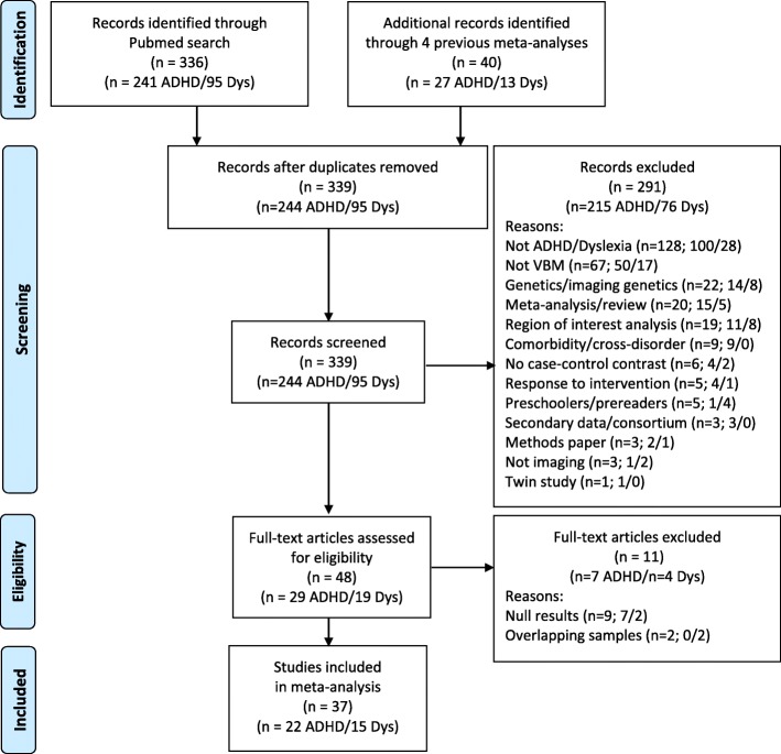
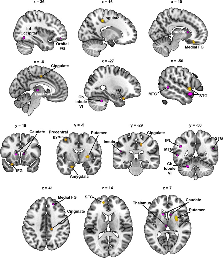
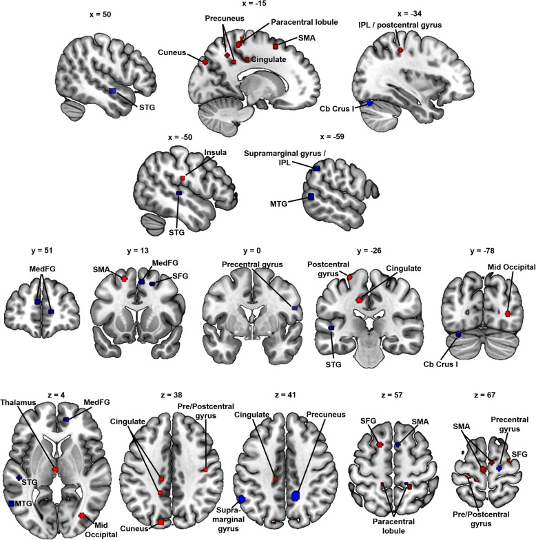
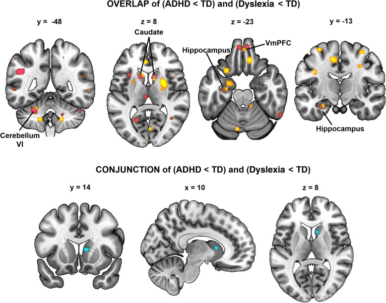
Methods: We conducted anatomic likelihood estimate (ALE) meta-analyses of voxel-based morphometry studies in which individuals with dyslexia (15 studies; 417 cases, 416 controls) or ADHD (22 studies; 898 cases, 763 controls) were compared to typically developing controls. We generated ALE maps for dyslexia vs. controls and ADHD vs. controls using more conservative (p < .001, k = 50) and more lenient (p < .005, k = 50) thresholds. To determine the overlap of gray matter correlates of dyslexia and ADHD, we examined the statistical conjunction between the ALE maps for dyslexia vs. controls and ADHD vs. controls (false discovery rate [FDR] p < .05, k = 50, 5000 permutations).
Results: Results showed largely distinct gray matter differences associated with dyslexia and ADHD. There was no evidence of statistically significant gray matter overlap at our conservative threshold, and only one region of overlap in the right caudate at our more lenient threshold. Reduced gray matter in the right caudate may be relevant to shared cognitive correlates in executive functioning and/or procedural learning. The more general finding of largely distinct regional differences in gray matter between dyslexia and ADHD suggests that other neuroimaging modalities may be more sensitive to overlapping neural correlates, and that current neuroimaging recruitment approaches may be hindering progress toward uncovering neural systems associated with comorbidity.
Conclusions: The current study is the first to meta-analyze overlap between gray matter differences in dyslexia and ADHD, which is a critical step toward constructing a multi-level understanding of this comorbidity that spans the genetic, neural, and cognitive levels of analysis.
Keywords: Attention-deficit/hyperactivity disorder; Caudate; Dyslexia; Meta-analysis; Voxel-based morphometry.
Conflict of interest statement
LMM receives book royalties from Guilford Press for the 2nd and 3rd editions of the textbook,
Figures
Similar articles
-
Seeking Overlapping Neuroanatomical Alterations between Dyslexia and Attention-Deficit/Hyperactivity Disorder: A Meta-Analytic Replication Study.Brain Sci. 2022 Oct 9;12(10):1367. doi: 10.3390/brainsci12101367. Brain Sci. 2022. PMID: 36291301 Free PMC article.
-
Global gray matter morphometry differences between children with reading disability, ADHD, and comorbid reading disability/ADHD.Brain Lang. 2018 Oct;185:54-66. doi: 10.1016/j.bandl.2018.08.004. Epub 2018 Sep 4. Brain Lang. 2018. PMID: 30189332 Free PMC article.
-
Shared grey matter correlates of reading and attention.Brain Lang. 2023 Feb;237:105230. doi: 10.1016/j.bandl.2023.105230. Epub 2023 Feb 1. Brain Lang. 2023. PMID: 36731345 Free PMC article.
-
A multimodal neuroimaging meta-analysis of functional and structural brain abnormalities in attention-deficit/hyperactivity disorder.Prog Neuropsychopharmacol Biol Psychiatry. 2025 Jan 10;136:111199. doi: 10.1016/j.pnpbp.2024.111199. Epub 2024 Nov 29. Prog Neuropsychopharmacol Biol Psychiatry. 2025. PMID: 39615871
-
Cortical and Subcortical Gray Matter Volume in Youths With Conduct Problems: A Meta-analysis.JAMA Psychiatry. 2016 Jan;73(1):64-72. doi: 10.1001/jamapsychiatry.2015.2423. JAMA Psychiatry. 2016. PMID: 26650724 Review.
Cited by
-
Facial expression recognition ability and its neuropsychological mechanisms in children with attention deficit and hyperactive disorder.Zhejiang Da Xue Xue Bao Yi Xue Ban. 2024 Apr 25;53(2):254-260. doi: 10.3724/zdxbyxb-2023-0390. Zhejiang Da Xue Xue Bao Yi Xue Ban. 2024. PMID: 38650447 Free PMC article. Review. Chinese, English.
-
Seeking Overlapping Neuroanatomical Alterations between Dyslexia and Attention-Deficit/Hyperactivity Disorder: A Meta-Analytic Replication Study.Brain Sci. 2022 Oct 9;12(10):1367. doi: 10.3390/brainsci12101367. Brain Sci. 2022. PMID: 36291301 Free PMC article.
-
Gray matter volume differences between early bilinguals and monolinguals: A study of children and adults.Hum Brain Mapp. 2022 Nov;43(16):4817-4834. doi: 10.1002/hbm.26008. Epub 2022 Jul 18. Hum Brain Mapp. 2022. PMID: 35848371 Free PMC article.
-
Interventions for children and adolescents with specific learning disability and co-occurring disorders.Pediatr Res. 2025 Jul 18. doi: 10.1038/s41390-025-04261-0. Online ahead of print. Pediatr Res. 2025. PMID: 40681698 Review.
-
The Polygenic Nature and Complex Genetic Architecture of Specific Learning Disorder.Brain Sci. 2021 May 14;11(5):631. doi: 10.3390/brainsci11050631. Brain Sci. 2021. PMID: 34068951 Free PMC article. Review.
References
-
- Willcutt E. Behavioral genetic approaches to understand the etiology of comorbidity. Behav Genet Psychopathol. 2014; [cited 2015 Jan 7]; Available from: http://link.springer.com/chapter/10.1007/978-1-4614-9509-3_8. - DOI
-
- Knopik VS, Neiderhiser JM, DeFries JC, Plomin R. Behavioral genetics. New York: Macmillan Higher Education; 2016.
Publication types
MeSH terms
Grants and funding
LinkOut - more resources
Full Text Sources
Medical
