. 2019 Nov 21;20(1):881.
doi: 10.1186/s12864-019-6086-2.

A qualitative transcriptional signature for the histological reclassification of lung squamous cell carcinomas and adenocarcinomas

Affiliations

A qualitative transcriptional signature for the histological reclassification of lung squamous cell carcinomas and adenocarcinomas

Xin Li et al. BMC Genomics. .

Abstract

Background: Targeted therapy for non-small cell lung cancer is histology dependent. However, histological classification by routine pathological assessment with hematoxylin-eosin staining and immunostaining for poorly differentiated tumors, particularly those from small biopsies, is still challenging. Additionally, the effectiveness of immunomarkers is limited by technical inconsistencies of immunostaining and lack of standardization for staining interpretation.

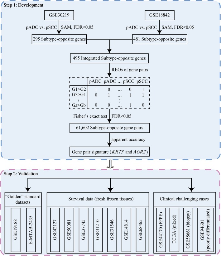
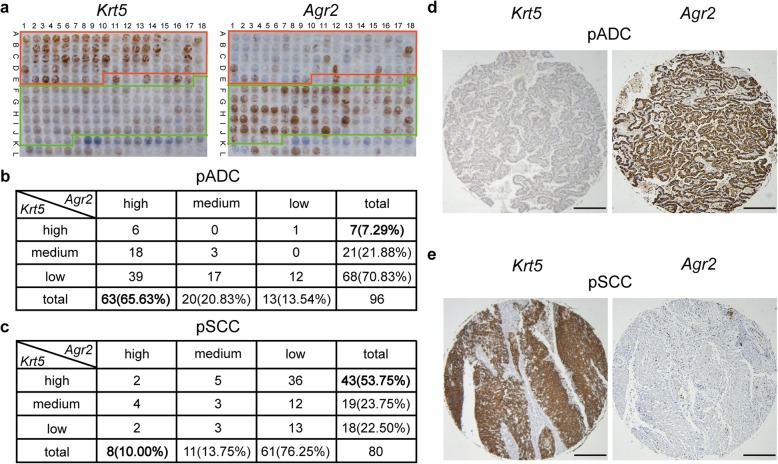
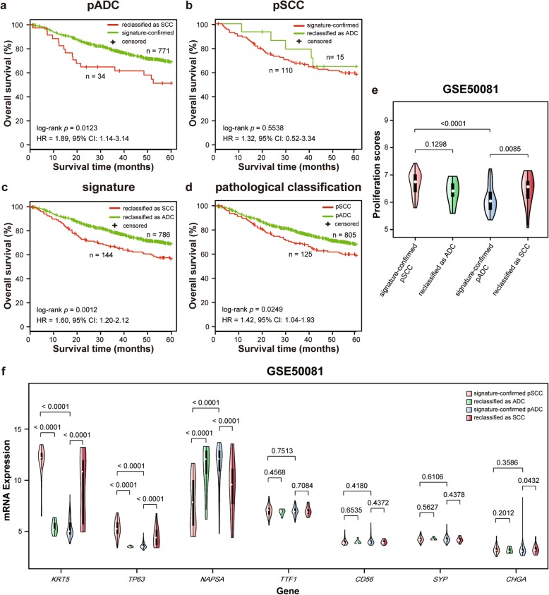
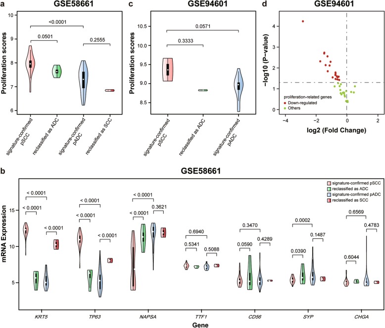
Results: Using gene expression profiles of pathologically-determined lung adenocarcinomas and squamous cell carcinomas, denoted as pADC and pSCC respectively, we developed a qualitative transcriptional signature, based on the within-sample relative gene expression orderings (REOs) of gene pairs, to distinguish ADC from SCC. The signature consists of two genes, KRT5 and AGR2, which has the stable REO pattern of KRT5 > AGR2 in pSCC and KRT5 < AGR2 in pADC. In the two test datasets with relative unambiguous NSCLC types, the apparent accuracy of the signature were 94.44 and 98.41%, respectively. In the other integrated dataset for frozen tissues, the signature reclassified 4.22% of the 805 pADC patients as SCC and 12% of the 125 pSCC patients as ADC. Similar results were observed in the clinical challenging cases, including FFPE specimens, mixed tumors, small biopsy specimens and poorly differentiated specimens. The survival analyses showed that the pADC patients reclassified as SCC had significantly shorter overall survival than the signature-confirmed pADC patients (log-rank p = 0.0123, HR = 1.89), consisting with the knowledge that SCC patients suffer poor prognoses than ADC patients. The proliferative activity, subtype-specific marker genes and consensus clustering analyses also supported the correctness of our signature.

Conclusions: The non-subjective qualitative REOs signature could effectively distinguish ADC from SCC, which would be an auxiliary test for the pathological assessment of the ambiguous cases.

Keywords: Histological subtype; Non-small cell lung cancer; Pathological assessment; Qualitative transcriptional signature; Relative gene expression orderings.


Conflict of interest statement

The authors declare that they have no competing interests.

Figures

Fig. 1
Fig. 1
The flowchart of this study. Using gene expression profiles of pADC and pSCC, we developed a qualitative transcriptional signature to individually distinguish ADC from SCC. The signature was tested in “golden” standard dataset, fresh frozen samples with survival data and clinical challenging cases, including FFPE specimens, mixed tumors, small biopsy specimens and poorly differentiated specimens. The pADC and pSCC represent pathologically-determined squamous cell carcinoma and pathologically-determined adenocarcinoma, respectively
Fig. 2
Fig. 2
Immunohistochemical analysis of Krt5 protein and Agr2 protein expressions in human lung cancer tissue microarray. a Krt5 and Agr2 proteins expression profile in lung cancer tissue array. The red frame containing samples from A1-E8 are pSCC. The green frame containing samples from E18-K5 are pADC. The remaining samples are the other subtypes of lung cancer and normal controls. b, c Inverse correlation between Krt5 protein and Agr2 protein expressions in pADC (b) and pSCC (c) samples. The protein expression score was quantified and considered as low, medium and high expression, basing on a multiplicative index of the average staining intensity and the extent of staining (see Methods). d, e Representative immunohistochemical staining results of Krt5 and Agr2 proteins in pADC (d) and pSCC (e) samples. Scale bar, 1 mm
Fig. 3
Fig. 3
The validation of the reclassifications by the signature for fresh frozen samples with survival data. a Kaplan-Meier curves of overall survival (OS) respectively for the pADC reclassified as SCC and the signature-confirmed pADC groups. b Kaplan-Meier curves of OS respectively for the pSCC reclassified as ADC and the signature-confirmed pSCC groups. c, d Kaplan-Meier curves of OS respectively for the SCC and ADC groups reclassified by the signature (c) and original pathological assessment (d). e The violin plot of proliferation scores in the reclassified and signature-confirmed samples, respectively, in the GSE50081 dataset with the higher reclassification rate in the fresh frozen samples. Wilcoxon rank sum test was used to test the difference of proliferation scores between two groups. f The violin plot of mRNA expressions of the seven subtype-specific marker genes in the GSE50081 dataset. The subtype-specific marker genes include ADC marker genes (NAPSA, TTF1), SCC marker genes (KRT5, TP63) and neuroendocrine marker genes (CD56, SYP, CHGA). The RankProd (RP) algorithm was used to test the difference of the subtype-specific marker genes between reclassified samples and signature-confirmed samples
Fig. 4
Fig. 4
The validation of the reclassifications by the signature for the FFPE and mixed tumor specimens. a The violin plot of proliferation scores and b mRNA expressions of the subtype-specific marker genes in the GSE44170 dataset derived from FFPE specimens. c The violin plot of proliferation scores and d mRNA expressions of the subtype-specific marker genes in the TCGA-ADC dataset derived from mixed tumor specimens. e The violin plot of proliferation scores and f mRNA expressions of the subtype-specific marker genes in the TCGA-SCC dataset derived from mixed tumor specimens
Fig. 5
Fig. 5
The validation of the reclassifications by the signature for small biopsy and poorly differentiated specimens. a The violin plot of proliferation scores and b mRNA expressions of the subtype-specific marker genes in the GSE58661 dataset derived from small biopsy specimens. c The violin plot of proliferation scores of 23 poorly differentiated specimens in the GSE94601 dataset. d The volcano plot of the differential expressions of the 44 proliferation-related genes in the pSCC samples reclassified as ADC when compared with the signature-confirmed pSCC samples. For the 44 proliferation-related genes, 20 genes were significantly differentially expressed and all the genes were down-regulated in the reclassified pSCC

Similar articles

Cited by

References

    1. Girard L, Rodriguez-Canales J, Behrens C, Thompson DM, Botros IW, Tang H, Xie Y, Rekhtman N, Travis WD, Wistuba II, et al. An expression signature as an aid to the histologic classification of non-small cell lung cancer. Clin Cancer Res. 2016;22(19):4880–4889. doi: 10.1158/1078-0432.CCR-15-2900. - DOI - PMC - PubMed
    1. Tian S. Classification and survival prediction for early-stage lung adenocarcinoma and squamous cell carcinoma patients. Oncol Lett. 2017;14(5):5464–5470. - PMC - PubMed
    1. Sun S, Schiller JH, Gazdar AF. Lung cancer in never smokers--a different disease. Nat Rev Cancer. 2007;7(10):778–790. doi: 10.1038/nrc2190. - DOI - PubMed
    1. Bishop JA, Benjamin H, Cholakh H, Chajut A, Clark DP, Westra WH. Accurate classification of non-small cell lung carcinoma using a novel microRNA-based approach. Clin Cancer Res. 2010;16(2):610–619. doi: 10.1158/1078-0432.CCR-09-2638. - DOI - PubMed
    1. Montezuma D, Azevedo R, Lopes P, Vieira R, Cunha AL, Henrique R. A panel of four immunohistochemical markers (CK7, CK20, TTF-1, and p63) allows accurate diagnosis of primary and metastatic lung carcinoma on biopsy specimens. Virchows Arch. 2013;463(6):749–754. doi: 10.1007/s00428-013-1488-z. - DOI - PubMed

MeSH terms