

Evaluation of microvascular changes in the perifoveal vascular network using optical coherence tomography angiography (OCTA) in type I diabetes mellitus: a large scale prospective trial
- PMID: 31752726
- PMCID: PMC6873669
- DOI: 10.1186/s12880-019-0391-8
Evaluation of microvascular changes in the perifoveal vascular network using optical coherence tomography angiography (OCTA) in type I diabetes mellitus: a large scale prospective trial
Abstract
Background: Diabetic retinopathy (DR) is the leading cause of blindness in type 1 Diabetes Mellitus (DM) patients, as a consequence of impaired blood flow in the retina. Optical coherence tomography angiography (OCTA) is a newly developed, non-invasive, retinal imaging technique that permits adequate delineation of the perifoveal vascular network. It allows the detection of paramacular areas of capillary non perfusion and/or enlargement of the foveal avascular zone (FAZ), representing an excellent tool for assessment of DR. The relationship of these microvascular changes with systemic factors such as metabolic control or duration of the disease still needs to be elucidated.
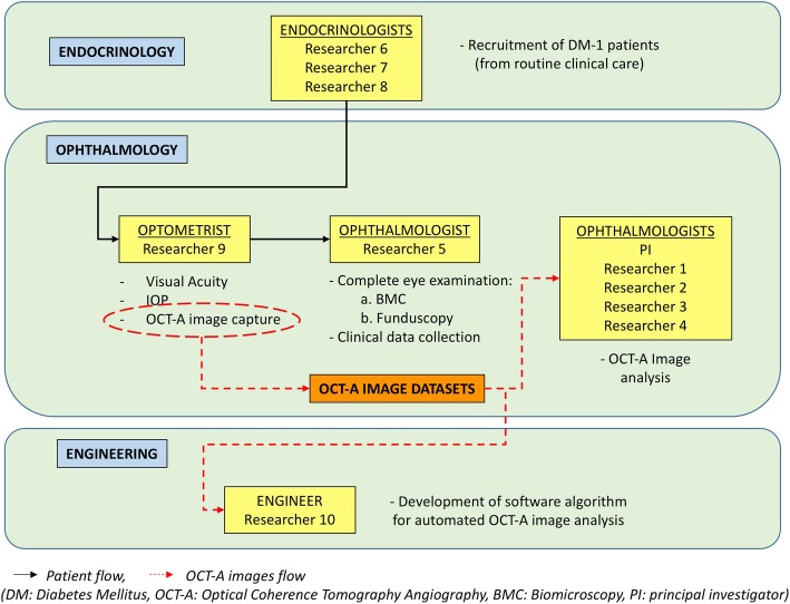
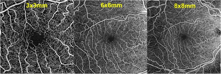
Methods: Prospective, consecutive, large-scale OCTA study. A complete ocular examination including a comprehensive series of OCTA images of different scan sizes captured with 2 OCT devices (Cirrus HD-OCT, Carl Zeiss Meditec, Dublin, CA, USA, and Triton Deep Range Imaging OCT, Topcon Corp, Topcon, Japan) will be obtained as part of the yearly routine follow up visits in type 1 DM patients seen in the Diabetes Unit of the Endocrinology department which give written informed consent to participate in the project. The aim of this study is to investigate the relationship between OCTA-derived parameters and systemic factors, as metabolic control (Hb1Ac, lipid profile, cholesterol, etc), and other relevant clinical factors as demographics or duration of the disease.
Discussion: This study is directed to investigate the relationship between the status of the perifoveal vascular network and systemic markers of the disease, and in particular to study whether these changes reflect those occurring elsewhere in the body affected by diabetic microvascular disease, as the kidneys or the brain. If these relationships were demonstrated, early detection of these microvascular changes by OCTA could lead to modifications in the pharmacological management of type 1 diabetic patients, as a way to reduce the risk of future complications in both the eye and other organs.
Trial registration: ClinicalTrials.gov, trial number NCT03422965.
Keywords: Diabetes mellitus; Diabetic retinopathy; Fovea; Macula; OCTA; Optical coherence tomography angiography; Type 1 Diabetes mellitus; Vascular network.
Conflict of interest statement
JZV and AA are speakers for Topcon and Zeiss. None of the authors have any financial interest in the devices employed in this study.
Figures
References
-
- Zhang K, Ferreyra HA, Grob S, et al. Diabetic retinopathy: genetics and etiologic mechanisms. In: Ryan SJ, Sadda SR, Hinton DR, et al., editors. Retina. London: Elsevier Saunders; 2013. pp. 925–939.
-
- Roser P, Kalscheuer H, Groener JB, Lehnhoff D, Klein R, Auffarth GU, Nawroth PP, Schuett F, Rudofsky G. Diabetic retinopathy screening ratio is improved when using a digital, nonmydriatic fundus camera onsite in a diabetes outpatient clinic. J Diabetes Res. 2016;2016:4101890. doi: 10.1155/2016/4101890. - DOI - PMC - PubMed
