. 2019 Nov 21;20(1):263.
doi: 10.1186/s12931-019-1235-3.

Analysis of key clinical features for achieving complete remission in stage III and IV non-small cell lung cancer patients

Affiliations

Analysis of key clinical features for achieving complete remission in stage III and IV non-small cell lung cancer patients

Takuya Aoki et al. Respir Res. .

Abstract

Background: Although development of immune checkpoint inhibitors and various molecular target agents has extended overall survival time (OS) in advanced non-small cell lung cancer (NSCLC), a complete cure remains rare. We aimed to identify features and treatment modalities of complete remission (CR) cases in stages III and IV NSCLC by analyzing long-term survivors whose OS exceeded 3 years.

Methods: From our hospital database, 1,699 patients, registered as lung cancer between 1st Mar 2004 and 30th Apr 2011, were retrospectively examined. Stage III or IV histologically or cytologically confirmed NSCLC patients with chemotherapy initiated during this period were enrolled. A Cox proportion hazards regression model was used. Data collection was closed on 13th Feb 2017.

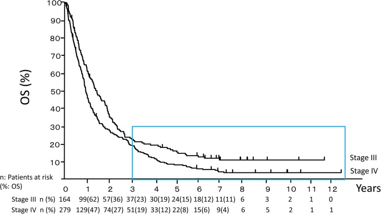
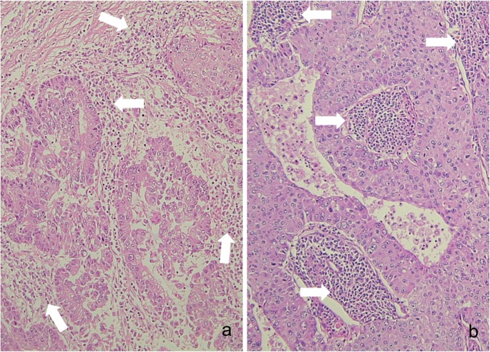
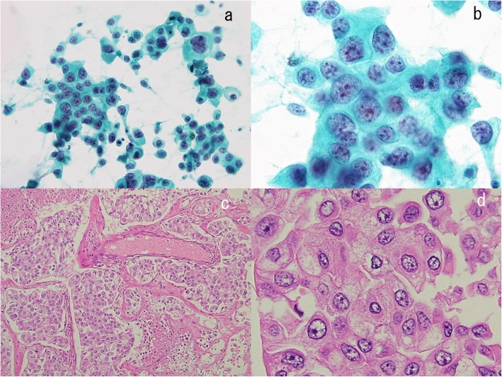
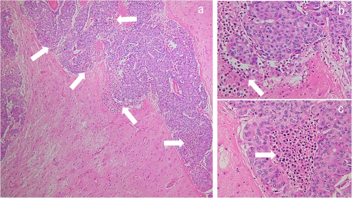
Results: There were 164 stage III and 279 stage IV patients, including 37 (22.6%) and 51 (18.3%) long-term survivors and 12 (7.3%) and 5 (1.8%) CR patients, respectively. The long-term survivors were divided into three groups: 3 ≤ OS < 5 years, 5 years ≤ OS with tumor, and 5 years ≤ OS without tumor (CR). The median OS of these groups were 1,405, 2,238, and 2,876 days in stage III and 1,368, 2,503, and 2,643 days in stage IV, respectively. The mean chemotherapy cycle numbers were 16, 20, and 10 in stage III and 24, 25, and 5 in stage IV, respectively. In the stage III CR group, all patients received chemoradiation, all oligometastases were controlled by radiation, and none had brain metastases. Compared with non-CR patients, the stage IV CR patients had smaller primary tumors and fewer metastases, which were independent prognostic factors for OS among long-term survivors. The 80% stage IV CR patients received radiation or surgery for controlling primary tumors, and the surgery rate for oligometastases was high. Pathological findings in the stage IV CR patients revealed that numerous inflammatory cells existed around and inside resected lung and brain tumors, indicating strong immune response.

Conclusions: Multiple line chemotherapies with primary and oligometastatic controls by surgery and/or radiation might achieve cure in certain advanced NSCLC. Cure strategies must be changed according to stage III or IV. This study was retrospectively registered on 16th Jun 2019 in UMIN Clinical Trials Registry (number UMIN000037078).

Keywords: advanced non-small cell lung cancer; complete remission; oligometastases; radiation; surgery.


Conflict of interest statement

The authors declare no conflicts of interest.

Figures

Fig. 1
Fig. 1
Overall survival (OS) in patients with stage III and IV non-small cell lung cancer. Overall survival was estimated by Kaplan-Meier analysis. Patients inside the square box were long-term survivors, whose OS exceeded 3 years
Fig. 2
Fig. 2
A representative complete remission (CR) patient with oligometastases in stage III. A CR patient with stage III (a-i), in whom successful control of primary and oligometastatic lesions was obtained, is shown. A male in his 50s consulted our hospital due to an abnormal shadow recognized on a chest X-ray (a). The computed tomography (CT) revealed the primary lesion to be in the right middle lobe (b), as well as swelling of a right hilar (c) and a mediastinal lymph node. Positron emission tomography (PET) / CT (d) and head magnetic resonance imaging (MRI) demonstrated the patient to have stage IIIA (cT1bN2M0) adenocarcinoma. The primary lesion showed scar formation after chemotherapy (e). However, PET/CT showed viable cells in the right hilar lymph node. Three-dimensional conformal radiation therapy was planned for the right hilar and mediastinal lymph nodes (f). On the day 380 since initial treatment, PET/CT revealed recurrence of the primary lesion and a new mediastinal lymph node metastasis (g). Three-dimensional conformal radiation therapy was administered for these lesions (h). The PET/CT (i) and the head MRI demonstrated CR
Fig. 3
Fig. 3
Pathological findings in the stage III patient with complete remission (CR), shown in Fig. 2. Cytology by bronchoscopy was class V according to the Papanicolaou staining (a). Histological examinations confirmed the diagnosis of adenocarcinoma, based on hematoxylin and eosin staining (b) and immunohistochemical staining for CEA (c) and TTF-1 (d). The carcinoma cells were positive for CEA (c) and TTF-1 (d)
Fig. 4
Fig. 4
Two representative complete remission (CR) patients with oligometastases in stage IV. The first CR patient in stage IV is shown in a-g. A male in his 50s presented with dyspnea, and bilateral malignant pleural and pericardial effusions were observed on the chest X-ray (a) and the CT (b). He was diagnosed as stage IVA (cT1aN2M1a) adenocarcinoma. After pericardial drainage followed by chemotherapy, the pleural and pericardial effusions disappeared (c). He complained of left femoral pain, and bone scintigraphy revealed bone metastases in the left femoral bone and left hip joint (d). After radiation therapy, he underwent joint replacement surgery. The primary lesion, located in the right upper lobe, showed gradual enlargement (e) and was surgically removed (f). The CR has since been maintained, based on head MRI and PET/CT (g). The second patient with brain metastases achieving CR is shown in h-o. A male in his 30s consulted our hospital due to loss of consciousness. A tumor was detected in the right upper lobe (h, i). There was no lymph node swelling, while a solitary brain metastasis was observed in the right temporal lobe (j). He was diagnosed as stage IVA (cT2aN0M1b) large cell carcinoma. Cyber-knife therapy for this lesion and surgery for the primary lesion were performed. After 4 courses of a first-line chemotherapy, three new brain metastases were detected in the left cerebellum, the right occipital lobe, and the left parietal lobe by head MRI. Cyber-knife treatments for these lesions were done. He subsequently complained of headache. Brain metastases in the left occipital lobe (k), the left cerebellum, and the right occipital lobe (l) were demonstrated by head MRI. Brain surgeries were performed for these lesions. The brain tumors and the lung tumor ultimately disappeared, as shown by the head MRI (m, n) and the PET/CT (o)
Fig. 5
Fig. 5
Cytological findings in a stage IV patient with complete remission (CR), shown in Fig. 4a-g. Carcinoma cells were confirmed by Papanicolaou staining of pericardial effusion (a, b). This carcinoma was a very aggressive type, based on the presence of numerous mitotic cells (b). Carcinoma cells stained positive for Ber-EP4 (c), indicating these cells to be adenocarcinoma, not mesothelioma
Fig. 6
Fig. 6
Pathological findings of the lung tumor resected from the right upper lobe, shown in Fig. 4e. Hematoxylin and eosin staining of the resected tumor is shown. Pathological examination revealed this tumor to be an adenocarcinoma showing a mixture of the acinar type (a) and the solid type (b). Numerous inflammatory cells around (a) and inside (b) the tumor are indicated by the white arrows. A lot of lymphoid follicles are visible inside the tumor (b). These inflammatory cells demonstrate marked immune response
Fig. 7
Fig. 7
Pathological findings in a stage IV patient with complete remission (CR), shown in Fig. 4h and i. Bronchoscopy was performed. Cytological examination revealed non-small cell type carcinoma, according to Papanicolaou staining (low power field; a, high power field; b). The surgically resected primary lung tumor (Fig. 4h and i) was stained with hematoxylin and eosin (low power field; c, high power field; d). Large cell carcinoma was confirmed by pathological examination
Fig. 8
Fig. 8
Pathological findings of the resected brain tumor in the right occipital lobe in Fig. 4n. This slide shows hematoxylin and eosin staining of the lesion resected from the right occipital lobe (low power field; a, high power field; b and c). Metastatic large cell carcinoma was confirmed. There are inflammatory cells around (b) and inside (c) the carcinoma, indicating strong immune response

References

    1. Chemotherapy in non-small cell lung cancer: a meta-analysis using updated data on individual patients from 52 randomised clinical trials. Non-small Cell Lung Cancer Collaborative Group. BMJ. 1995;311:899–909. - PMC - PubMed
    1. Schiller JH, Harrington D, Belani CP, Langer C, Sandler A, Krook J, Zhu J, Johnson DH. Comparison of four chemotherapy regimens for advanced non-small-cell lung cancer. N Engl J Med. 2002;346:92–98. doi: 10.1056/NEJMoa011954. - DOI - PubMed
    1. Ohe Y, Ohashi Y, Kubota K, Tamura T, Nakagawa K, Negoro S, Nishiwaki Y, Saijo N, Ariyoshi Y, Fukuoka M. Randomized phase III study of cisplatin plus irinotecan versus carboplatin plus paclitaxel, cisplatin plus gemcitabine, and cisplatin plus vinorelbine for advanced non-small-cell lung cancer: Four-Arm Cooperative Study in Japan. Ann Oncol. 2007;18:317–323. doi: 10.1093/annonc/mdl377. - DOI - PubMed
    1. Paez JG, Janne PA, Lee JC, Tracy S, Greulich H, Gabriel S, Herman P, Kaye FJ, Lindeman N, Boggon TJ, et al. EGFR mutations in lung cancer: correlation with clinical response to gefitinib therapy. Science. 2004;304:1497–1500. doi: 10.1126/science.1099314. - DOI - PubMed
    1. Lynch TJ, Bell DW, Sordella R, Gurubhagavatula S, Okimoto RA, Brannigan BW, Harris PL, Haserlat SM, Supko JG, Haluska FG, et al. Activating mutations in the epidermal growth factor receptor underlying responsiveness of non-small-cell lung cancer to gefitinib. N Engl J Med. 2004;350:2129–2139. doi: 10.1056/NEJMoa040938. - DOI - PubMed