. 2019 Nov 21;14(1):375.
doi: 10.1186/s13018-019-1437-x.

The effect of robot-navigation-assisted core decompression on early stage osteonecrosis of the femoral head

Affiliations

The effect of robot-navigation-assisted core decompression on early stage osteonecrosis of the femoral head

Benjun Bi et al. J Orthop Surg Res. .

Abstract

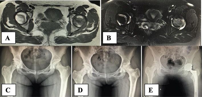
Background: The aim of the current paper is to evaluate the effects of robot-navigation-assisted core decompression compared with conventional core decompression surgery for early-stage osteonecrosis of the femoral head.

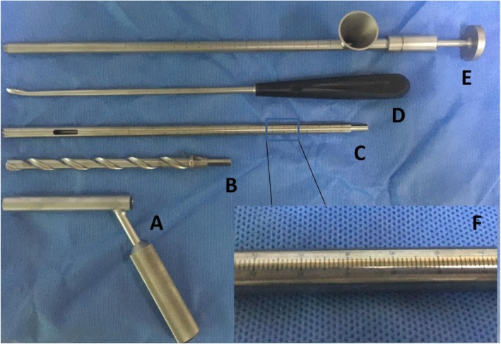
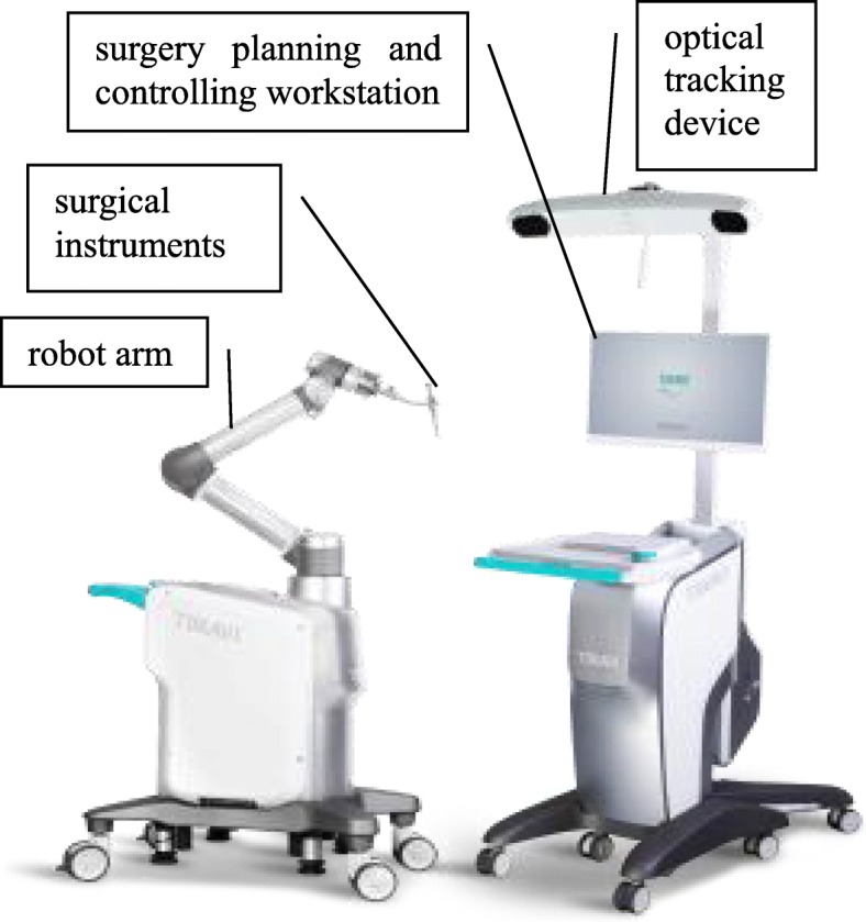
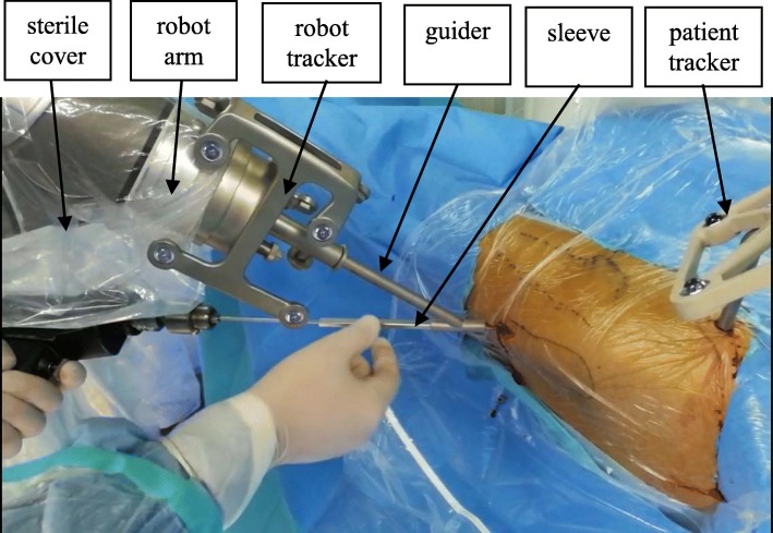
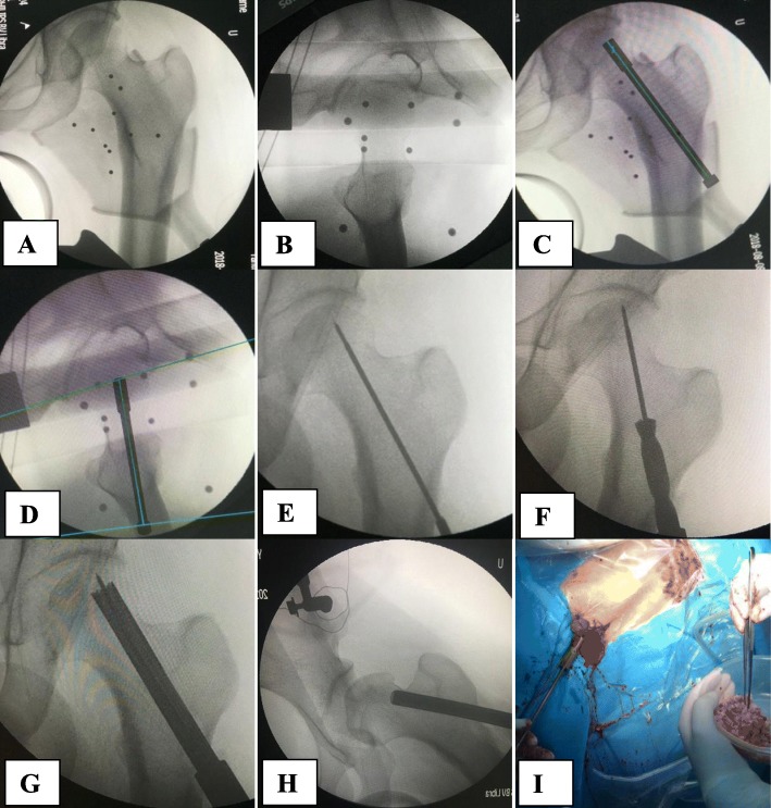
Methods: Twenty patients with a total of 36 hips who were diagnosed with Association Research Circulation Osseous stage 2 avascular necrosis of the femoral head and who received core decompression with or without robotic assistance were reviewed. The Harris hip score and visual analog scale score were used to assess clinical function. Intraoperative radiation exposure and operation time were used to evaluate the effectiveness of the robot-assisted system.

Results: At a mean follow-up of 26.4 months (24-36 months), the Harris hip score, visual analog scale score, and survival rate of the patients were similar between the conventional and robot-assisted groups. The guidewire insertion time, number of guidewire attempts, and radiation exposure during guidewire insertion were all significantly lower in the robot-assisted group than in the conventional group.

Conclusions: Robot-assisted core decompression of the femoral head is as safe and effective as a conventional core decompression surgery. It can reduce operation time and decrease intraoperative radiation exposure.

Keywords: Avascular necrosis of the femoral head; Core decompression; Robot navigation.


Conflict of interest statement

The authors declare that they have no competing interests.

Figures

Fig. 1
Fig. 1
Surgical instruments used for core decompression and bone graft of the femoral head. a Soft tissue protecting sleeve with an inner diameter of 10 mm. b Solid trephine used to penetrate to lateral cortex of the femur and make a short tunnel within the proximal femur. c, f Hollow trephine with a diameter of 10 mm used for core decompression of the necrotic area. The scale on the hollow trephine is used for monitoring the location of the trephine in the femoral head with an accuracy of 1 mm. d Long curette used for removing necrotic bone of the femoral head. e Grafting pipe with a funnel at one end through which cancellous bone can be easily put into the pipe and then impacted into the femoral head through the long pipe
Fig. 2
Fig. 2
Main components of TiRobot™: a robot arm, an optical tracking device, a surgical planning and controlling workstation, and some surgical instruments
Fig. 3
Fig. 3
Surgical instruments: robot arm is isolated by the sterile cover, and robot tracker and patient tracker are fixed, respectively, at the distal end of the robot arm and on the patient, guider attaches to the robot arm and firmly holds the sleeve; the sleeve can slide along the guider and invade patient
Fig. 4
Fig. 4
Illustration of the robot-assisted core decompression and bone graft. a, b Anteroposterior and lateral intraoperative fluoroscopic images of the hip were taken and input to the workstation. c, d Surgical trajectory for guidewire insertion on anteroposterior and lateral view of the hip. e Guide wire insertion. f Solid trephine insertion along the guidewire. g Hollow trephine insertion along the guidewire into the necrotic area of the femoral head. h, i Grafting of the cancellous bone using the grafting pipe with a funnel
Fig. 5
Fig. 5
Clinical case presentation. A 29-year-old woman, bilateral hip pain for 3 months with glucocorticoid use for 3 years before hospital admission. MRI T1WI (a), T2WI (b), and anteroposterior X-ray film (c) showed stage 2c osteonecrosis of two hips according to the ARCO classification. Two years after robot-assisted core decompression of two femoral heads, X-ray film showed the collapse of the left femoral head (d) with the right side intact. Finally, the patient received a total hip replacement of the left hip joint (e)

References

    1. Adelani MA, Keeney JA, Palisch A, Fowler SA, Clohisy JC. Has total hip arthroplasty in patients 30 years or younger improved? A systematic review. Clin Orthop Relat Res. 2013;471:2595–2601. doi: 10.1007/s11999-013-2975-x. - DOI - PMC - PubMed
    1. Lai YS, Wei HW, Cheng CK. Incidence of hip replacement among national health insurance enrollees in Taiwan. J Orthop Surg. 2008;3:42. doi: 10.1186/1749-799X-3-42. - DOI - PMC - PubMed
    1. Mankin HJ. Nontraumatc necrosis of bone (osteonecrosis) N Engl J Med. 1992;326:1473–1479. doi: 10.1056/NEJM199205283262206. - DOI - PubMed
    1. Kurtz S, Ong K, Lau E, Mowat F, Halpern M. Projections of primary and revision hip and knee arthroplasty in the United States from 2005 to 2030. J Bone Joint Surg Am. 2007;89:780–785. doi: 10.2106/JBJS.F.00222. - DOI - PubMed
    1. Ficat RP, Arlet J. Forage-biopsie de la tete femorale dans I’osteonecrose primative. Observations histo-pathologiques portant sur huit forages. Rev Rhum. 1964;31:257–264.