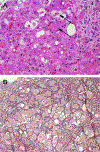
Liver Histology: Diagnostic and Prognostic Features
- PMID: 31753251
- PMCID: PMC6986808
- DOI: 10.1016/j.cld.2019.09.004
Liver Histology: Diagnostic and Prognostic Features
Abstract
When patients with suspected drug-induced liver injury (DILI) undergo liver biopsy, the pathologist can provide a wealth of information on the morphologic changes. The most common histologic patterns of DILI include mimics of acute and chronic hepatitis as well as acute cholestasis, chronic cholestasis, and a mixed pattern that combines hepatitis with cholestasis. The pattern may suggest etiologies of injury or correlate with reported patterns of injury for specific agents. Biopsy may exonerate or indict particular drugs as causal agents of injury and provide specific information on severity of injury and specific types of changes related to various outcomes.
Keywords: Acute hepatitis; Cholestatic hepatitis; Hepatic necrosis; Hepatotoxicity; Nodular regenerative hyperplasia.
Published by Elsevier Inc.
Figures


Similar articles
-
The pathology of drug-induced liver injury.Semin Liver Dis. 2009 Nov;29(4):364-72. doi: 10.1055/s-0029-1240005. Epub 2009 Oct 13. Semin Liver Dis. 2009. PMID: 19826970 Review.
-
Hepatic histological findings in suspected drug-induced liver injury: systematic evaluation and clinical associations.Hepatology. 2014 Feb;59(2):661-70. doi: 10.1002/hep.26709. Epub 2013 Dec 18. Hepatology. 2014. PMID: 24037963 Free PMC article.
-
Histological patterns in drug-induced liver disease.J Clin Pathol. 2009 Jun;62(6):481-92. doi: 10.1136/jcp.2008.058248. J Clin Pathol. 2009. PMID: 19474352 Review.
-
Comparative histologic features among liver biopsies with biliary-pattern injury and confirmed clinical diagnoses.Hum Pathol. 2024 Apr;146:8-14. doi: 10.1016/j.humpath.2024.03.003. Epub 2024 Mar 11. Hum Pathol. 2024. PMID: 38479481
-
Persistent liver biochemistry abnormalities are more common in older patients and those with cholestatic drug induced liver injury.Am J Gastroenterol. 2015 Oct;110(10):1450-9. doi: 10.1038/ajg.2015.283. Epub 2015 Sep 8. Am J Gastroenterol. 2015. PMID: 26346867 Free PMC article.
Cited by
-
A novel mouse model of hepatocyte-specific apoptosis-induced myeloid cell-dominant sterile liver injury and repair response.Am J Physiol Gastrointest Liver Physiol. 2024 Oct 1;327(4):G499-G512. doi: 10.1152/ajpgi.00005.2024. Epub 2024 Aug 6. Am J Physiol Gastrointest Liver Physiol. 2024. PMID: 39104322
-
A Rare Disease Presenting Postpartum: Acute Fatty Liver of Pregnancy.Case Rep Obstet Gynecol. 2021 Sep 30;2021:1143470. doi: 10.1155/2021/1143470. eCollection 2021. Case Rep Obstet Gynecol. 2021. PMID: 34631182 Free PMC article.
-
Development of a Novel Ex Vivo Porcine Hepatic Segmental Perfusion Proof-of-Concept Model Towards More Ethical Translational Research.Cureus. 2023 Feb 18;15(2):e35143. doi: 10.7759/cureus.35143. eCollection 2023 Feb. Cureus. 2023. PMID: 36949973 Free PMC article.
-
Comparison of DL-Methionine and L-Methionine levels in liver metabolism activity in commercial broilers fed a diet without antibiotic growth promoters.Vet World. 2025 Mar;18(3):598-605. doi: 10.14202/vetworld.2025.598-605. Epub 2025 Mar 18. Vet World. 2025. PMID: 40342738 Free PMC article.
-
Detailed Molecular Mechanisms Involved in Drug-Induced Non-Alcoholic Fatty Liver Disease and Non-Alcoholic Steatohepatitis: An Update.Biomedicines. 2022 Jan 17;10(1):194. doi: 10.3390/biomedicines10010194. Biomedicines. 2022. PMID: 35052872 Free PMC article. Review.
References
-
- Kleiner DE, Gaffey MJ, Sallie R, et al. Histopathologic changes associated with fialuridine hepatotoxicity. Mod Pathol 1997;10(3):192–9. - PubMed
-
- McKenzie R, Fried MW, Sallie R, et al. Hepatic failure and lactic acidosis due to fialuridine (FIAU), an investigational nucleoside analogue for chronic hepatitis B. N Engl J Med 1995;333(17):1099–105. - PubMed
-
- Popper H, Rubin E, Cardiol D, et al. Drug-induced liver disease: a penalty for progress. Arch Intern Med 1965;115:128–36. - PubMed
-
- Zen Y, Yeh MM. Hepatotoxicity of immune checkpoint inhibitors: a histology study of seven cases in comparison with autoimmune hepatitis and idiosyncratic drug-induced liver injury. Mod Pathol 2018;31(6):965–73. - PubMed
Publication types
MeSH terms
Grants and funding
LinkOut - more resources
Full Text Sources
Medical

