Review
. 2020 Jan 6;219(1):e201909033.
doi: 10.1083/jcb.201909033.

Autophagy and cancer: Modulation of cell death pathways and cancer cell adaptations

Affiliations
Review

Autophagy and cancer: Modulation of cell death pathways and cancer cell adaptations

Christina G Towers et al. J Cell Biol. .

Abstract

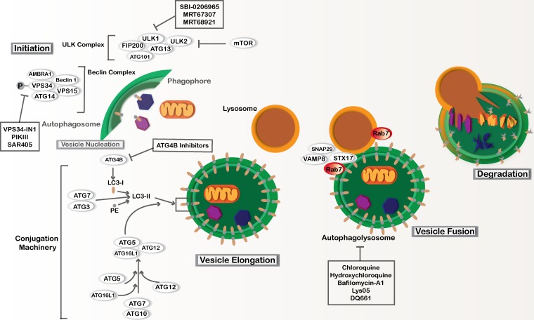
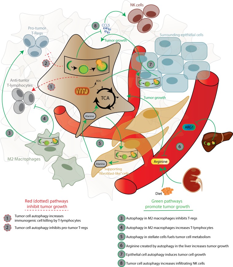
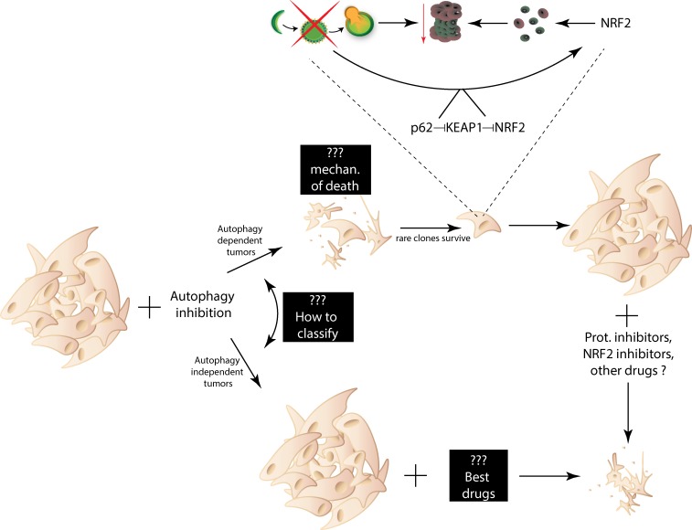
Autophagy is intricately linked with many intracellular signaling pathways, particularly nutrient-sensing mechanisms and cell death signaling cascades. In cancer, the roles of autophagy are context dependent. Tumor cell-intrinsic effects of autophagy can be both tumor suppressive and tumor promotional. Autophagy can therefore not only activate and inhibit cell death, but also facilitate the switch between cell death mechanisms. Moreover, autophagy can play opposing roles in the tumor microenvironment via non-cell-autonomous mechanisms. Preclinical data support a tumor-promotional role of autophagy in established tumors and during cancer therapy; this has led to the launch of dozens of clinical trials targeting autophagy in multiple cancer types. However, many questions remain: which tumors and genetic backgrounds are the most sensitive to autophagy inhibition, and which therapies should be combined with autophagy inhibitors? Additionally, since cancer cells are under selective pressure and are prone to adaptation, particularly after treatment, it is unclear if and how cells adapt to autophagy inhibition. Here we review recent literature addressing these issues.


Figures

Figure 1.
Figure 1.
Macro-autophagy. Macro-autophagy involves core autophagy proteins or ATGs and is subdivided into different stages, including phagophore initiation, vesicle nucleation, vesicle elongation, and autophagosome fusion with lysosomes. The ULK complex involves Unc-51–like autophagy activating kinases 1 and 2 (ULK1 and ULK2), ATG13, ATG101, and FAK family kinase interacting protein of 200 kD (FIP200). This complex can be regulated by nutrient availability via mTOR regulation as well as other signaling pathways to induce phagophore initiation. The Beclin complex is activated downstream of the ULK complex and is also necessary for phagophore initiation. The Beclin complex includes coiled-coil, moesin-like, BCL2 interacting protein (Beclin-1), activating molecule in Beclin-1 regulated autophagy (AMBRA-1), phosphatidylinositol 3-kinase catalytic subunit type 3 and regulatory subunit 4 (VPS34 and VPS15, respectively), and ATG14. Vesicle elongation depends on two ubiquitin-like conjugation systems. ATG5 is conjugated to ATG12 with the help of the E1-like enzyme, ATG7, and the E2-like enzyme, ATG10. The ATG5–ATG12 conjugate binds to ATG16L1, and together they act as a E3-like enzyme to facilitate the conjugation of microtubule-associated protein 1A/1B LC3 to PE. This second conjugation is also aided by ATG7 as well as the E1-like enzyme, ATG3. Prior to LC3-PE conjugation, LC3 is cleaved by the cysteine protease ATG4B. LC3-PE is incorporated into the autophagosome membrane. SNARE proteins including syntaxin-17 (STX17), synaptosome-associated protein 29 (SNAP29), and vesicle-associated membrane protein 8 (VAMP8) facilitate fusion between fully formed autophagosomes and lysosomes. The GTPase Rab7 is also important during fusion. After fusion occurs, the cytoplasmic material within the autolysosome as well as the intravesicular LC3-II is degraded by pH-sensitive enzymes found within the acidic compartments. Pharmacological agents that are currently used to inhibit autophagy in preclinical models are annotated.
Figure 2.
Figure 2.
Non–tumor cell-autonomous roles of autophagy in cancer. The red dotted lines indicate pathways that have been implicated in the tumor-suppressive roles of autophagy in cancer. These mechanisms include how autophagy in the tumor cells can increase the presence of infiltrating antitumor T-lymphocytes and decrease protumor regulatory T cells in the TME. Green lines indicate pathways that have been implicated in the tumor-promotional roles of autophagy in cancer. Autophagy in the tumor cells can inhibit the antitumorigenic NK cells via inhibition of the cytokine, CCL5. Autophagy in nontumor cells within the TME can also affect the tumor growth. Autophagy in the supporting fibroblast-like cells and surrounding epithelial cells can support metabolism and tumor cell proliferation. Autophagy in macrophages can increase protumor regulatory T cells and decrease infiltrating tumor-suppressive T cells. In CD8+ T cells, loss of autophagy increases the memory effector phenotype and also increases the antitumor cytokine IFNγ. Circulating arginine supplied from the kidneys and diet can also support tumor cell proliferation. Autophagy in hepatocytes is important for regulating the arginine-degrading enzyme arginase-1 (ARG1).
Figure 3.
Figure 3.
Cancer cells can adapt to autophagy inhibition. There are still a number of “black boxes” when it comes to targeting autophagy as a cancer therapeutic. While we know that some cancers are particularly sensitive to autophagy inhibition, the exact biomarkers that dictate autophagy dependence remain at large. It is also unclear if autophagy-independent cells may be exquisitely sensitive to other targeted agents. Recently, it was shown that in autophagy-dependent cancer cell lines that die after acute autophagy inhibition, rare clones can survive by up-regulating NRF2 to maintain protein homeostasis. Consequently, the cells with acquired autophagy independence gained new targetable susceptibilities, i.e., proteasome inhibitors. There are likely additional mechanisms cells can use to circumvent autophagy inhibition and corresponding novel susceptibilities that have yet to be discovered.

References

    1. Akin D., Wang S.K., Habibzadegah-Tari P., Law B., Ostrov D., Li M., Yin X.M., Kim J.S., Horenstein N., and Dunn W.A. Jr. 2014. A novel ATG4B antagonist inhibits autophagy and has a negative impact on osteosarcoma tumors. Autophagy. 10:2021–2035. 10.4161/auto.32229 - DOI - PMC - PubMed
    1. Anding A.L., and Baehrecke E.H.. 2017. Cleaning House: Selective Autophagy of Organelles. Dev. Cell. 41:10–22. 10.1016/j.devcel.2017.02.016 - DOI - PMC - PubMed
    1. Audesse A.J., Dhakal S., Hassell L.A., Gardell Z., Nemtsova Y., and Webb A.E.. 2019. FOXO3 directly regulates an autophagy network to functionally regulate proteostasis in adult neural stem cells. PLoS Genet. 15:e1008097 10.1371/journal.pgen.1008097 - DOI - PMC - PubMed
    1. Azad M.B., Chen Y., Henson E.S., Cizeau J., McMillan-Ward E., Israels S.J., and Gibson S.B.. 2008. Hypoxia induces autophagic cell death in apoptosis-competent cells through a mechanism involving BNIP3. Autophagy. 4:195–204. 10.4161/auto.5278 - DOI - PMC - PubMed
    1. Baginska J., Viry E., Berchem G., Poli A., Noman M.Z., van Moer K., Medves S., Zimmer J., Oudin A., Niclou S.P., et al. . 2013. Granzyme B degradation by autophagy decreases tumor cell susceptibility to natural killer-mediated lysis under hypoxia. Proc. Natl. Acad. Sci. USA. 110:17450–17455. 10.1073/pnas.1304790110 - DOI - PMC - PubMed

Publication types

LinkOut - more resources