. 2019 Dec 2;218(12):3967-3976.
doi: 10.1083/jcb.201909028. Epub 2019 Nov 21.

The RNA export factor Mex67 functions as a mobile nucleoporin

Affiliations

The RNA export factor Mex67 functions as a mobile nucleoporin

Carina Patrizia Derrer et al. J Cell Biol. .

Abstract

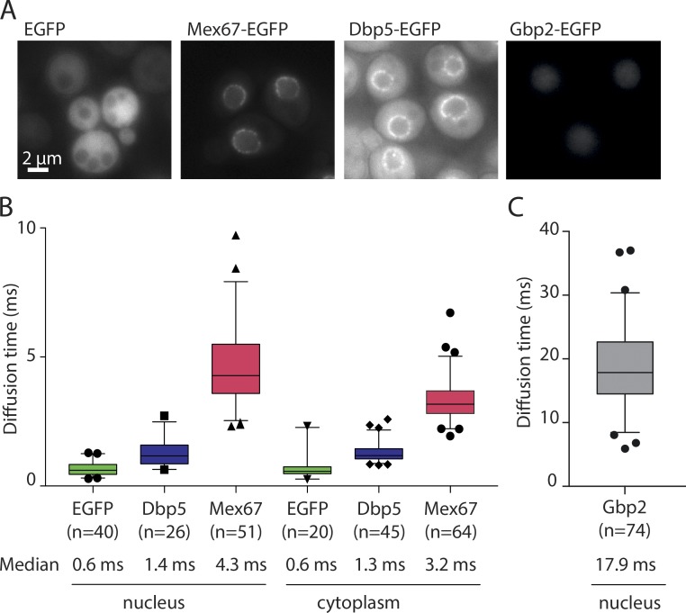
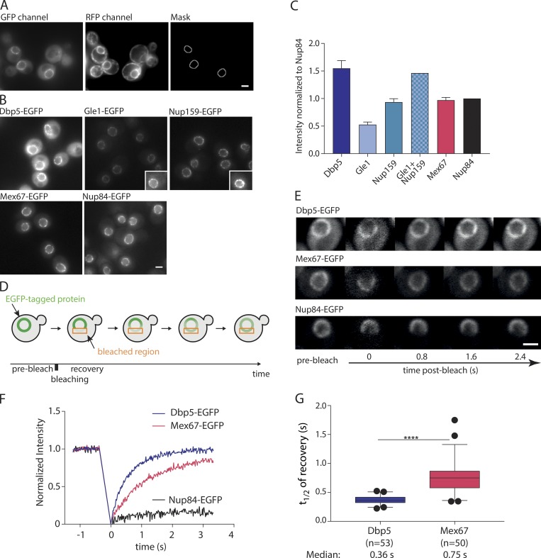
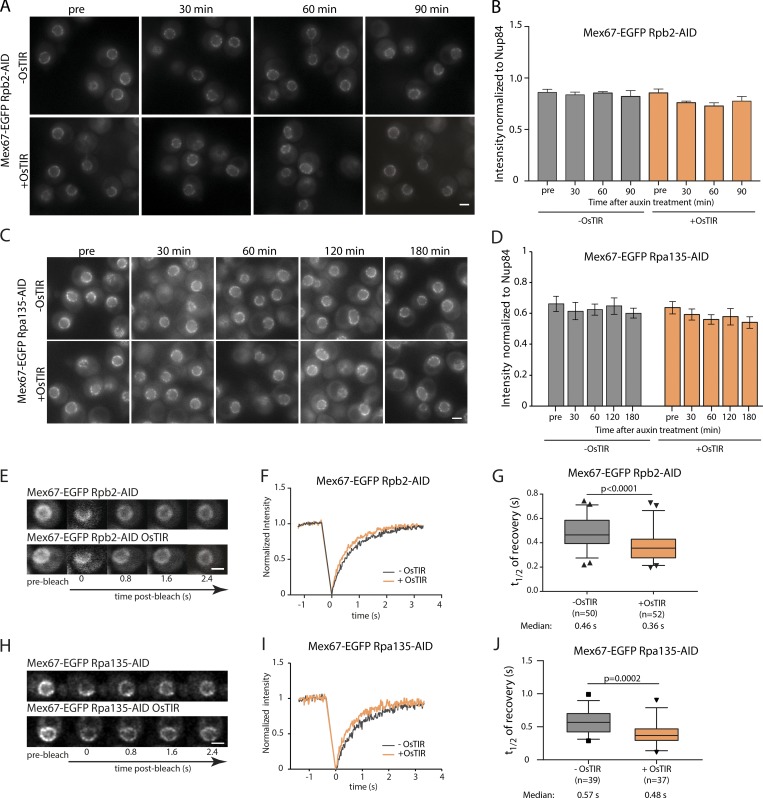
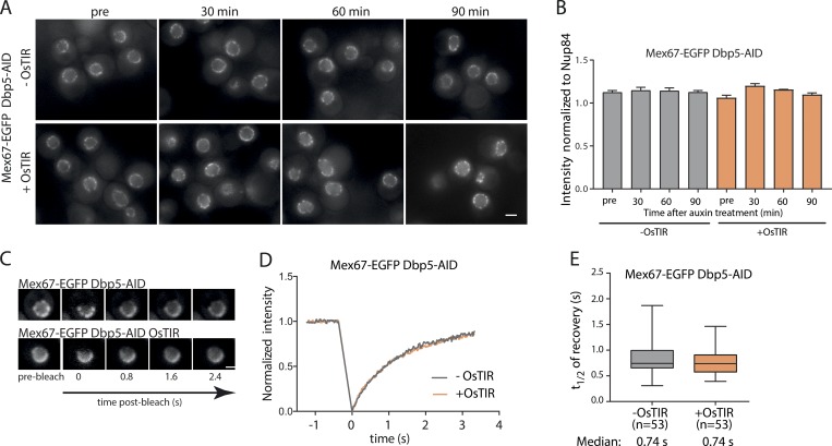
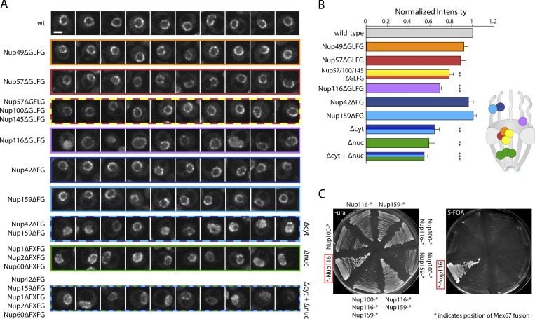
The RNA export factor Mex67 is essential for the transport of mRNA through the nuclear pore complex (NPC) in yeast, but the molecular mechanism of this export process remains poorly understood. Here, we use quantitative fluorescence microscopy techniques in live budding yeast cells to investigate how Mex67 facilitates mRNA export. We show that Mex67 exhibits little interaction with mRNA in the nucleus and localizes to the NPC independently of mRNA, occupying a set of binding sites offered by FG repeats in the NPC. The ATPase Dbp5, which is thought to remove Mex67 from transcripts, does not affect the interaction of Mex67 with the NPC. Strikingly, we find that the essential function of Mex67 is spatially restricted to the NPC since a fusion of Mex67 to the nucleoporin Nup116 rescues a deletion of MEX67 Thus, Mex67 functions as a mobile NPC component, which receives mRNA export substrates in the central channel of the NPC to facilitate their translocation to the cytoplasm.


Figures

Figure 1.
Figure 1.
Diffusion of mRNA export factors measured by FCS. (A) Representative fluorescence wide-field microscopy images of yeast strains expressing GFP-tagged mRNA export factors. Scale bar, 2 µm. (B and C) Diffusion times measured by FCS for the indicated GFP-fusion proteins. Data are pooled from three independent biological replicates. In the boxplots, the line represents the median, the box represents the 25th to 75th percentile, whiskers extend to the 5th to 95th percentile, and black symbols represent data points outside this range.
Figure 2.
Figure 2.
Mex67 and Dbp5 bind dynamically to the NPC. (A) Example for the production of the NuRIM analysis mask for quantification of GFP intensity on the nuclear envelope using DsRed-HDEL signal for nuclear envelope segmentation. (B) Representative fluorescence images of mRNA export factors and related Nups used for intensity analysis. Insets: Example cells with increased contrast settings for better visualization. (C) Quantification of nuclear envelope intensities by NuRIM. Bars show the means of three biological replicates, and the error bars represent the SEM. At least 1,000 cells were analyzed per condition and replicate. (D) Schematic illustration of FRAP at the nuclear envelope. (E–G) FRAP of Dbp5, Mex67, and Nup84 tagged with GFP. (E) Representative FRAP images. (F) FRAP curves normalized to prebleach and postbleach values. Mean curves of ≥50 cells are shown. (G) Half time of recovery retrieved from fitting individual FRAP curves with a single component model are plotted in boxplots. The line represents the median, the box represents the 25th to 75th percentile, and whiskers extend to the 5th to 95th percentile. ****, P < 0.001, Mann–Whitney test. Scale bars, 2 µm.
Figure 3.
Figure 3.
Mex67 dynamics, but not its amount at the NPC, is affected by the presence of cargo molecules. (A–D) NuRIM quantification of Mex67-EGFP intensity at the nuclear envelope upon depletion of RNA Pol II (A and B) or RNA Pol I (C and D) via auxin induced degradation. (A and C) Representative wide-field fluorescence microscopy images at different times after auxin treatment. (B and D) Intensity of Mex67-EGFP at the nuclear envelope. Mean of three biological replicates is shown. Error bars represent SEM. At least 300 cells were analyzed per condition and replicate. (E–J) FRAP analysis of Mex67-EGFP dynamics at the nuclear envelope upon depletion of RNA Pol II or RNA Pol I via auxin induced degradation. (E and H) Representative images of FRAP experiments. (F and I) FRAP curves normalized to prebleach and postbleach values. Mean curves of ≥50 cells are shown. (G and J) Half time of recovery retrieved from fitting FRAP. The line represents the median, the box represents the 25th to 75th percentile, whiskers extend to the 5th to 95th percentile, and black dots represent data points outside this range. Data in F and G are pooled from three biological replicates, and data in I and J from two. P values are from a Mann–Whitney test. Scale bars, 2 µm.
Figure 4.
Figure 4.
Mex67 binding to the NPC is independent of Dbp5. (A and B) NuRIM quantification of Mex67-EGFP intensity at the nuclear envelope upon depletion of Dbp5 via auxin induced degradation. (A) Representative wide field fluorescence microscopy images at different times after auxin treatment. (B) Intensity of Mex67-EGFP at the nuclear envelope. Mean of three biological replicates is shown. Error bars represent SEM. At least 300 cells were analyzed per condition and replicate. (C) FRAP analysis of Mex67-EGFP dynamics at the nuclear envelope upon depletion of Dbp5 via auxin-induced degradation. (C) Representative images of FRAP experiments. (D) FRAP curves normalized to prebleach and postbleach values. Mean curves of ≥50 cells are shown. (E) Half time of recovery retrieved from fitting individual FRAP curves. The line represents the median, the box represents the 25th to 75th percentile, and the whiskers extend to the 5th to 95th percentile. Data in D and E are pooled from three biological replicates. Scale bars, 2 µm.
Figure 5.
Figure 5.
Mex67 relies on multiple FG repeats for binding the NPC and fulfills its essential function only at the NPC. (A) Example nuclei are shown for different strains expressing Nups that have deleted FG/GFLG/FxFG regions as indicated. Images are single slices of z-stacks acquired on a spinning disk microscope. Slices were selected for being equatorial planes using an ER marker channel. Scale bar, 2 µm. (B) Intensity of Mex67 at the nuclear envelope using NuRIM in different strains with deletions of various FG repeats. Shown is the mean of three biological replicates normalized to wild-type cells. Error bars represent the SEM. Stars indicate significant P values of a paired t test compared with wild type (**, P < 0.01; ***, P < 0.001). Schematic indicates approximate positions of the NPC components in the NPC structure. Note that this applies to the body of the protein and not necessarily to its FG repeat region. (C) N-terminal fusion of Mex67 to Nup116 rescues deletion of MEX67 locus. Strains in which a deletion of the MEX67 locus was covered with Mex67 expressed from a plasmid containing the URA3 selection marker gene were engineered to express fusions of Mex67 to different nucleoporins. Streaks of these strains on 5-FOA–containing plate indicate rescue only by the N-terminal Nup116 fusion. Asterisk indicates position of Mex67 fusion.

References

    1. Adams R.L., Mason A.C., Glass L., Aditi, and Wente S.R.. 2017. Nup42 and IP6 coordinate Gle1 stimulation of Dbp5/DDX19B for mRNA export in yeast and human cells. Traffic. 18:776–790. 10.1111/tra.12526 - DOI - PMC - PubMed
    1. Bancaud A., Huet S., Rabut G., and Ellenberg J.. 2010. Fluorescence perturbation techniques to study mobility and molecular dynamics of proteins in live cells: FRAP, photoactivation, photoconversion, and FLIP. Cold Spring Harb. Protoc. 10.1101/pdb.top90 - DOI - PubMed
    1. Baudin A., Ozier-Kalogeropoulos O., Denouel A., Lacroute F., and Cullin C.. 1993. A simple and efficient method for direct gene deletion in Saccharomyces cerevisiae. Nucleic Acids Res. 21:3329–3330. 10.1093/nar/21.14.3329 - DOI - PMC - PubMed
    1. Ben-Yishay R., Mor A., Shraga A., Ashkenazy-Titelman A., Kinor N., Schwed-Gross A., Jacob A., Kozer N., Kumar P., Garini Y., and Shav-Tal Y.. 2019. Imaging within single NPCs reveals NXF1's role in mRNA export on the cytoplasmic side of the pore. J. Cell Biol. 218:2962–2981. 10.1083/jcb.201901127 - DOI - PMC - PubMed
    1. Cole C.N., and Scarcelli J.J.. 2006a Transport of messenger RNA from the nucleus to the cytoplasm. Curr. Opin. Cell Biol. 18:299–306. 10.1016/j.ceb.2006.04.006 - DOI - PubMed

Publication types

MeSH terms