. 2019 Dec 27;294(52):20097-20108.
doi: 10.1074/jbc.RA119.011506. Epub 2019 Nov 21.

High-resolution genome-wide expression analysis of single myofibers using SMART-Seq

Affiliations

High-resolution genome-wide expression analysis of single myofibers using SMART-Seq

Darren M Blackburn et al. J Biol Chem. .

Abstract

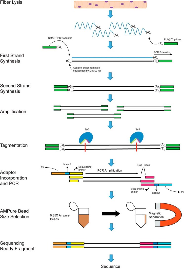
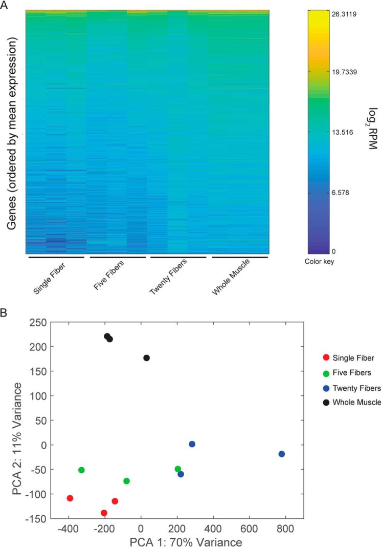
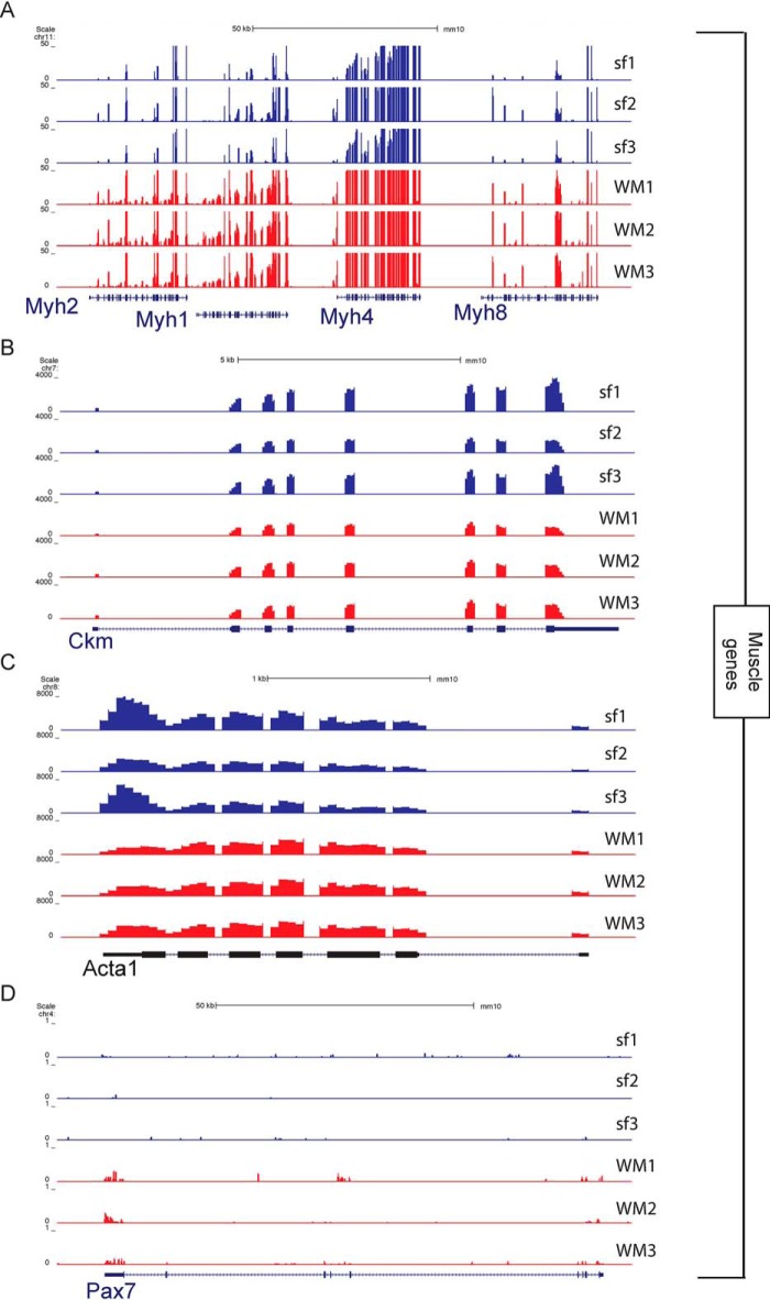
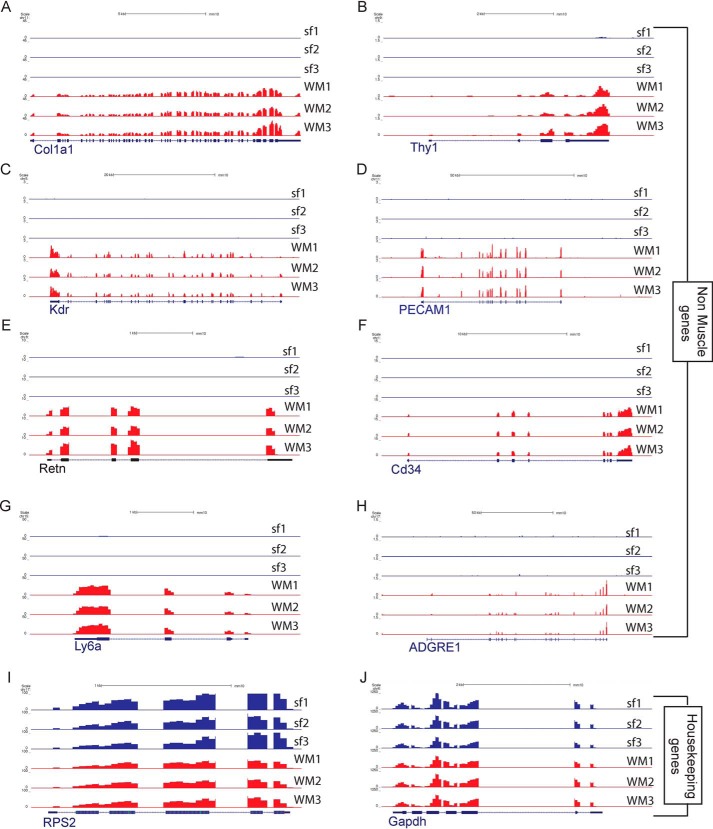
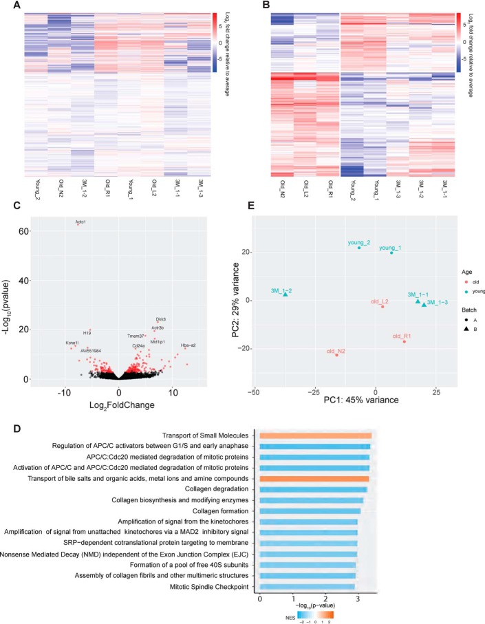
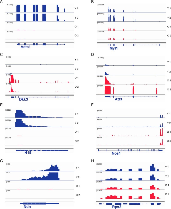
Skeletal muscle is a heterogeneous tissue. Individual myofibers that make up muscle tissue exhibit variation in their metabolic and contractile properties. Although biochemical and histological assays are available to study myofiber heterogeneity, efficient methods to analyze the whole transcriptome of individual myofibers are lacking. Here, we report on a single-myofiber RNA-sequencing (smfRNA-Seq) approach to analyze the whole transcriptome of individual myofibers by combining single-fiber isolation with Switching Mechanism at 5' end of RNA Template (SMART) technology. Using smfRNA-Seq, we first determined the genes that are expressed in the whole muscle, including in nonmyogenic cells. We also analyzed the differences in the transcriptome of myofibers from young and old mice to validate the effectiveness of this new method. Our results suggest that aging leads to significant changes in the expression of metabolic genes, such as Nos1, and structural genes, such as Myl1, in myofibers. We conclude that smfRNA-Seq is a powerful tool to study developmental, disease-related, and age-related changes in the gene expression profile of skeletal muscle.

Keywords: RNA; SMART-Seq; gene expression; molecular biology; molecular cell biology; muscle.


Conflict of interest statement

The authors declare that they have no conflicts of interest with the contents of this article

Figures

Figure 1.
Figure 1.
SMART technology and incorporation of Illumina adaptors to the fiber mRNA. Shown is a schematic displaying the steps and biochemical reactions involved in the generation of sequence ready cDNA fragments. M-MLV, Moloney murine leukemia virus; RT, reverse transcription.
Figure 2.
Figure 2.
Comparative analysis of whole transcriptome from single myofibers and whole muscle. A, heat map of gene expression in single fibers, groups of five fibers, groups of twenty fibers, and whole muscle, each with three replicates. Colors represent mean gene expression within each sample, from highest expression (yellow) to lowest expression (dark blue). Genes are ordered from top to bottom by their average expression across all samples. B, projection of samples along the first two principal components found by PCA applied to log reads-per-million gene expression.
Figure 3.
Figure 3.
University of California, Santa Cruz snapshots showing expression of myogenic genes in single myofibers and whole muscle. A, part of the myosin heavy chain (Myh) gene cluster located on chromosome 11. B, muscle creatine kinase (Ckm). C, actin α1 (Acta1) gene. D, paired box 7 (Pax7) gene expressed in the associated satellite cells.
Figure 4.
Figure 4.
University of California, Santa Cruz snapshots showing expression of nonmyogenic genes between single myofibers and whole muscle. A, collagen type 1 α1 chain (Col1a1) gene expressed in fibroblasts. B, CD90 (Thy1) gene expressed in fibroblasts. C, kinase insert domain receptor gene (Kdr) as a marker for endothelial cells. D, CD31 gene (Pecam1) as a marker for endothelial cells. E, resistin (Retn) as a marker for adipocytes. F, Cd34 as a marker for hematopoietic cells. G, Ly6a to detect the presence of FAPs. H, adhesion G protein–coupled receptor E1 (Adgre1) gene for macrophages. I, housekeeping gene Rps2. J, housekeeping gene Gapdh.
Figure 5.
Figure 5.
Comparative analysis of whole transcriptome between single myofibers from young and old mice. A, heat map of all genes expressed in old and young myofibers. B, heat map of differentially expressed genes between the old and young myofibers. Colors indicate Log2 fold change relative to average per gene, with red indicating a higher expression and blue as a lower expression between young (1 and 3 months) and old (19 months) myofibers. C, volcano plot of the differentially expressed genes between young and old myofibers. Points in red indicate there is a significant difference between the two groups. D, GO term analysis of the top 15 differentially regulated pathways. E, projection of samples along first two principal components found by PCA applied to log reads-per-million gene expression of young (1 and 3 months) and old (19 months) myofibers.
Figure 6.
Figure 6.
IGV snapshots showing the expression of selected differentially expressed genes between young and old myofibers. Young myofiber tracks are labeled in blue, and old are in red. A, actin α cardiac muscle 1 (Actc1). B, myosin light chain 1 (Myl1). C, dickkopf WNT signaling pathway inhibitor 3 (Dkk3). D, activating transcription factor 3 (Atf3). E, H19, imprinted maternally expressed transcript (H19). F, nitric-oxide synthase 1 (Nos1). G, necdin (Ndn). H, housekeeping gene ribosomal protein S2 (Rps2). IGV, Integrative Genomics Viewer.

References

    1. Giordani L., He G. J., Negroni E., Sakai H., Law J. Y. C., Siu M. M., Wan R., Corneau A., Tajbakhsh S., Cheung T. H., and Le Grand F. (2019) High-dimensional single-cell cartography reveals novel skeletal muscle-resident cell populations. Mol. Cell 74, 609–621.e6 10.1016/j.molcel.2019.02.026 - DOI - PubMed
    1. Joe A. W., Yi L., Natarajan A., Le Grand F., So L., Wang J., Rudnicki M. A., and Rossi F. M. (2010) Muscle injury activates resident fibro/adipogenic progenitors that facilitate myogenesis. Nat. Cell Biol. 12, 153–163 10.1038/ncb2015 - DOI - PMC - PubMed
    1. Arnold L., Henry A., Poron F., Baba-Amer Y., van Rooijen N., Plonquet A., Gherardi R. K., and Chazaud B. (2007) Inflammatory monocytes recruited after skeletal muscle injury switch into antiinflammatory macrophages to support myogenesis. J. Exp. Med. 204, 1057–1069 10.1084/jem.20070075 - DOI - PMC - PubMed
    1. Christov C., Chrétien F., Abou-Khalil R., Bassez G., Vallet G., Authier F. J., Bassaglia Y., Shinin V., Tajbakhsh S., Chazaud B., and Gherardi R. K. (2007) Muscle satellite cells and endothelial cells: close neighbors and privileged partners. Mol. Biol. Cell 18, 1397–1409 10.1091/mbc.e06-08-0693 - DOI - PMC - PubMed
    1. Pette D., and Staron R. S. (1997) Mammalian skeletal muscle fiber type transitions. Int. Rev. Cytol. 170, 143–223 10.1016/S0074-7696(08)61622-8 - DOI - PubMed

Publication types