. 2019 Nov 22;366(6468):1024-1028.
doi: 10.1126/science.aax8780.

Computational design of a modular protein sense-response system

Affiliations

Computational design of a modular protein sense-response system

Anum A Glasgow et al. Science. .

Abstract

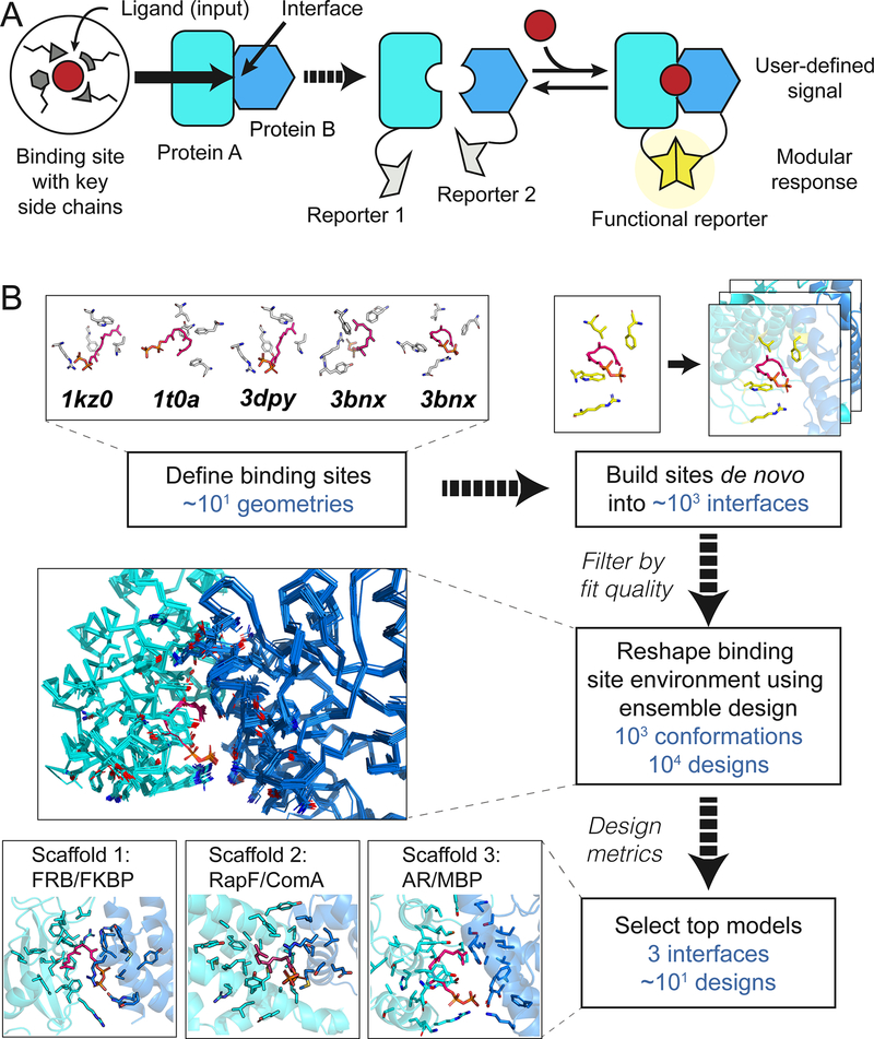
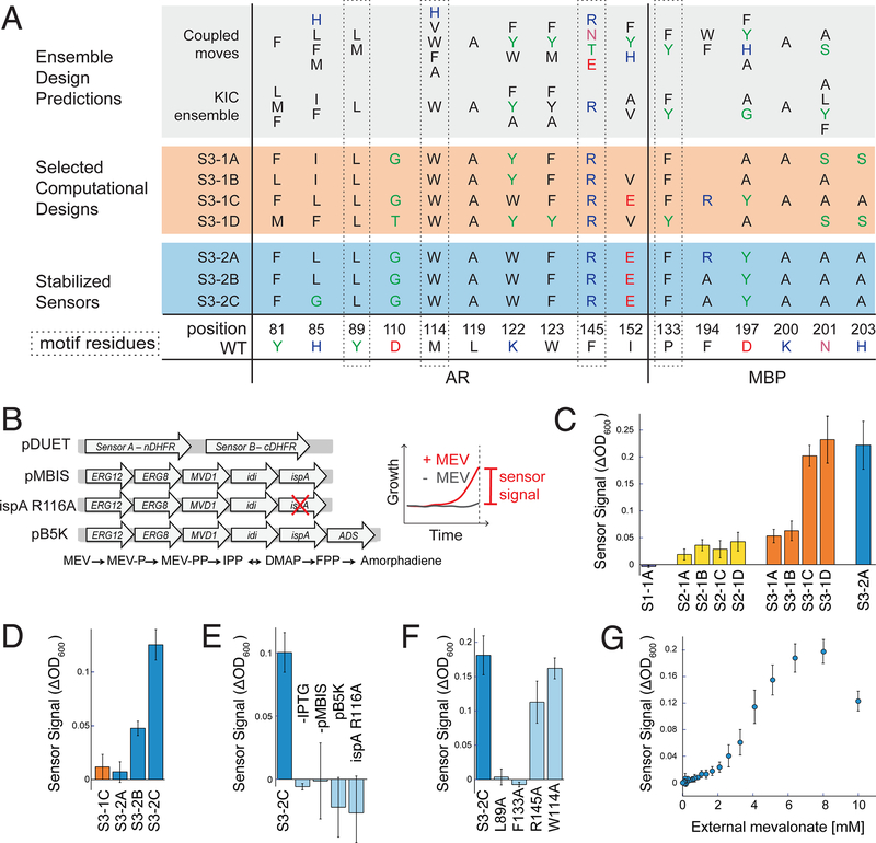
Sensing and responding to signals is a fundamental ability of living systems, but despite substantial progress in the computational design of new protein structures, there is no general approach for engineering arbitrary new protein sensors. Here, we describe a generalizable computational strategy for designing sensor-actuator proteins by building binding sites de novo into heterodimeric protein-protein interfaces and coupling ligand sensing to modular actuation through split reporters. Using this approach, we designed protein sensors that respond to farnesyl pyrophosphate, a metabolic intermediate in the production of valuable compounds. The sensors are functional in vitro and in cells, and the crystal structure of the engineered binding site closely matches the design model. Our computational design strategy opens broad avenues to link biological outputs to new signals.


Conflict of interest statement

Competing interests: The authors declare no competing interests.

Figures

Fig. 1.
Fig. 1.. Computational design.
(A) Cartoon of the design strategy. A small-molecule binding site is built de novo into protein-protein interfaces (left) to create synthetic chemically inducible dimerization systems (CIDs, right). Linking the designed sensor proteins to split reporters yields modular CID systems, in which different reporter outputs can be coupled to user-defined small molecule input signals. (B) Steps in the design of a synthetic CID system sensing FPP. Top: Binding site geometries with key interacting side chains selected from FPP-binding proteins (pdb codes indicated) are computationally modeled into a large number of protein-protein interfaces. Middle: Binding sites with feasible geometries are reshaped and optimized by flexible backbone design; shown is a conformational ensemble for a single sequence. Bottom: Top designs from 3 different scaffolds (bottom) selected for experimental tests (Fig. 2).
Fig. 2.
Fig. 2.. Sensor function in bacteria.
(A) Designed sequences at key positions for scaffold 3. Grey shading: preferred residues from flexible backbone reshaping by kinematic closure (KIC(22, 24)) or coupled moves(25); orange shading: individual computational designs selected based on ligand burial (S3–1A), consensus (S3–1B), optimized ligand packing (S3–1C) and predicted ligand binding score (S3–1D); blue shading: sensors stabilized by 2 additional mutations from SSM (S3–2B and S3–2C also contained 2 mutations from epPCR that were not in the designed FPP binding site, Fig. S1). (B) Constructs (left, details in Appendix 1) used in the split mDHFR reporter assay (right). pDUET, sensor proteins linked to the split mDHFR reporter; pMBIS, engineered pathway of 5 enzymes to convert mevalonate (MEV) into FPP(20); ispA R116A, pMBIS containing R116A mutation in ispA that reduces catalytic activity 13-fold(27); pB5K, pMBIS with amorphadiene synthase (ADS)(20). Sensor signal is quantified as change in OD600 in the presence and absence of mevalonate. (C) Sensor signal in the split mDHFR assay for computational designs based on scaffold 1 (FKBP-FRB12, purple bar), scaffold 2 (RapF- ComA, yellow bars) and scaffold 3 (AR-MBP, orange bars). Sensor S3–2A (identified from library 2 with 2 mutations distal from the designed FPP binding site (Table S1)), is shown for comparison (blue bar). (D) Improvement of sensor signal by stability-enhancing mutations in S3– 2B and S3–2C at increased stringency (trimethoprim concentration 6 μM versus 1 μM in panel C). (E) Dependence of S3–2C sensor signal on sensor expression (-IPTG) and FFP production (- pMBIS, pB5K, ispA R116A). (F) Dependence of S3–2C sensor signal on motif residues. (G) Dependence of the S3–2C sensor signal on concentration of the FPP precursor mevalonate added extracellularly. Error bars are standard deviation from at least 4 biological replicates and 8 technical replicates for each biological replicate.
Fig. 3.
Fig. 3.. In vitro sensor characterization and output modularity.
(A) Sequence changes in sensor constructs tested in vitro. Motif residues are also shown. The starting construct, S3–2D (blue), is identical to S3–2C in the engineered FPP binding site but contains additional previously published stabilizing mutations in AR(31) (shown in Table S1). (B-H) In vitro binding measurements from biolayer interferometry (BLI) using purified protein (panels B-E) or FPP titrations with sensors expressed using in vitro transcription / translation (TxTl) (panels F-H). (B) Apparent AR interaction with immobilized MBP in the presence (closed circles) or absence (open squares) of 200 μM FPP, comparing designs S3–2D (blue) and S3–3A containing the Y197A mutation (orange). (C) Summary of BLI results for apparent AR-MBP dimerization with and without FPP. (D) Summary of BLI results for FPP binding to the individual designed AR and MBP proteins comprising design S3–2D (Table S1). (E) Apparent AR interaction with immobilized MBP for a computationally designed variant using the S3–2D crystal structure as the input, with (purple, S3–3C) or without (red, S3–3B) the Y197A mutation. (F) Apparent affinity of the S3–2D and S3–3A sensors for FPP using luminescent or fluorescent reporters in TxTl experiments. (G, H) FPP titrations in TxTl using the luminescent reporter (G) or the fluorescent reporter (H). Error bars are standard deviations for n ≥ 3.
Fig. 4.
Fig. 4.. The S3–2D crystal structure closely matches the computational design model.
(A) Overlay of the design model (grey) with the crystal structure (designed AR: cyan, designed MBP: blue, FPP: pink) showing FPP binding in the computationally designed binding site at the AR-MBP interface (circle). The design crystallized in the closed MBP conformation while MBP was in the open conformation in the original scaffold on which the model was based, leading to a difference in rigid-body orientation (arrow) of one lobe of MBP distal to the FPP binding site. (B) FPP overlaid with 2mFo-DFc electron density map (1.2σ, cyan) and ligand 2mFo-DFc omit map (1.0σ, dark blue). Strong density peaks were present in both maps for the phosphates and several anchoring hydrophobic groups. (C) Open-book representation of the FPP binding site on AR, showing close match of designed side chain conformations to the crystal structure. (D) Open-book representation of the FPP binding site on MBP, indicating a clash between the position of MBP Y197 in the crystal structure (blue) and the designed FPP orientation in the model (grey), causing slight rearrangements of FPP and F133 (arrows). (E) Alignment of the holo (cyan) and apo (yellow) structures of S3–2D, showing overall agreement with the exception of the side chain of W114 (arrows). In panels (C-E), residues are labeled black when designed and green/blue when present in the original scaffold complex.

Comment in

  • Designer sense-response systems.
    Chica RA. Chica RA. Science. 2019 Nov 22;366(6468):952-953. doi: 10.1126/science.aaz8085. Science. 2019. PMID: 31753985 No abstract available.

References

    1. Huang PS et al., High thermodynamic stability of parametrically designed helical bundles. Science 346, 481–485 (2014). - PMC - PubMed
    1. Jacobs TM et al., Design of structurally distinct proteins using strategies inspired by evolution. Science 352, 687–690 (2016). - PMC - PubMed
    1. Thomson AR et al., Computational design of water-soluble alpha-helical barrels. Science 346, 485–488 (2014). - PubMed
    1. Hill RB, Raleigh DP, Lombardi A, DeGrado WF, De novo design of helical bundles as models for understanding protein folding and function. Acc Chem Res 33, 745–754 (2000). - PMC - PubMed
    1. Harbury PB, Plecs JJ, Tidor B, Alber T, Kim PS, High-resolution protein design with backbone freedom. Science 282, 1462–1467 (1998). - PubMed

Publication types