. 2019 Dec 17;116(51):25677-25687.
doi: 10.1073/pnas.1910733116. Epub 2019 Nov 21.

Mammalian germ cells are determined after PGC colonization of the nascent gonad

Affiliations

Mammalian germ cells are determined after PGC colonization of the nascent gonad

Peter K Nicholls et al. Proc Natl Acad Sci U S A. .

Abstract

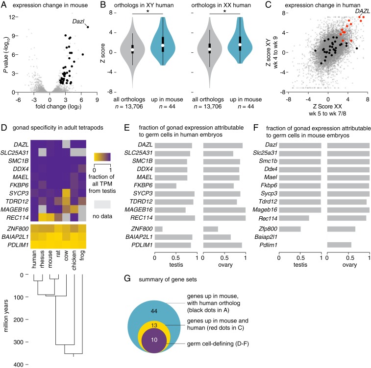
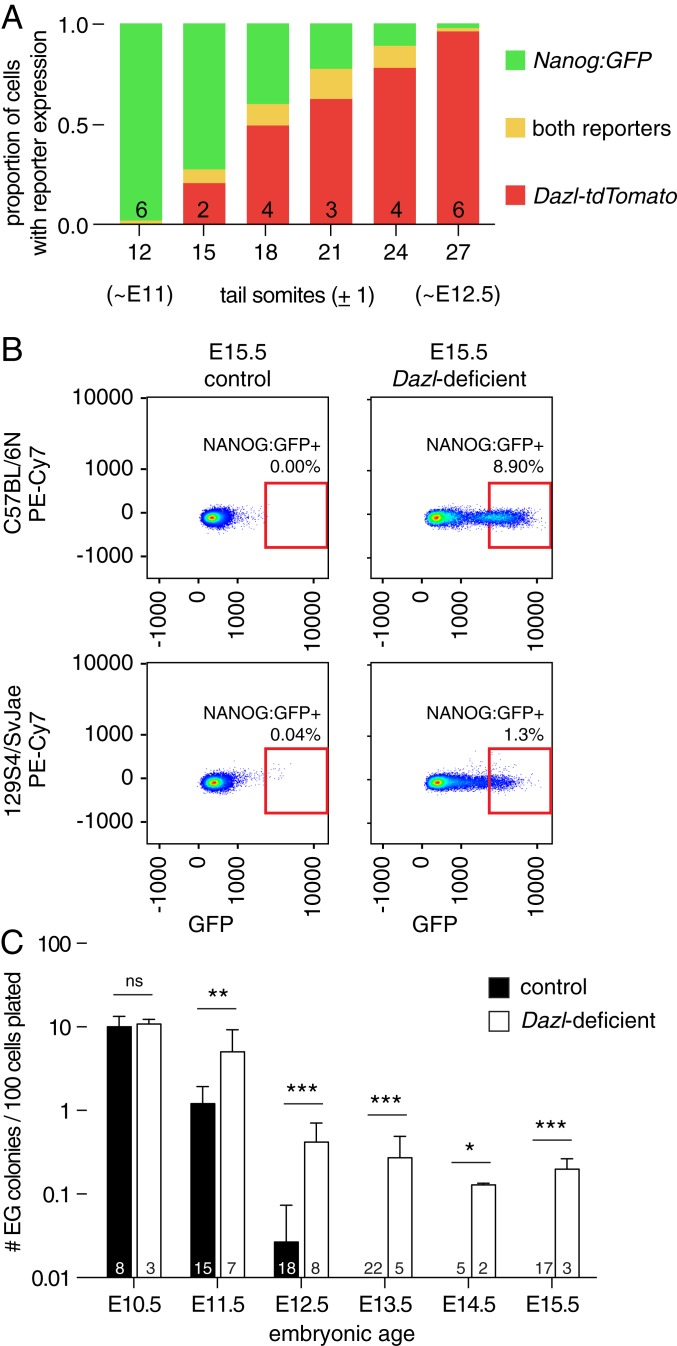
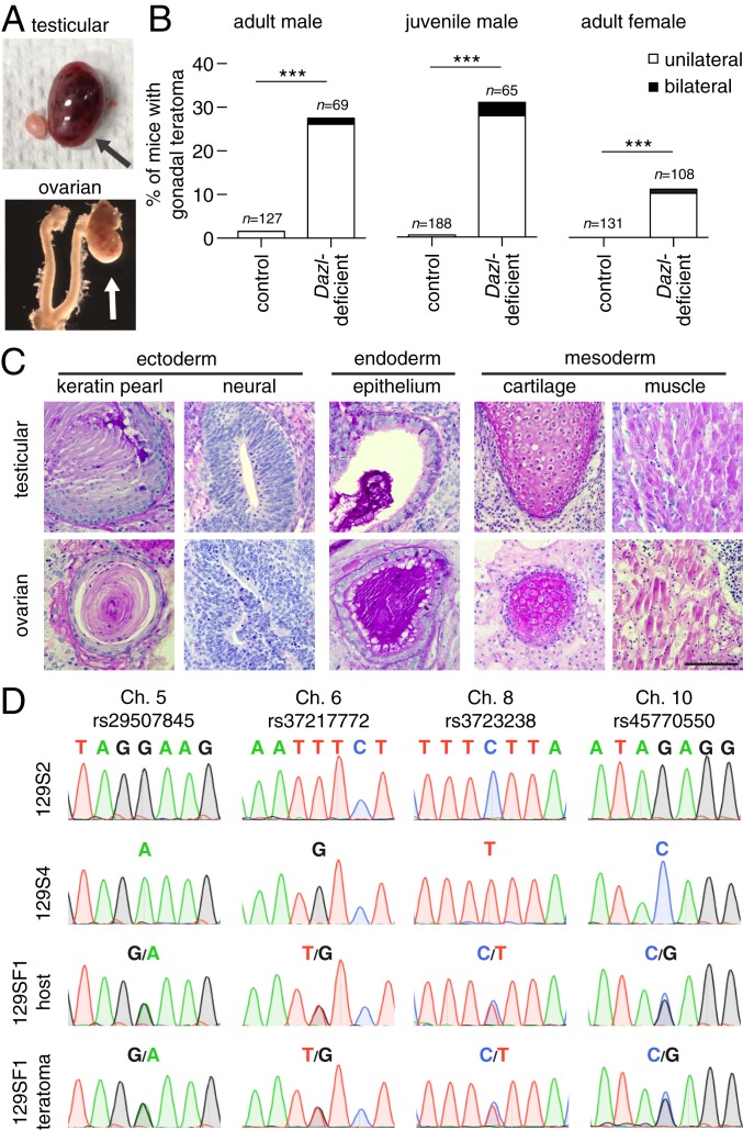
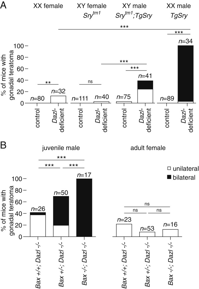
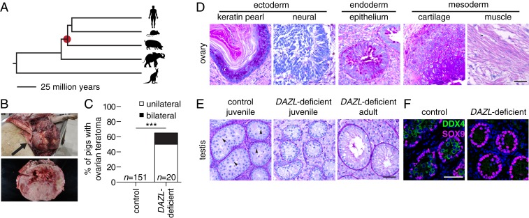
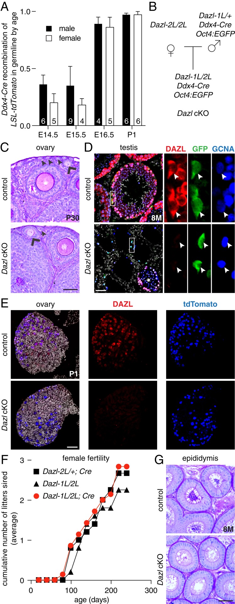
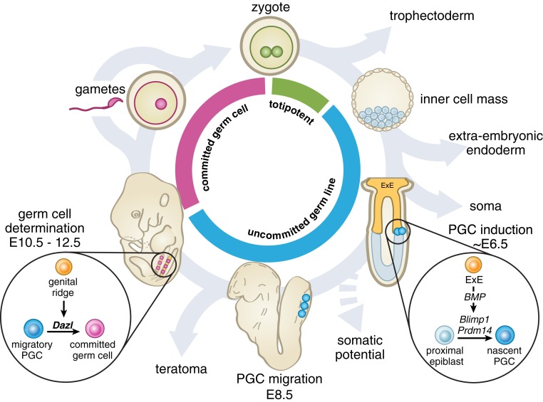
Mammalian primordial germ cells (PGCs) are induced in the embryonic epiblast, before migrating to the nascent gonads. In fish, frogs, and birds, the germline segregates even earlier, through the action of maternally inherited germ plasm. Across vertebrates, migrating PGCs retain a broad developmental potential, regardless of whether they were induced or maternally segregated. In mammals, this potential is indicated by expression of pluripotency factors, and the ability to generate teratomas and pluripotent cell lines. How the germline loses this developmental potential remains unknown. Our genome-wide analyses of embryonic human and mouse germlines reveal a conserved transcriptional program, initiated in PGCs after gonadal colonization, that differentiates germ cells from their germline precursors and from somatic lineages. Through genetic studies in mice and pigs, we demonstrate that one such gonad-induced factor, the RNA-binding protein DAZL, is necessary in vivo to restrict the developmental potential of the germline; DAZL's absence prolongs expression of a Nanog pluripotency reporter, facilitates derivation of pluripotent cell lines, and causes spontaneous gonadal teratomas. Based on these observations in humans, mice, and pigs, we propose that germ cells are determined after gonadal colonization in mammals. We suggest that germ cell determination was induced late in embryogenesis-after organogenesis has begun-in the common ancestor of all vertebrates, as in modern mammals, where this transition is induced by somatic cells of the gonad. We suggest that failure of this process of germ cell determination likely accounts for the origin of human testis cancer.

Keywords: Dazl; commitment; germ cell; pluripotency; teratoma.


Conflict of interest statement

Competing interest statement: A.L.W., D.F.C., and S.C.F. are employees and shareholders of Recombinetics Inc. I.D. is a member of the scientific advisory board of Recombinetics Inc. The remaining authors have declared that no competing interests exist.

Figures

Fig. 1.
Fig. 1.
A conserved program of germ cell transcription is induced upon PGC colonization of nascent gonads in mice and humans. (A) Gene expression changes in mouse germline between E9.5 and E11.5, as measured by RNA-seq. Black dots denote 44 genes that are up-regulated and have single human orthologs (fold change > 4, FDR value < 0.05); gray dots denote all other expressed genes (n = 11,282). (B and C) Gene expression changes in XY and XX human embryonic germlines between weeks 4 and 9, as measured by single cell RNA-seq. (B) Violin plots; as a set, genes induced in mouse germline (from A, n = 44) show greater expression increases in XY (Left) and XX (Right) human germline after PGCs colonize the gonads than do the set of all expressed orthologs (n = 13,706; *P value < 0.0007 by Wilcoxon rank sum test; black bar, interquartile range; circle, median value). (C) Scatter plot; black and red dots denote genes robustly up-regulated in mice, and possessing a single human ortholog (from A, n = 44); red dot genes are also significantly up-regulated in both XY and XX human germlines (n = 13). Gray dots denote all other expressed genes. (D) Heatmap; summary of gonad specificity of commonly up-regulated genes (red dots in C, n = 13), by RNA-seq, in 9 adult tissues from 7 tetrapods. Specificity fraction is determined by dividing testis expression (in TPM, transcripts per million) by sum of expression in all analyzed adult tissues, for each species. Genes with no annotated ortholog are shown in gray. (E and F) Germ cell expression of commonly up-regulated factors (red dots in C, n = 13) in (E) human embryonic testis and ovary and (F) mouse E14.5 testis and ovary by RNA-seq (SI Appendix, SI Materials and Methods). Ratio of 1 indicates germ cell-specific expression; 0 indicates somatic cell expression. (G) Euler diagram of gene sets identified through analyses in AF.
Fig. 2.
Fig. 2.
Dazl is required for restricting developmental potential of the germline in diverse strains of mice. (A) Developmental time course of germline using Dazl-tdTomato and Nanog:GFP reporters, detected by flow cytometry. At ∼12 ts (∼E11), most cells expressed only the Nanog:GFP reporter (green). As development proceeds, the proportions of cells expressing both fluorescent reporters (orange), or only the Dazl-tdTomato reporter (red), change. Numbers of embryos tested are listed in each column, and the fraction of cells expressing each reporter is shown as an average. (B) Flow cytometry for Nanog:GFP-positive cells of E15.5 control and Dazl-deficient ovaries of indicated strains. Autofluorescence in PE-Cy7 channel is shown on y axis. Red box indicates area in which Nanog:GFP-positive cells were counted. (C) Derivation of EG cell lines from control and B6.Dazl-deficient embryos. Cells were collected by fluorescence-activated cell sorting (FACS) at embryonic age indicated on x axis, and cultured under defined conditions. After 10 d, EG cell colonies were counted, and rate of EG cell derivation (per 100 EGFP-positive cells plated) was calculated. Number of embryos tested is listed in each column, mean + SD, *P value < 0.05, ** <0.01, *** <0.001, ns = not significant, using t test or Fisher’s exact test as appropriate.
Fig. 3.
Fig. 3.
Spontaneous gonadal teratomas occur in Dazl-deficient mice, and arise from mitotic cells. (A) Appearance of unilateral gonadal teratomas (arrows) in 129S4.Dazl-deficient mice. (B) Incidence of gonadal teratomas in mice. Males were dissected by 4 mo (adult male) or at 4 wk of age (juvenile male), and adult females were dissected at 2 mo of age; n = number of animals examined, ***P value ≤ 0.0001 using Fisher’s exact test. (C) Representative histology of teratomas from testis (Upper) and ovary (Lower) stained with periodic acid-Schiff reagent (PAS). (Scale bar, 100 µm.) (D) Representative Sanger sequencing at 4 SNP loci from 129S2 and 129S4 mice (Upper), and from 129SF1 host and teratoma (Lower). Teratomas are heterozygous at each SNP locus.
Fig. 4.
Fig. 4.
Teratoma formation in Dazl-deficient mice is affected by sex reversal, and by ablation of Bax-mediated cell death. (A) Incidence of gonadal teratomas in sex-reversed mice. Mutation of Sry (Srytm1) causes XY embryos to develop as anatomic females. Expression of Sry transgene (TgSry) causes XX embryos to develop as anatomic males. (B) Incidence of testicular teratomas (Left) and ovarian teratomas (Right) in Dazl-deficient mice (−/−) who were either homozygous wild-type (+/+), heterozygous (+/−), or deficient (−/−) for Bax; n = number of animals examined, **P value < 0.01, *** <0.0001, ns = not significant using Fisher’s exact test.
Fig. 5.
Fig. 5.
Spontaneous ovarian teratomas in DAZL-deficient pigs. (A) The pig is an outgroup to rodents and primates, sharing a common ancestor 95 million years ago (red dot). (B) Ovarian teratoma (arrow) in DAZL-deficient pig. The tumor measured 46 × 23 × 28 cm and weighed 15.4 kg. (C) Incidence of ovarian teratomas in control and DAZL-deficient pigs. ***P value < 0.0001 using Fisher’s exact test. (D) Representative histology of teratoma from ovary, stained with PAS. (E) Histology of control (Left, 18 wk, spermatocytes marked with arrow head) and DAZL-deficient testis at 11 wk (Center) and 9 mo of age (Right), stained with PAS. (F) Immunofluorescence of control (Left, 18 wk) and DAZL-deficient pig testis (Right, 11 wk). Germ cells were stained with DDX4 (green), and somatic cells were stained with SOX9 (magenta). DNA was stained with DAPI (blue). (Scale bars, 50 µm.)
Fig. 6.
Fig. 6.
A brief period of Dazl expression is sufficient for germ cell commitment, and for completion of oogenesis. (A) Time course of Ddx4-Cre (MvhCre-mOrange) recombinase activity in embryonic germline using a fluorescent Cre reporter mouse line, LSL-tdTomato. Cre-mediated recombination (resulting in tdTomato expression) in Oct4:EGFP-positive cells is assayed by flow cytometry. Numbers of embryos tested are listed in each column, mean + SD. (B) Breeding scheme for Dazl conditional knockout (Dazl cKO) mice. (C) Histology of control (Upper) and B6.Dazl cKO ovary (Lower) stained with PAS at 20 d of age, with primary (arrow head) and secondary (chevron) follicles marked. (D) Immunofluorescence of control (Upper) and B6.Dazl cKO testis (Lower) at 8 mo of age. Germ cells are immunostained by GCNA (blue), and undifferentiated spermatogonia are immunostained by GFP (green) expressed from the Oct4:EGFP reporter. Cre recombination is confirmed by immunostaining for DAZL (red). DNA is stained with DAPI (gray). Insets show each marker in Oct4:EGFP-positive spermatogonia (arrow head); * denotes tubule with incomplete Dazl recombination in cKO testis. (E) Immunofluorescence of control (Upper) and B6.Dazl cKO ovary (Lower) at postnatal day 1. Germ cells are immunostained by tdTomato protein (expressed following recombination of tdTomato-LSL allele by Ddx4-Cre; blue). Cre recombination is confirmed by immunostaining for DAZL (red). DNA is stained with DAPI (gray). (F) B6.Dazl cKO females remained fertile for at least 8 mo. (G) B6.Dazl cKO males were sterile, with no spermatozoa in epididymal ducts. All data are mean + SD. (Scale bars, 50 µm.)
Fig. 7.
Fig. 7.
A proposed model for germ cell commitment in mammals. The germline comprises all cells whose descendants include gametes. At fertilization, the totipotent zygote has the capacity to give rise to all cell lineages. As development proceeds, extraembryonic and somatic lineages differentiate away from the germline. PGCs (blue cells) are induced from the epiblast, preserving the germline, but also maintaining a broad developmental potential. At PGC colonization of the gonads, expression of a germ cell program—marked by DAZL—is induced in germline cells by the genital ridge. Our studies show that DAZL is necessary for the restriction of developmental potential in the germline, resulting in the determination of germ cells (pink cells). Determined germ cells then undertake gametogenesis and must cycle through fertilization to reestablish totipotency and continue the germline cycle in a new diploid individual. ExE, extraembryonic ectoderm.

Comment in

References

    1. Lawson K. A., et al. , Bmp4 is required for the generation of primordial germ cells in the mouse embryo. Genes Dev. 13, 424–436 (1999). - PMC - PubMed
    1. Ohinata Y., et al. , A signaling principle for the specification of the germ cell lineage in mice. Cell 137, 571–584 (2009). - PubMed
    1. Extavour C. G., Akam M., Mechanisms of germ cell specification across the metazoans: Epigenesis and preformation. Development 130, 5869–5884 (2003). - PubMed
    1. Strome S., Updike D., Specifying and protecting germ cell fate. Nat. Rev. Mol. Cell Biol. 16, 406–416 (2015). - PMC - PubMed
    1. Wylie C. C., Heasman J., Snape A., O’Driscoll M., Holwill S., Primordial germ cells of Xenopus laevis are not irreversibly determined early in development. Dev. Biol. 112, 66–72 (1985).

Publication types

MeSH terms

Supplementary concepts