Review
. 2019 Nov 21;132(22):jcs229286.
doi: 10.1242/jcs.229286.

Multiscale dynamics of tight junction remodeling

Affiliations
Review

Multiscale dynamics of tight junction remodeling

Saranyaraajan Varadarajan et al. J Cell Sci. .

Abstract

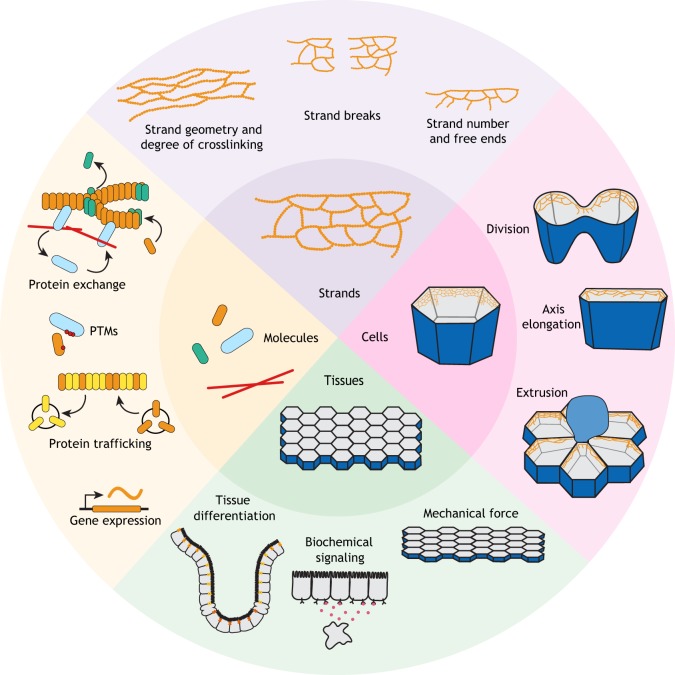
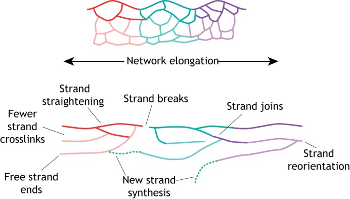
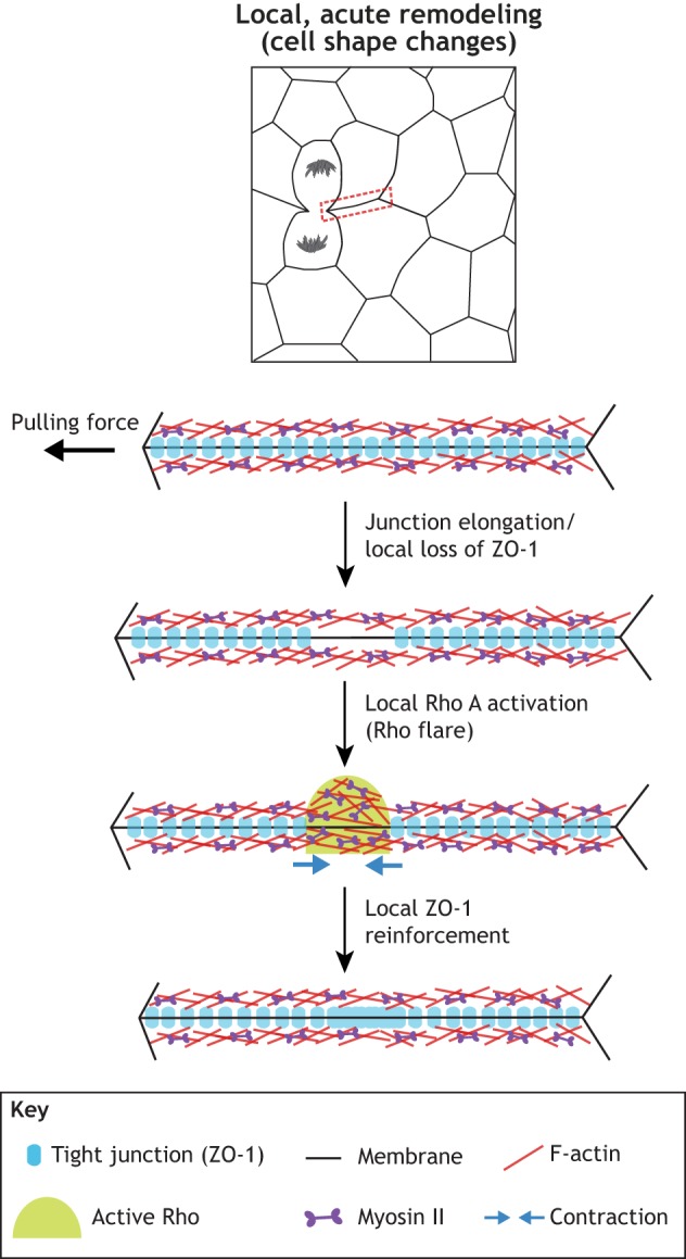
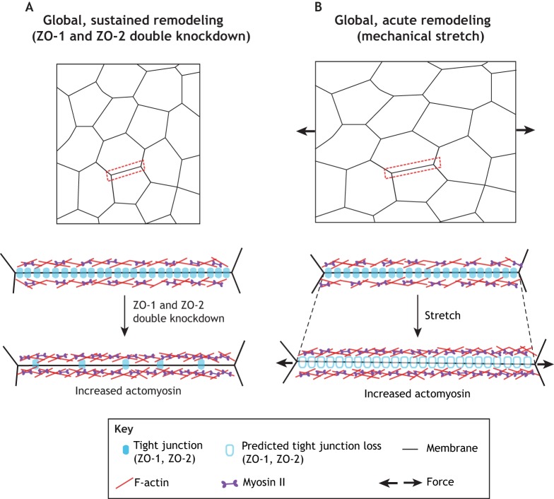
Epithelial cells form tissues that generate biological barriers in the body. Tight junctions (TJs) are responsible for maintaining a selectively permeable seal between epithelial cells, but little is known about how TJs dynamically remodel in response to physiological forces that challenge epithelial barrier function, such as cell shape changes (e.g. during cell division) or tissue stretching (e.g. during developmental morphogenesis). In this Review, we first introduce a framework to think about TJ remodeling across multiple scales: from molecular dynamics, to strand dynamics, to cell- and tissue-scale dynamics. We then relate knowledge gained from global perturbations of TJs to emerging information about local TJ remodeling events, where transient localized Rho activation and actomyosin-mediated contraction promote TJ remodeling to repair local leaks in barrier function. We conclude by identifying emerging areas in the field and propose ideas for future studies that address unanswered questions about the mechanisms that drive TJ remodeling.

Keywords: Actomyosin; Barrier function; Epithelia; Morphogenesis; Paracellular permeability; Tight junction.


Conflict of interest statement

Competing interestsThe authors declare no competing or financial interests.

Figures

Fig. 1.
Fig. 1.
TJs are dynamic at the molecular, strand, cellular and tissue scale. Molecular dynamics include the trafficking of proteins to the TJ, the formation of protein complexes and incorporation of claudins into strands. Strand dynamics include the assembly of strands into networks, dynamic reorganization of these networks at baseline and in response to stimuli, change in network geometry, and the incorporation and removal of claudins from strands. At the cellular scale, TJs undergo local alterations as cell–cell boundaries elongate, shrink or otherwise experience a change in shape, such as during cell division, extrusion or axis elongation. At the tissue scale, TJs change in response to tissue-scale forces (e.g. mechanical forces associated with development or organ function), biochemical signals (e.g. immune response), and differentiation. In turn, the state of the tissue influences the expression and trafficking of different TJ components, ultimately altering the molecular, strand and cellular dynamics of TJs.
Fig. 2.
Fig. 2.
Potential mechanisms to elongate the strand network. These include strand straightening and reduction of interstrand crosslinks through strand breakage, reorientation and joining. In addition, network elongation may involve new strand synthesis by incorporation of claudins at free strand ends or de novo strand assembly.
Fig. 3.
Fig. 3.
TJs are remodeled in response to local changes in tension. Elongation of a junction near a dividing cell (boxed area, magnification shown below) causes local loss of ZO-1 and a breach in barrier function (not shown). Activation of RhoA (green dome) at the site of ZO-1 loss promotes actomyosin-mediated contraction of junctions (blue arrows), which concentrates TJ proteins to repair the barrier leak. Modified with permission from Stephenson et al., 2019.
Fig. 4.
Fig. 4.
TJs are remodeled in response to tissue-scale changes in tension over longer and shorter periods of time. (A) Sustained global remodeling of TJs through genetic manipulation – specifically ZO-1 and ZO-2 double knockdown. In epithelial cells, double knockdown of ZO-1 and ZO-2 increases the accumulation of perijunctional actomyosin, and cells exhibit increased paracellular flux of large solutes (not illustrated). A magnification of the boxed area is shown below, showing this region before (top) and after (bottom) double knockdown of ZO-1 and ZO-2. (B) Acute global remodeling of TJs through mechanical stimuli. Mechanical stretching of epithelial tissue (indicated by black arrows) also increases accumulation of perijunctional actomyosin, and cells exhibit barrier leaks (not shown). A magnification of the boxed area is shown below, showing this region before (top) and after (bottom) stretch. See text for additional details.

References

    1. Acharya B. R., Nestor-Bergmann A., Liang X., Gupta S., Duszyc K., Gauquelin E., Gomez G. A., Budnar S., Marcq P., Jensen O. E. et al. (2018). A mechanosensitive RhoA pathway that protects epithelia against acute tensile stress. Dev. Cell 47, 439-452.e6. 10.1016/j.devcel.2018.09.016 - DOI - PubMed
    1. Anderson J. M. and Van Itallie C. M. (2009). Physiology and function of the tight junction. Cold Spring Harb. Perspect Biol. 1, a002584 10.1101/cshperspect.a002584 - DOI - PMC - PubMed
    1. Arnold T. R., Stephenson R. E. and Miller A. L. (2017). Rho GTPases and actomyosin: partners in regulating epithelial cell-cell junction structure and function. Exp. Cell Res. 358, 20-30. 10.1016/j.yexcr.2017.03.053 - DOI - PMC - PubMed
    1. Balda M. S., Whitney J. A., Flores C., Gonzalez S., Cereijido M. and Matter K. (1996). Functional dissociation of paracellular permeability and transepithelial electrical resistance and disruption of the apical-basolateral intramembrane diffusion barrier by expression of a mutant tight junction membrane protein. J. Cell Biol. 134, 1031-1049. 10.1083/jcb.134.4.1031 - DOI - PMC - PubMed
    1. Bartle E. I., Rao T. C., Urner T. M. and Mattheyses A. L. (2018). Bridging the gap: Super-resolution microscopy of epithelial cell junctions. Tissue Barriers 6, e1404189 10.1080/21688370.2017.1404189 - DOI - PMC - PubMed

Publication types

LinkOut - more resources