

Fulminant amoebic colitis in the era of computed tomography scan: A case report and review of the literature
- PMID: 31754504
- PMCID: PMC6837831
- DOI: 10.4102/sajr.v22i1.1354
Fulminant amoebic colitis in the era of computed tomography scan: A case report and review of the literature
Abstract
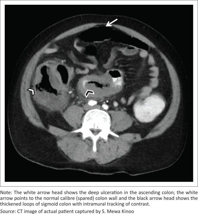
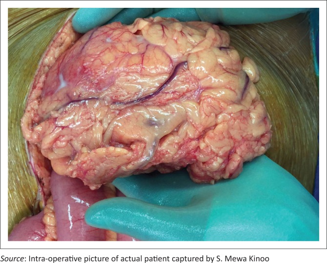
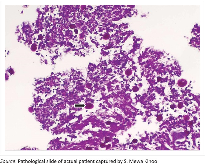
Amoebic colitis, caused by ingestion of water or food contaminated with the protozoan Entamoeba histolytica, can progress to a fulminant colitis. Computed tomography (CT) findings reported in the literature on this type of colitis are sparse. We present a 59-year-old male patient with a one-week history of progressive abdominal pain, abdominal distension and associated watery and bloody diarrhoea. A CT scan revealed deep ulcerations with submucosal and intramural tracking of contrast. Colonoscopy and biopsy confirmed a diagnosis of Amoebic colitis. The patient required a laparotomy and demised. Deep ulcerations with submucosal and intramural tracking of contrast on CT are diagnostic of fulminant amoebic colitis. Although not demonstrated at CT in this case, discontinuous bowel necrosis, omental wrapping (seen at laparotomy in our case) and neovascularisation of the bowel wall may be other features to look out for.
© 2018. The Authors.
Conflict of interest statement
The authors declare that they have no financial or personal relationships which may have inappropriately influenced them in writing this article.
Figures

References
-
- Ng DC, Kwok SY, Cheng Y, Chung CC, Li MK. Colonic amoebic abscess mimicking carcinoma of the colon. Hong Kong Med J. 2006;12(1):71–73. - PubMed
-
- Luvuno FM, Mtshali Z. Amoebic colitis and the surgeon. Contin Med Educ. 1983;1:76–79.
Publication types
LinkOut - more resources
Full Text Sources